Products
Pricing
Testimonials
Blog
Resources
About
News
Contact Us
Login
Book a Demo
BLOG
Blog Home
>
Case Study
Case Study
Case Study
|
April 21, 2026
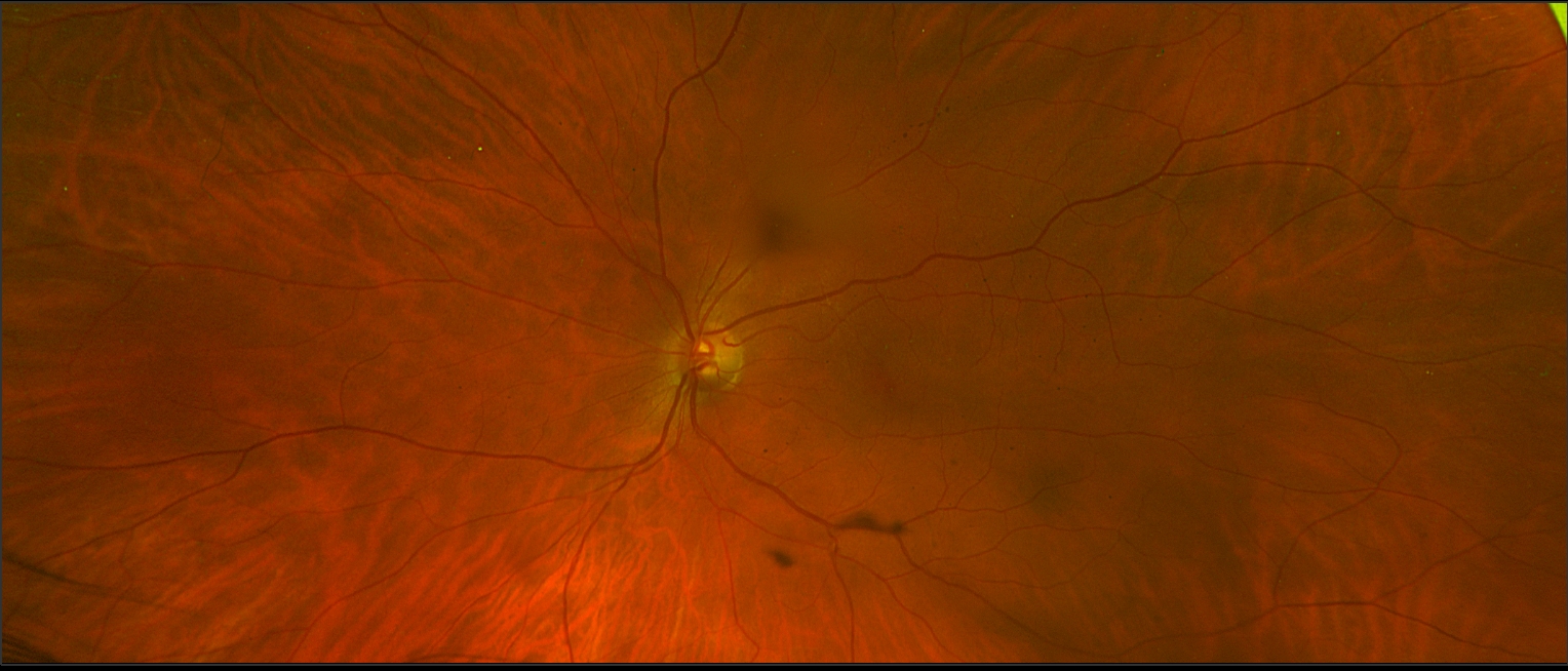
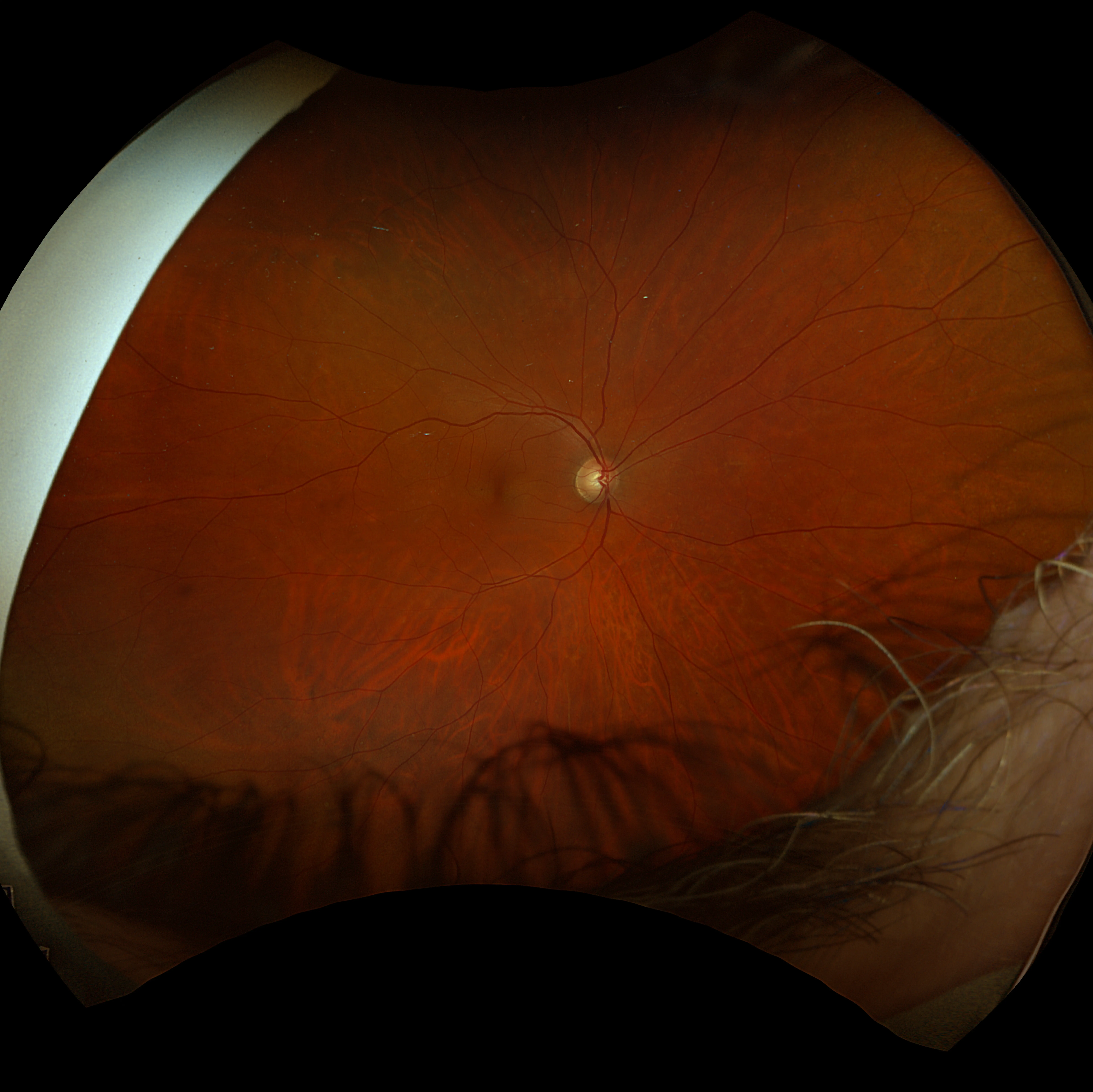
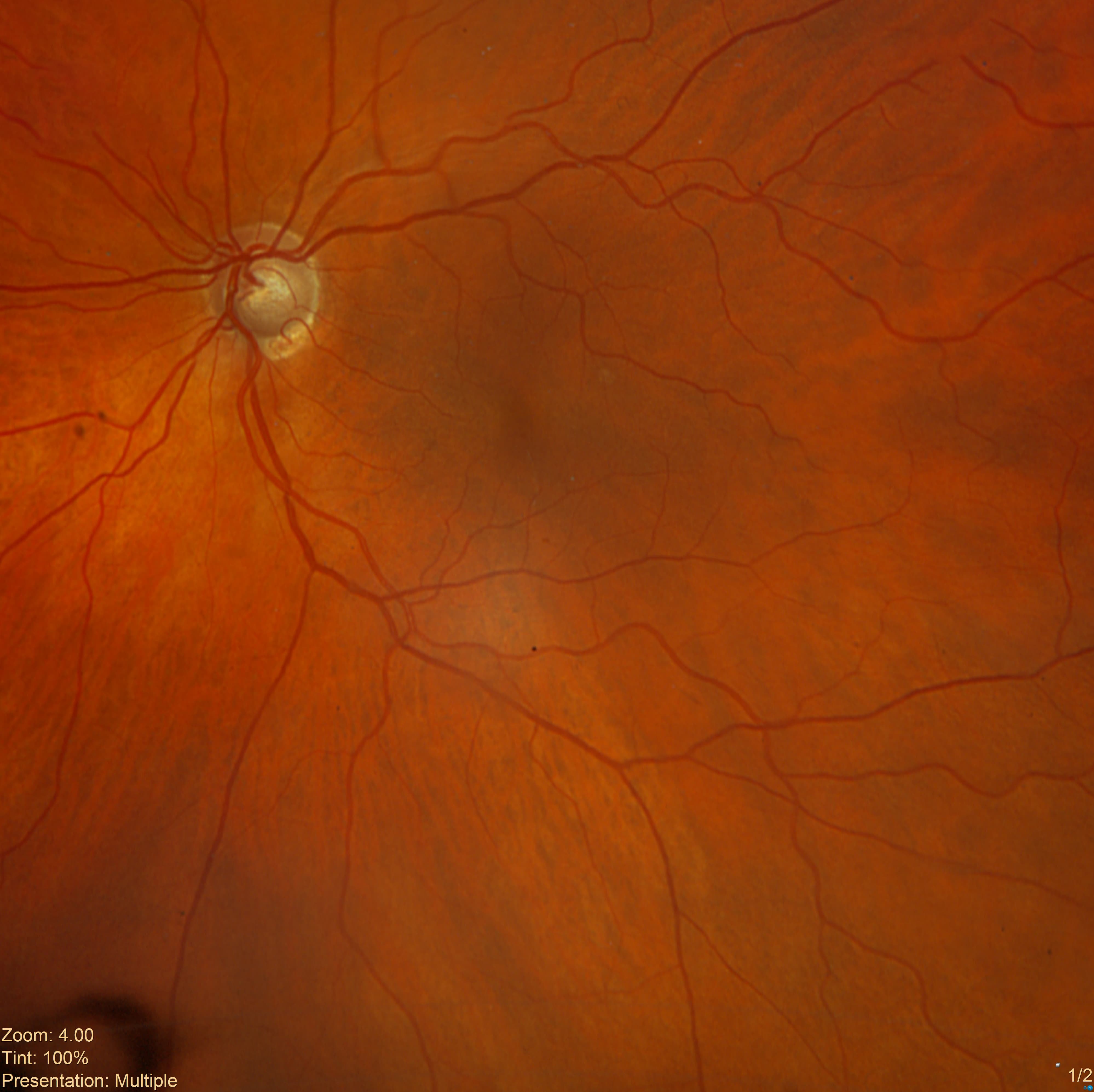
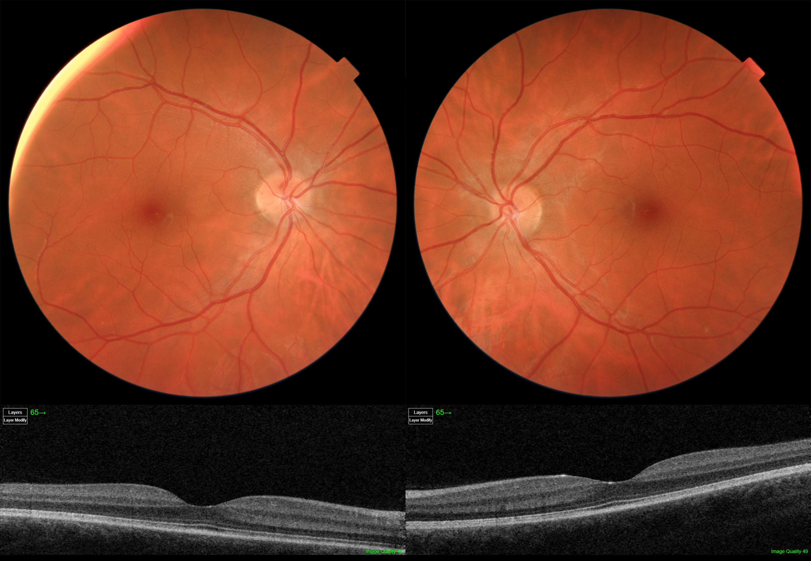
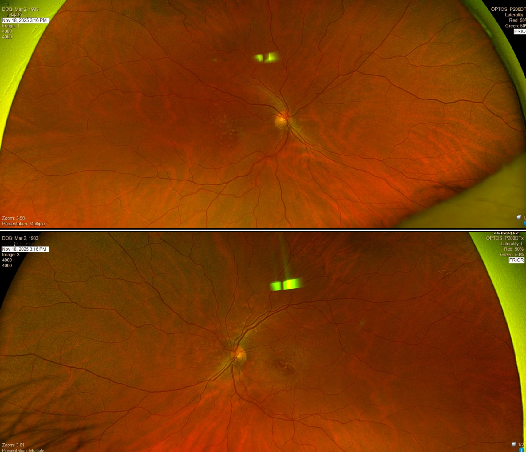
Case Study: Blurred Vision With Macular Elevation
73-year-old with blurred vision and macular elevation on OCT showing central changes and subretinal features requiring further assessment.
Read More
Case Study
|
April 21, 2026
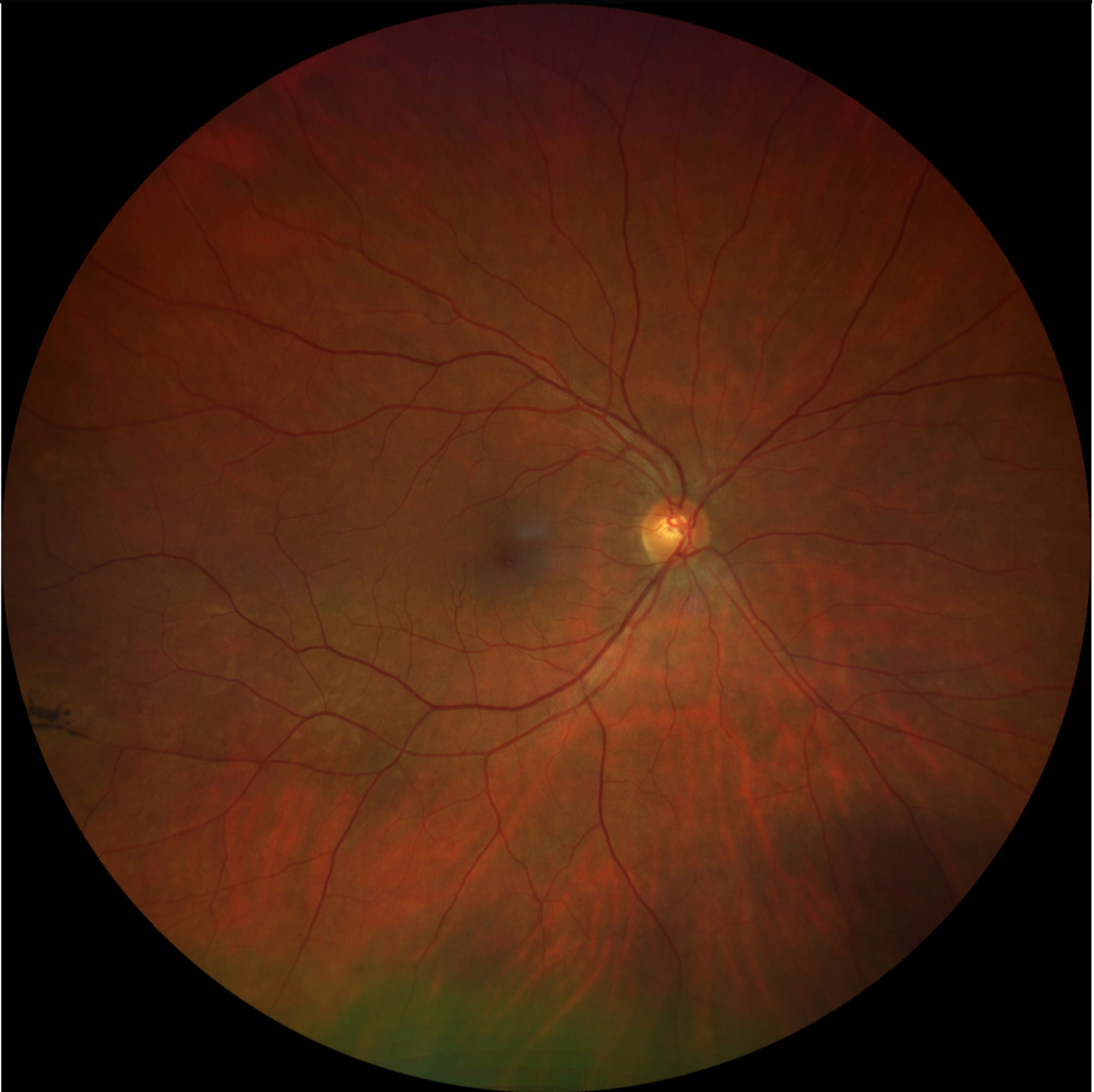
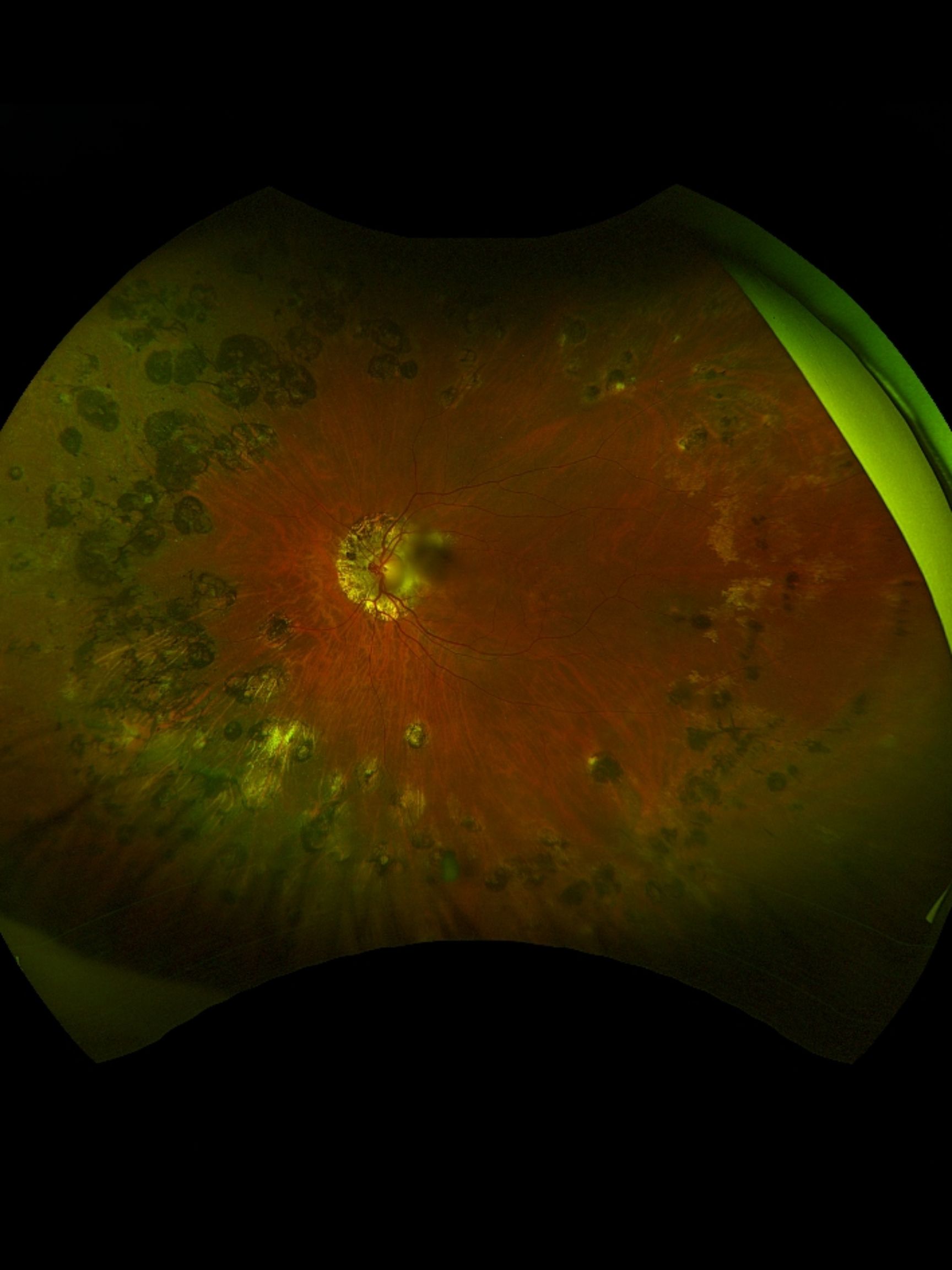
Case Study: Bilateral Drusen in Young Adult
43-year-old with bilateral drusen and normal vision raises concern for early macular changes and genetic risk.
Read More
Case Study
|
April 21, 2026
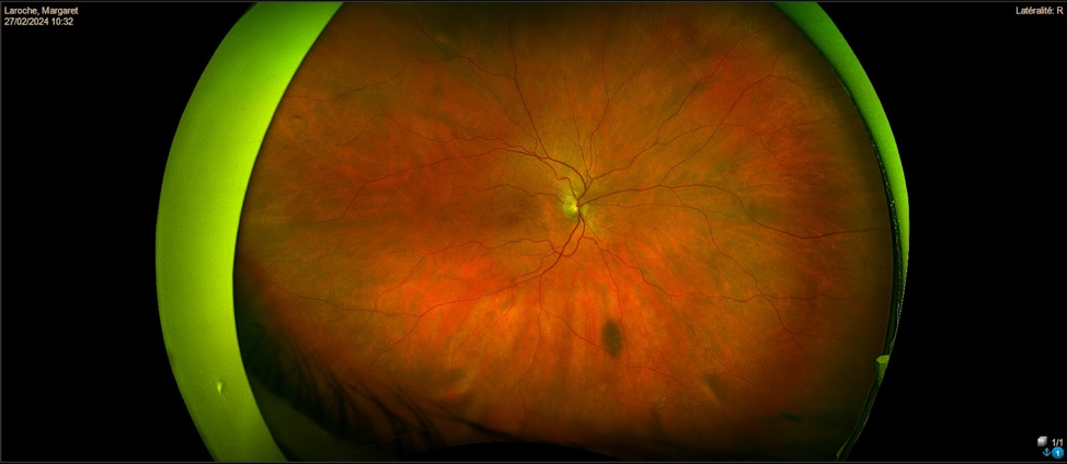
Case Study: Nasal Retinal Change Progression
63-year-old with asymptomatic nasal retinal changes and suspected progression on imaging, raising questions about monitoring vs referral.
Read More
Case Study
|
April 17, 2026
Case Study: Unilateral Retinal Hemorrhages
78-year-old diabetic with unilateral retinal hemorrhages raises concern for vascular occlusion versus retinopathy.
Read More
Case Study
|
April 16, 2026
Case Study: Persistent Floaters Blurred Vision
37-year-old with floaters and blurred vision, high myopia, and vitreous changes requiring specialist evaluation.
Read More
Case Study
|
April 16, 2026
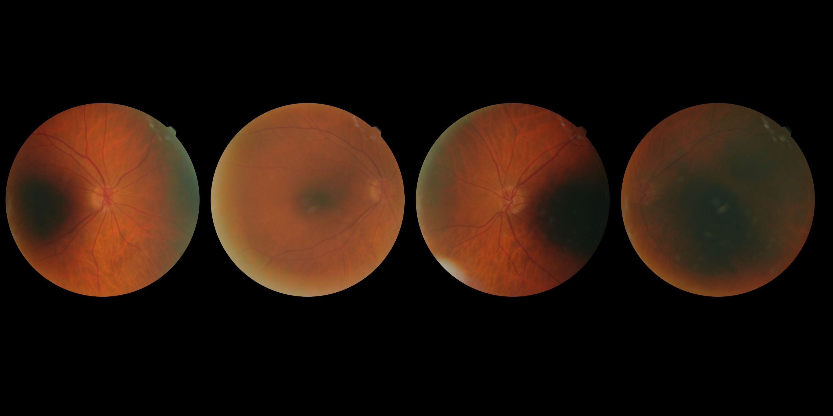
Case Study: Peripheral Retinal Lesion Concern
76-year-old with stable vision and peripheral retinal lesion concern on imaging, asymptomatic, requiring specialist evaluation guidance.
Read More
Case Study
|
April 14, 2026
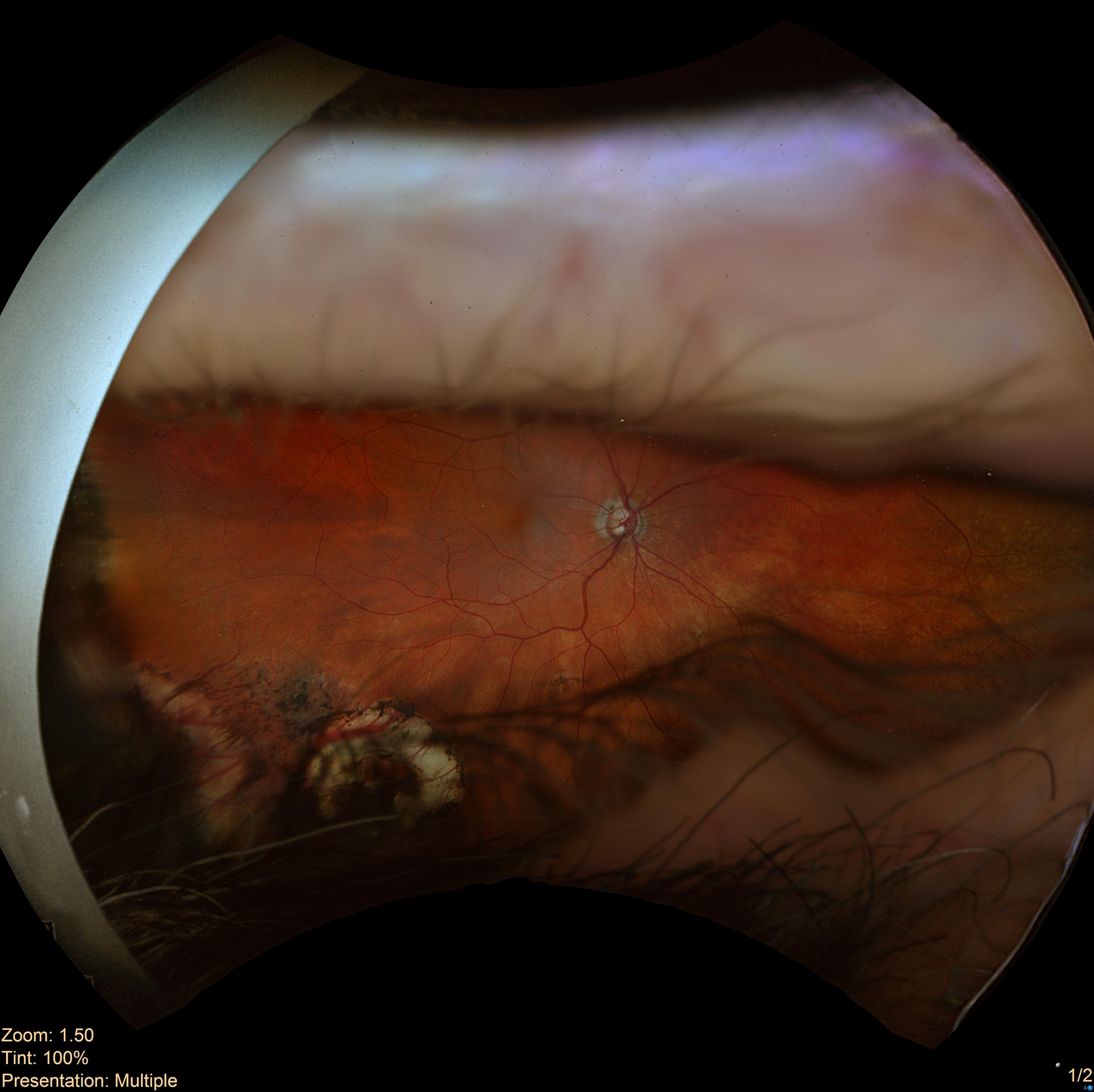
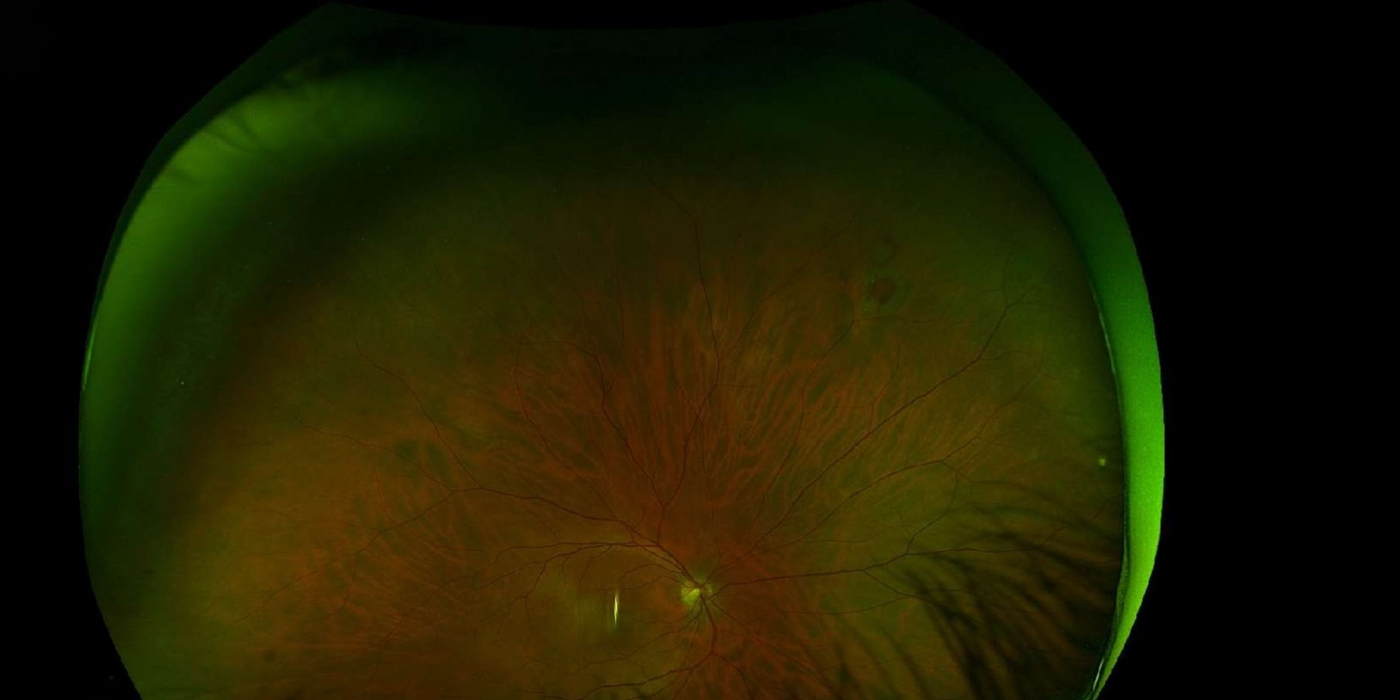
Case Study: Large Temporal Vitreous Tuft
37-year-old male with incidental vitreous traction OS and normal exam findings, raising management and referral considerations.
Read More
Case Study
|
April 14, 2026
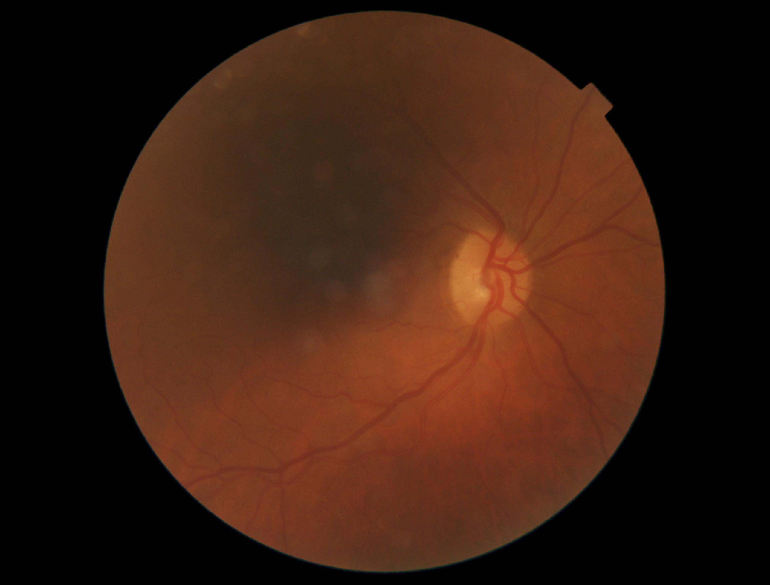
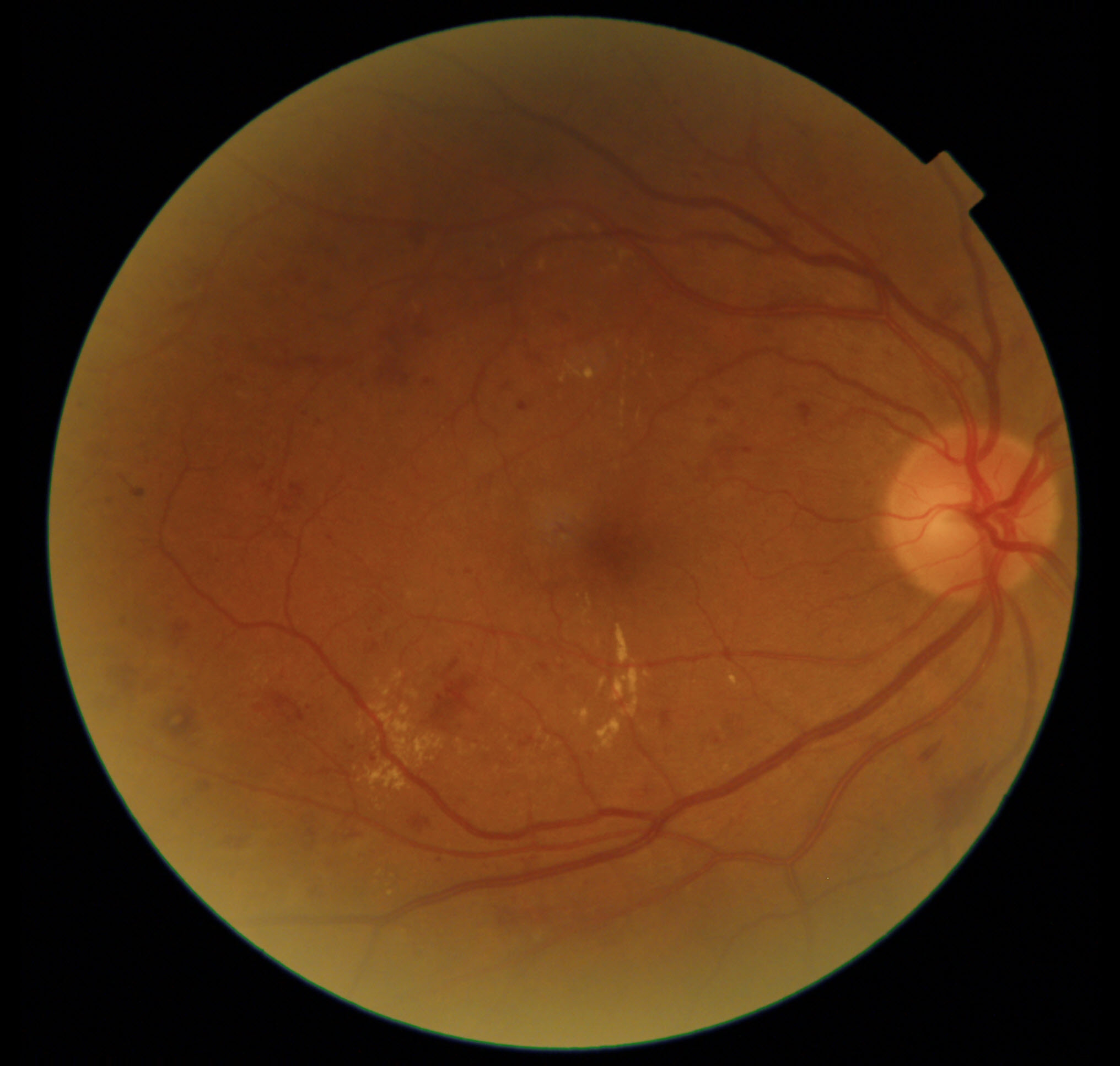
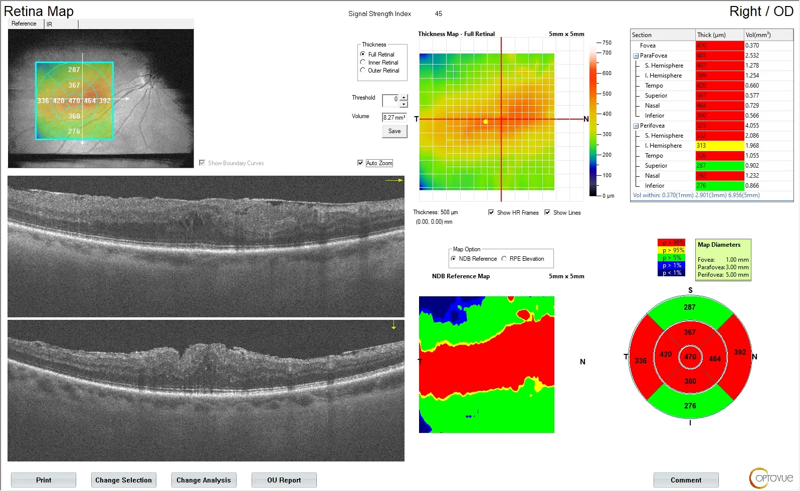
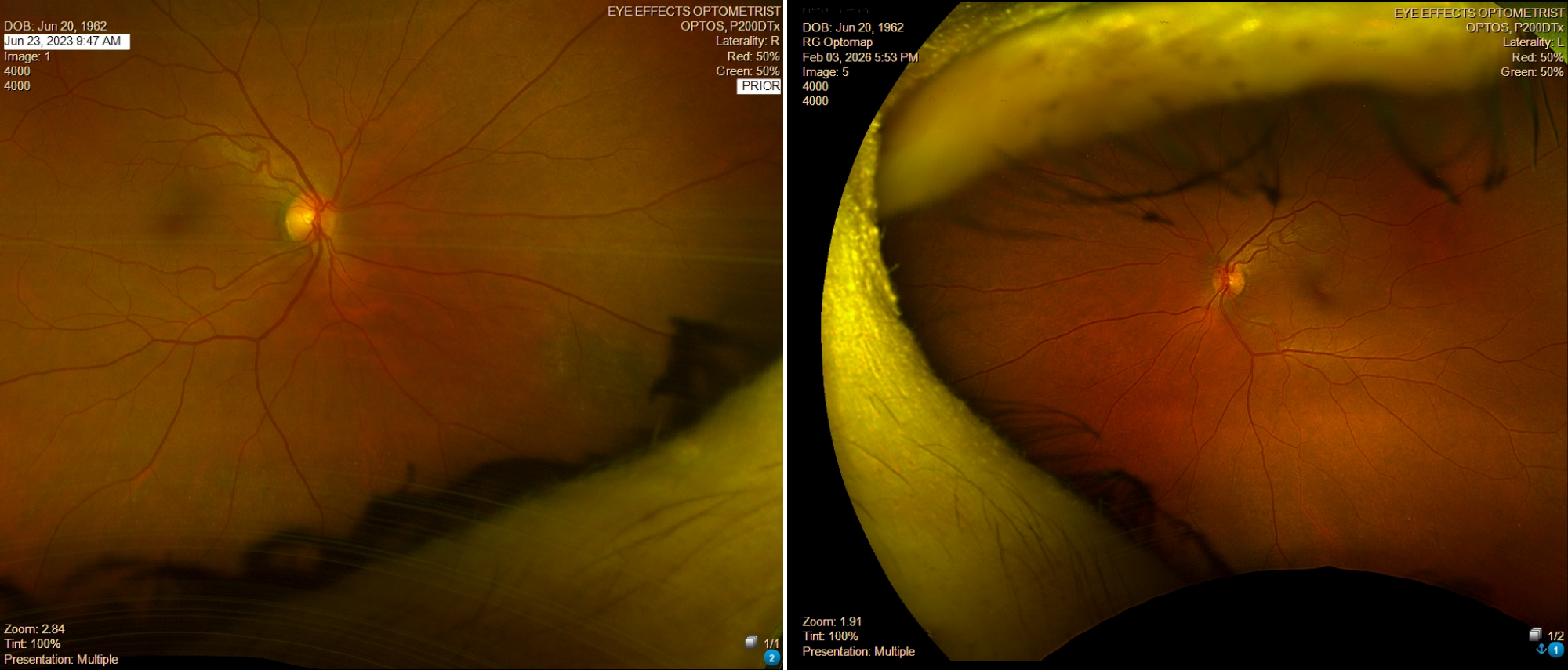
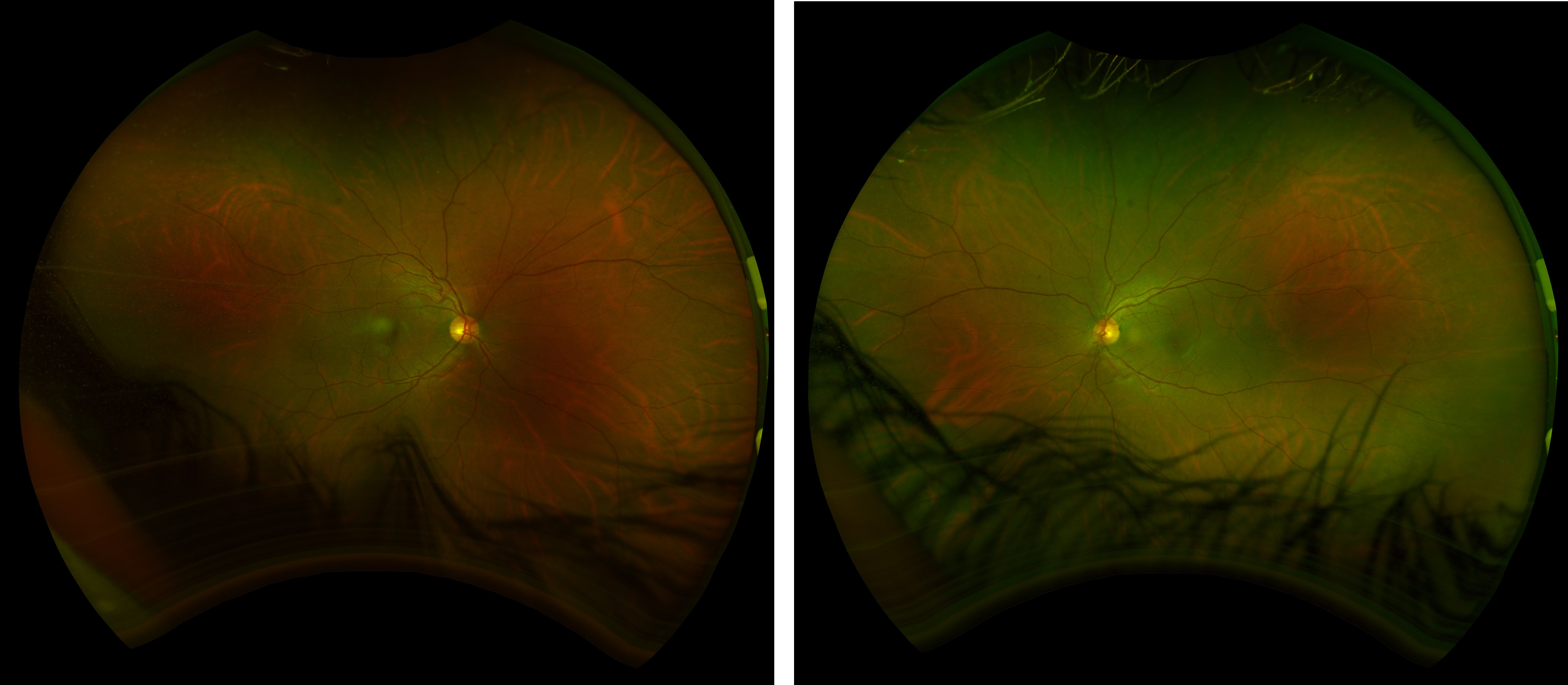
Case Study: Unilateral Macular PED With Edema
66-year-old with unilateral macular PED and reduced vision OS on a background of peripheral retinal changes. Explore evaluation and referral urgency.
Read More
Case Study
|
March 19, 2026
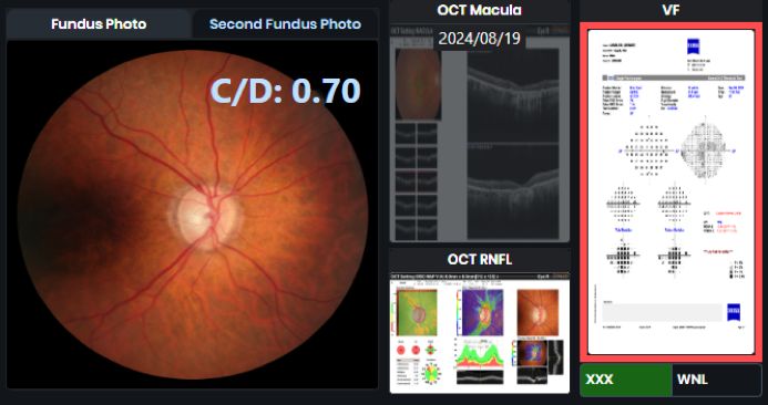
Case Study: Glaucoma Suspect with Strong Family History and Thick Corneas
A 52-year-old patient presents with optic nerve changes and subtle visual field defects; evaluation insights included.
Read More
Case Study
|
March 11, 2026
Case Study: Longstanding Operculated Retinal Hole – Observe or Laser?
48-year-old male with asymptomatic longstanding superonasal operculated retinal hole OD, slightly larger than prior exam; question of prophylactic laser retinopexy.
Read More
Case Study
|
March 11, 2026
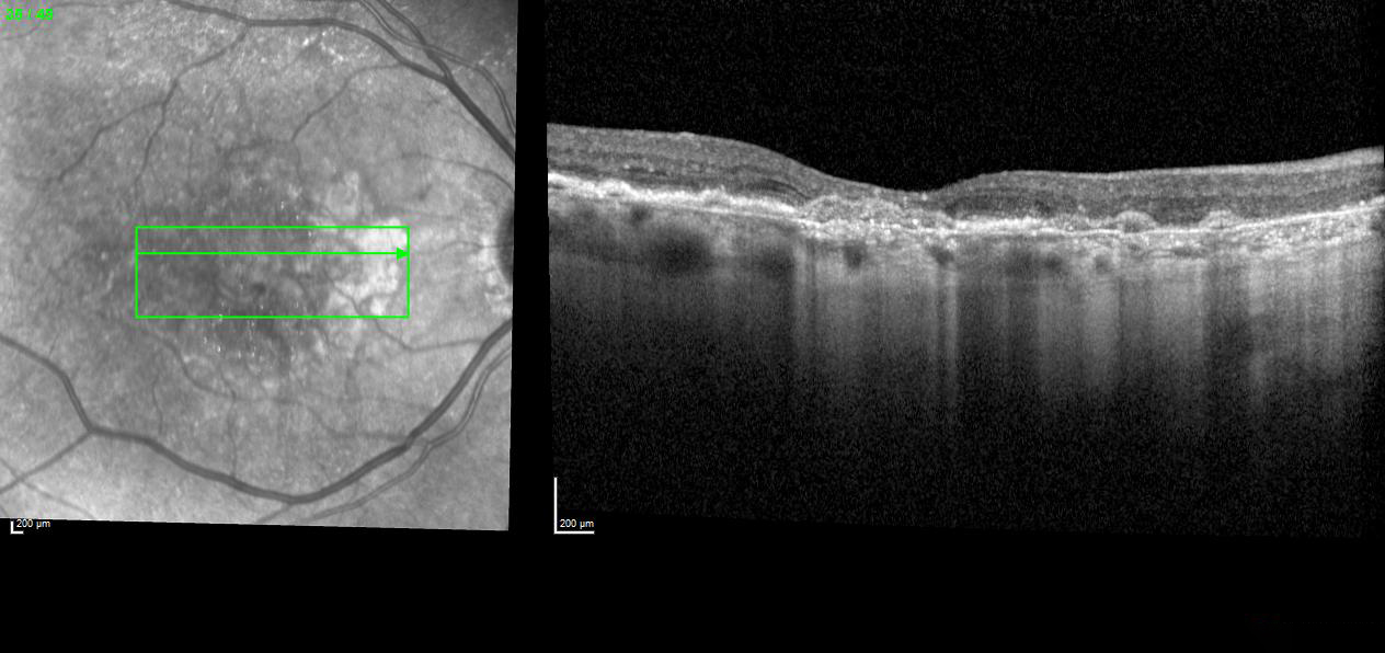
Case Study: Central Serous Retinopathy
A 37-year-old patient presented with central serous retinopathy (CSR); mild subretinal fluid noted, visual acuity minimally affected.
Read More
Case Study
|
March 10, 2026
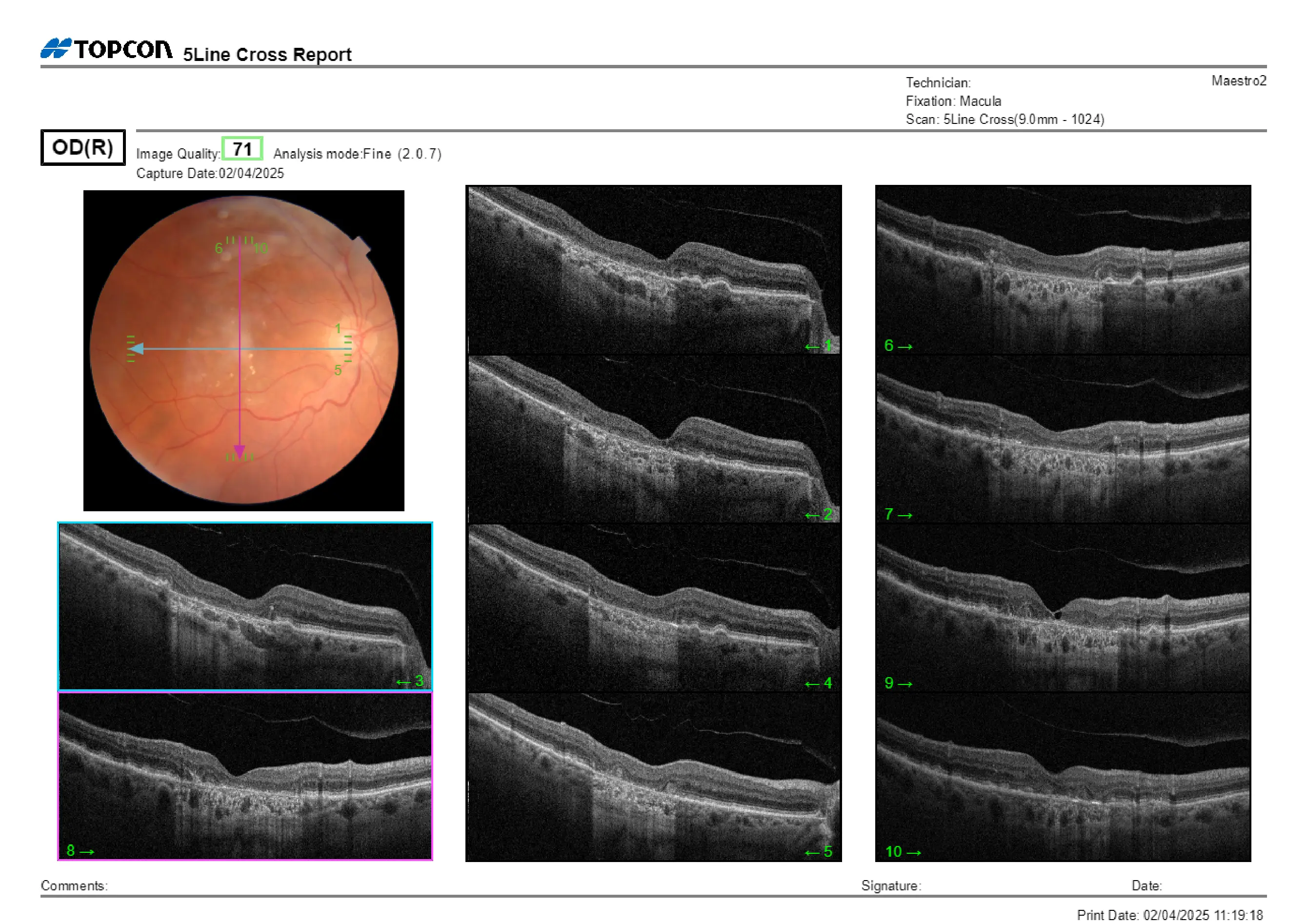
Case Study: Reduced Visual Acuity OD with Foveal Irregularity and Early Cataract
67-year-old female with reduced BCVA OD, mild cataract changes, and foveal irregularity; history of lupus and prior plaquenil exposure.
Read More
Case Study
|
March 10, 2026
Case Study: Temporal Blot Hemorrhage with Possible Microaneurysms
A 46-year-old male with isolated temporal blot hemorrhage OD and possible surrounding microaneurysms; systemic versus myopic etiology considered.
Read More
Case Study
|
February 24, 2026
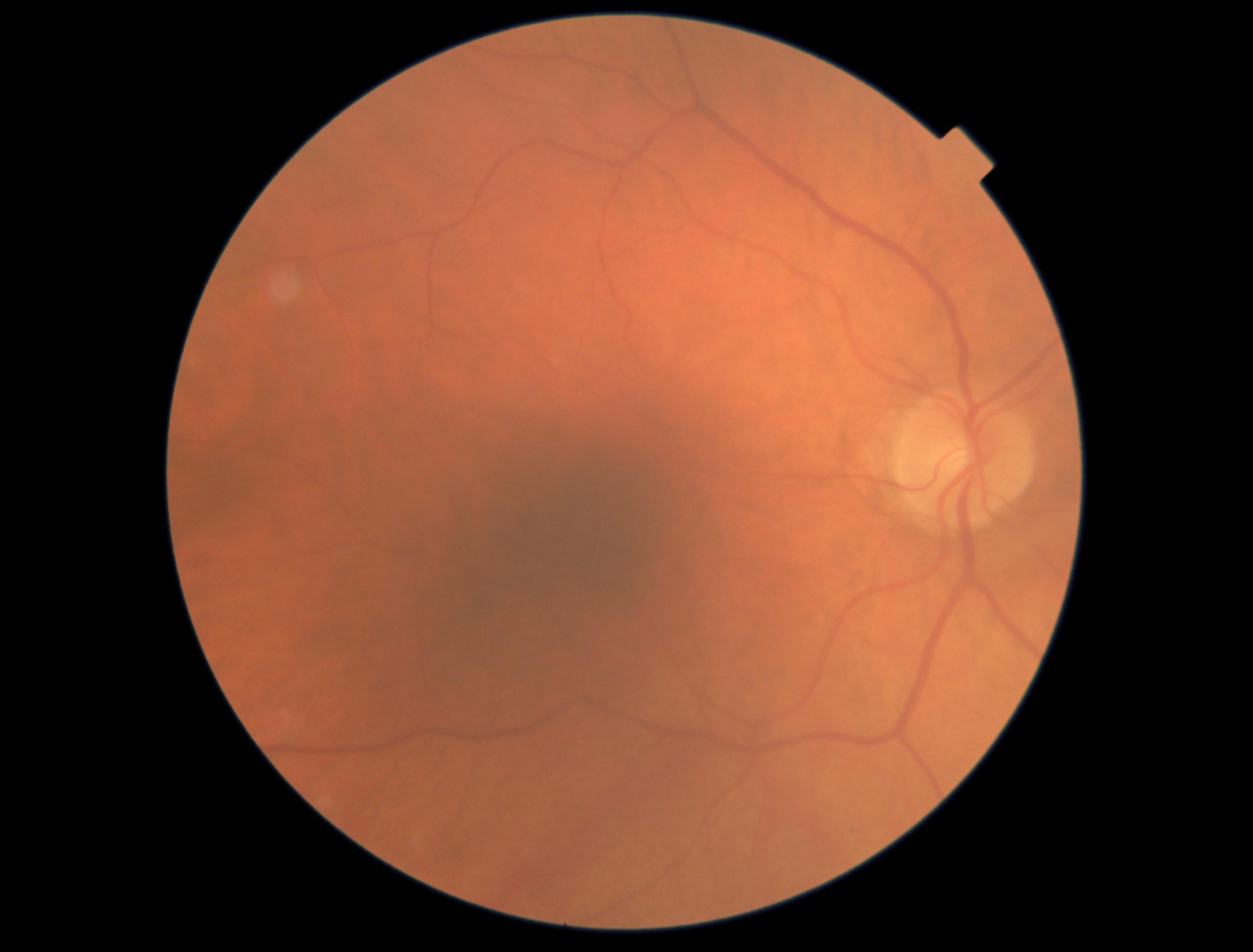
Case Study: Mild Cystic Macular Changes with Good Vision
20/20 vision with mild cystic macular changes; possible occult RVO versus VMT-related change reviewed.
Read More
Case Study
|
February 24, 2026
Case Study: Stable Vision with Lamellar Hole
20/20–20/25 vision with ERM OD and lamellar macular hole OS; observation versus surgery reviewed.
Read More
Case Study
|
February 17, 2026
Case Study: Elevated Eye Pressures and Optic Changes
90-year-old male follow-up shows mild epiretinal changes, optic nerve cupping, and slight RNFL thinning; monitoring interval under review.
Read More
Case Study
|
January 20, 2026
Case Study: Elevated Pressures with Myopic Discs
66-year-old with high eye pressures, myopic discs, mild RNFL thinning, and normal visual fields; monitoring and follow-up decisions under consideration.
Read More
Case Study
|
January 19, 2026
Case Study: Mild Macular Changes
59-year-old male with excellent vision and mild CSDME OS reviewed via Care1 to confirm appropriate observation and follow-up.
Read More
Case Study
|
January 16, 2026
Case Study: Retinal Changes and Vision Monitoring
79F follow-up: Macular changes stable, vision excellent (20/20 OD, 20/25 OS). Is it time for closer monitoring or keep up with Amsler testing?
Read More
Case Study
|
December 31, 2025
Case Study: Large Cupping with Visual Defects
Patient follow-up shows large optic cups and pressure changes; therapy adjusted. Is close monitoring enough or are further changes needed?
Read More
Case Study
|
December 27, 2025
Case Study: Chronic Retinal Changes in Diabetic Patient
60-year-old female with diabetes. Vision stable, scattered macular exudates. Ongoing monitoring recommended. Follow-up interval?
Read More
Case Study
|
December 18, 2025
Case Study: Longstanding Field Defect With Stable Low Pressures
Stable IOP and visual fields with a longstanding defect. Should this 70-YO continue routine monitoring or adjust management?
Read More
Case Study
|
December 11, 2025
Case Study: Vision Changes with Subretinal Atrophy
75-year-old female with liver disease and alcohol history. Vision stable, possible intervention needed. What follow-up interval is recommended?
Read More
Case Study
|
December 3, 2025
Case Study: Stable Optic Nerve with Mild Field Loss
79-year-old female with stable optic nerve and visual field changes. Should current therapy continue, or is adjustment needed?
Read More
Case Study
|
November 20, 2025
Case Study: Progressive Vision Loss and Macular Changes in One Eye
70-year-old with vision loss in one eye shows lens and macular changes; is retina referral needed or safe to monitor?
Read More
Case Study
|
November 17, 2025
Case Study: Monitoring Advanced Macular Changes in an 85-Year-Old Patient
85-year-old with macular changes shows stable vision. When is in-person referral needed versus continued monitoring?
Read More
Case Study
|
November 4, 2025
Case Study: Vision Changes Near the Optic Nerve
Assessing peripapillary atrophy and visual field defects to distinguish stable anatomical changes from active glaucomatous pathology.
Read More
Case Study
|
September 15, 2025
Case Study: Patient With Past Retinal Fluid, Normal Vision Today
Triage and monitoring protocols for asymptomatic patients with a history of subretinal fluid. Ensuring long-term stability via OCT review.
Read More
Case Study
|
September 11, 2025
Case Study: Trauma-Linked Flashes
Critical protocol for screening retinal tears and PVD following blunt ocular trauma. Vital diagnostic triage insights for primary eye care providers.
Read More
Case Study
|
September 11, 2025
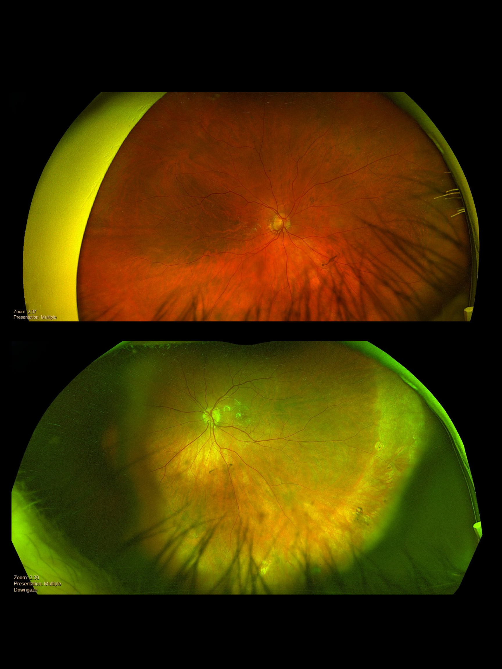
Case Study: Atrophic Scar from Resolved Choroidal Neovascularization (CNV)
Identifying inactive peripapillary CNV scars and implementing long-term monitoring for potential reactivation using multi-modal imaging.
Read More
Case Study
|
September 11, 2025
Case Study: Pigmented Paravenous Retinochoroidal Atrophy (PPRCA)
Clinical review of rare Pigmented Paravenous Retinochoroidal Atrophy, focusing on characteristic venous-linked pigment and retinal thinning.
Read More
Case Study
|
September 11, 2025
Case Study: Peripheral CNVM in 60-Year-Old Female
Managing stable peripheral CNVM and vascular signs in a 60-year-old patient with drusen. Specialist referral guidelines and clinical insights.
Read More
Case Study
|
September 11, 2025
Case Study: Rare Pediatric Case: Optic Nerve Pit
Expert insights on congenital optic nerve pits and preventing vision loss from serous macular detachment in pediatric patients.
Read More
Case Study
|
September 10, 2025
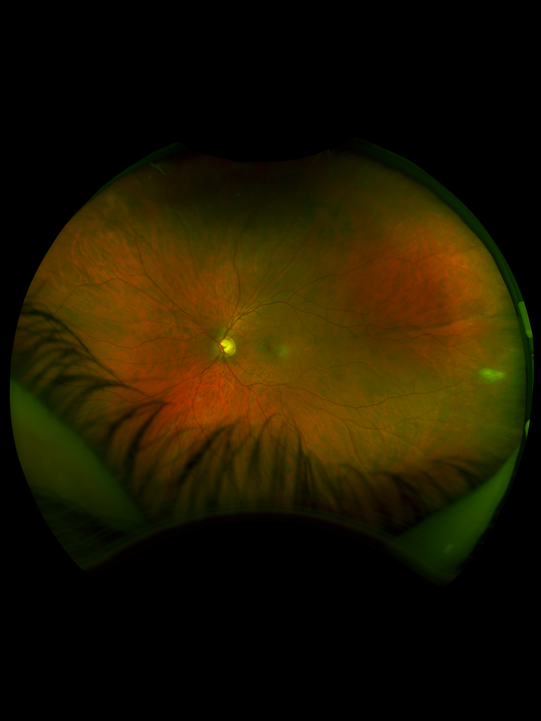
Case Study: Bilateral Macular Edema
Multi-modal imaging analysis of bilateral macular edema to differentiate systemic versus ocular etiologies for precise patient management.
Read More


.png)
.jpg)
.jpg)