75-year-old patient with an isolated retinal flame hemorrhage and normal vision. Explore the clinical workup and referral considerations.
Case Study: Flame Hemorrhage Along Arcade
An optometrist uploaded this case to Care1.
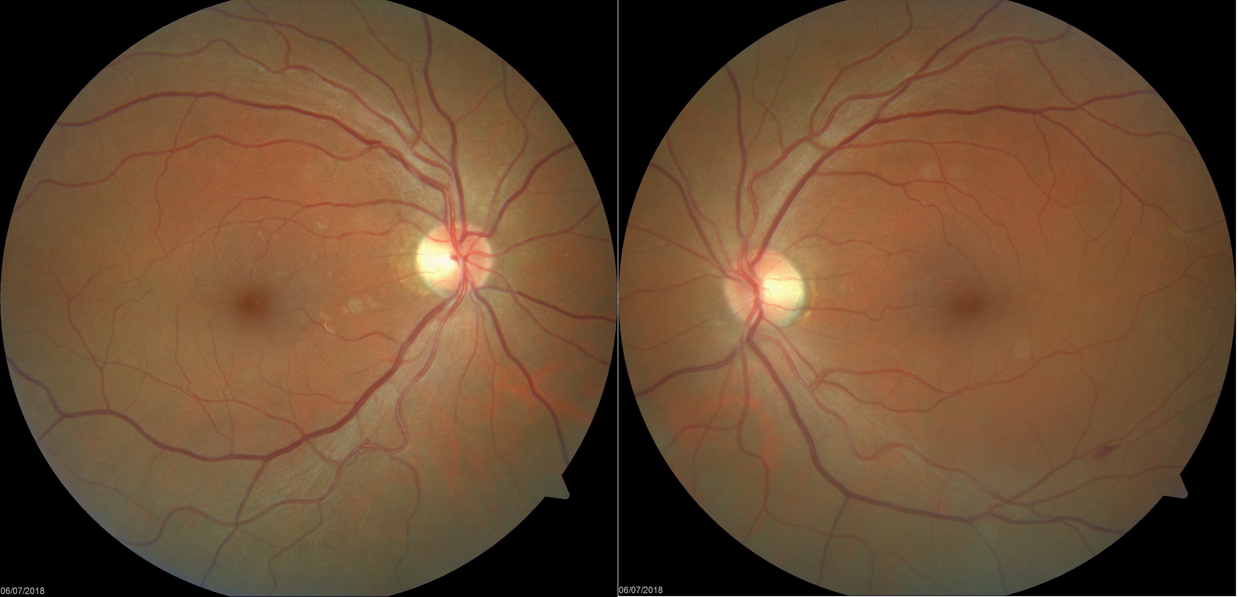
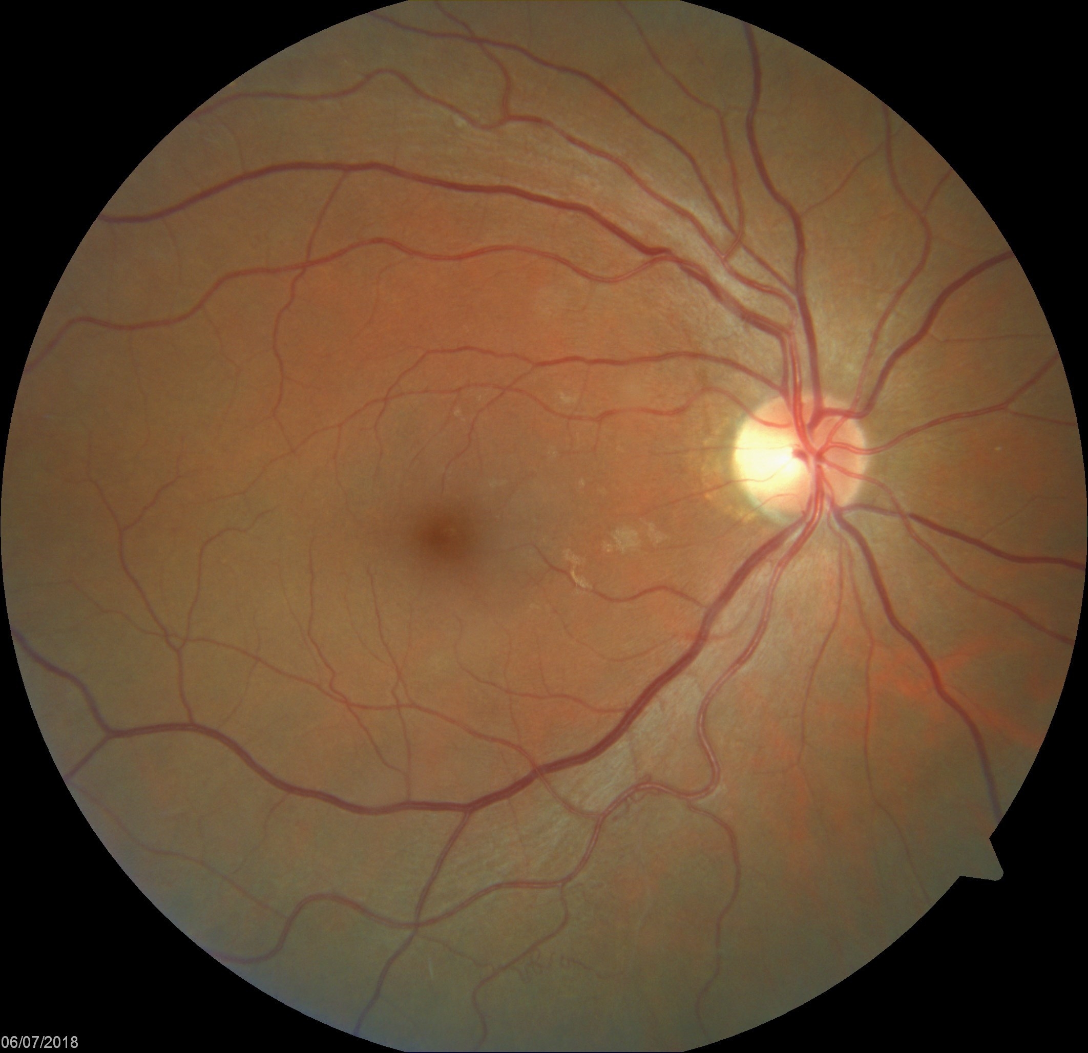
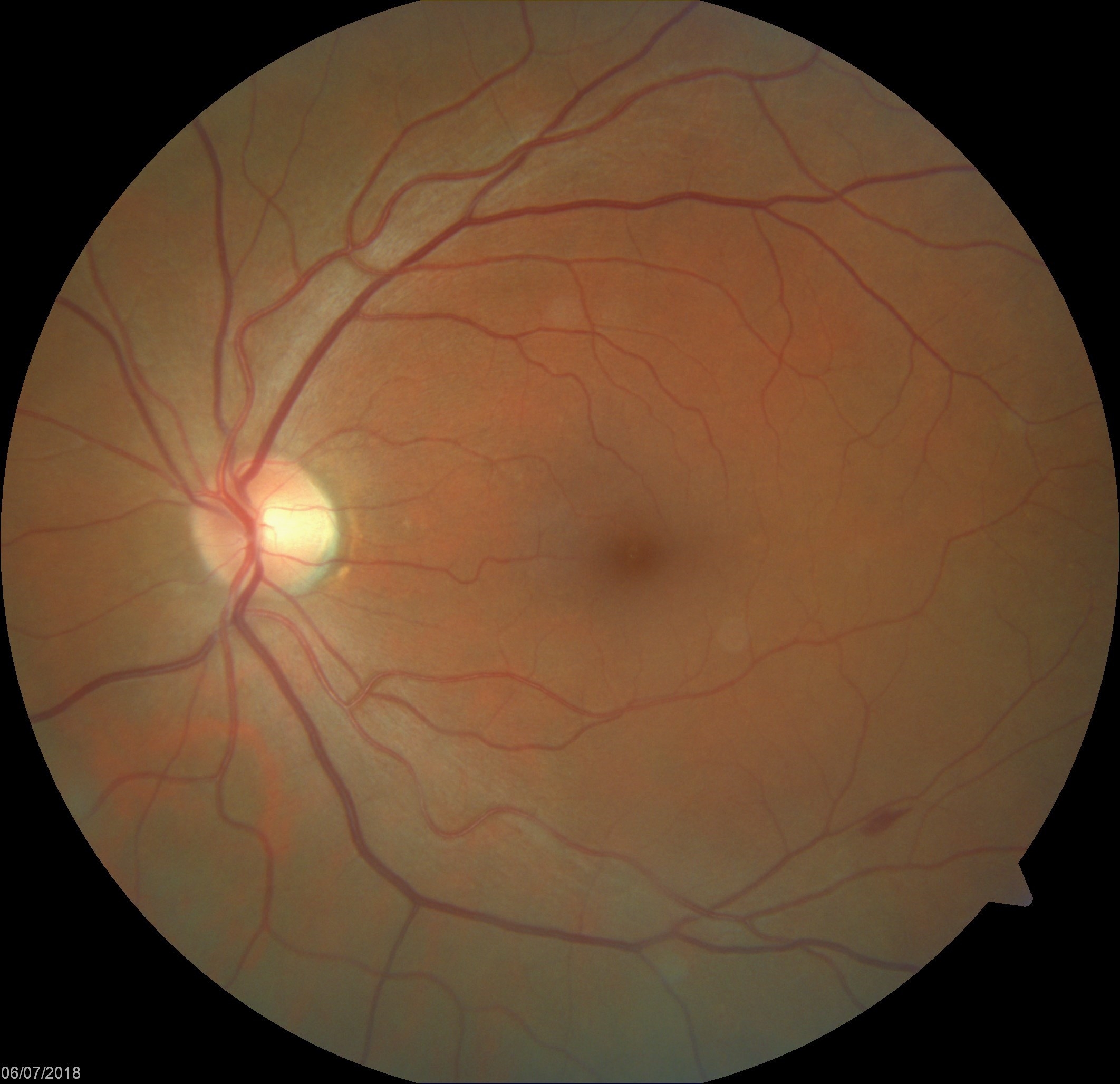
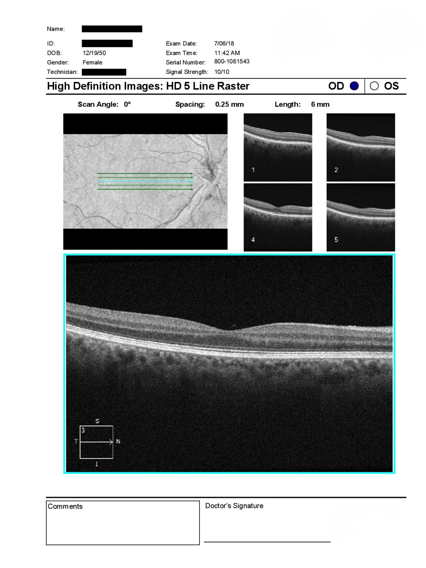
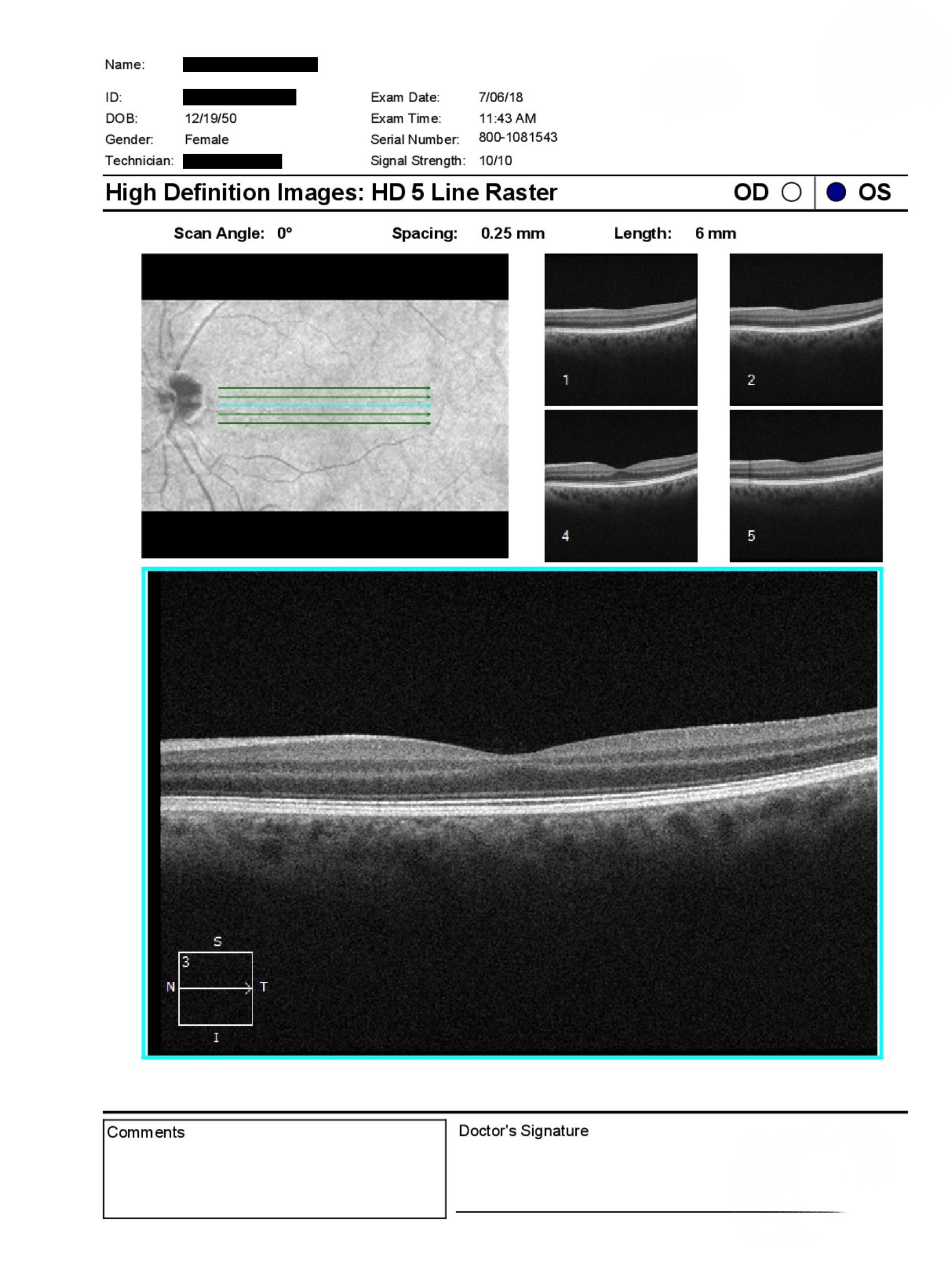
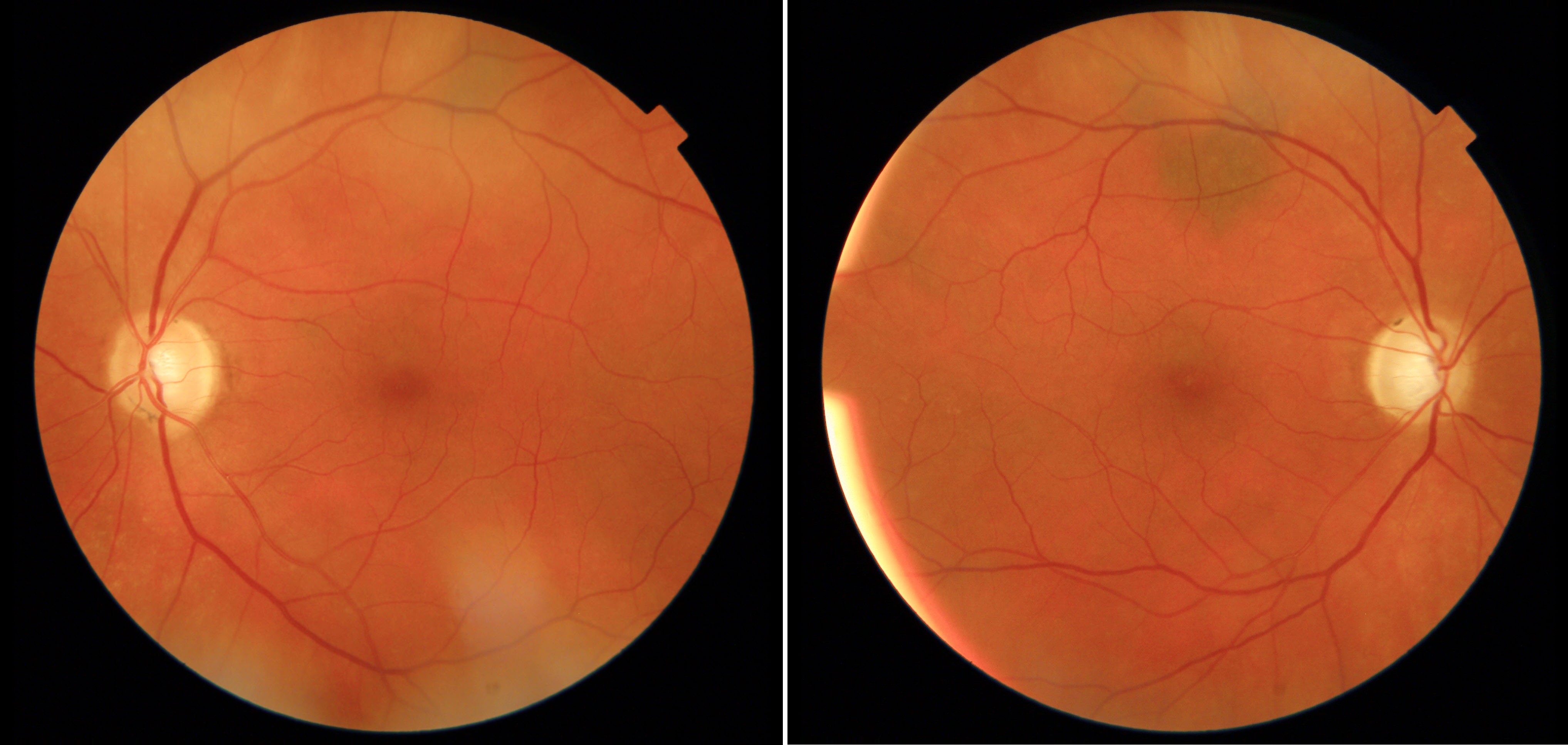
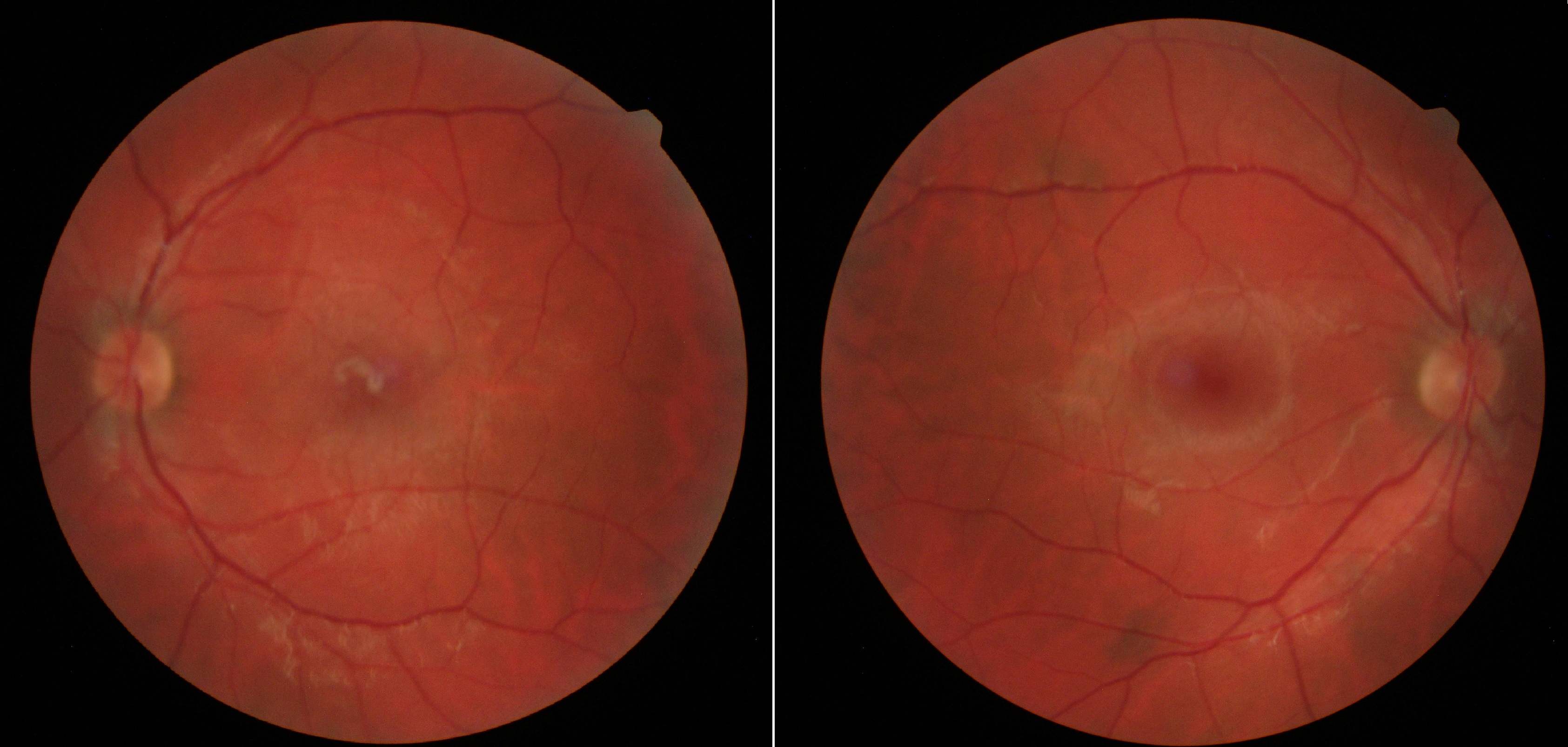
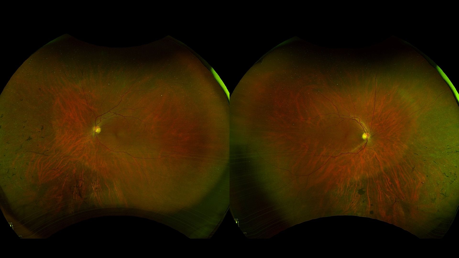
A 75-year-old woman was referred for evaluation of a retinal flame hemorrhage in the left eye. BCVA was 20/20 OU, and IOP measured 16 mmHg OD and 17 mmHg OS. The anterior segment examination showed clear corneas, open angles, and clear posterior chamber intraocular lenses bilaterally. Posterior segment examination was normal OD, with an isolated flame hemorrhage along the inferior arcade OS. Macular OCT was stable.
What additional investigation or management would you recommend for this isolated retinal finding?
A retina specialist provided a virtual consult within 1-2 weeks through Care1. Scroll below to see their diagnosis.
Care1 Ophthalmologist's Teleconsult
The macular OCT remained stable, with no evidence of structural retinal abnormalities. In the setting of an isolated retinal hemorrhage and otherwise reassuring ocular findings, observation was considered appropriate. Because retinal hemorrhages may reflect underlying systemic vascular disease, communication with the patient's primary care physician regarding assessment of vascular risk factors, including hypertension and diabetes, was recommended. Routine follow-up with the referring optometrist was advised.
Care1 AI's Clinical Insight
Retinal flame-shaped hemorrhages occur within the retinal nerve fiber layer and are commonly associated with retinal vascular disorders or systemic vascular disease. Careful clinical evaluation includes assessment of retinal location, associated retinal findings, and systemic risk factors to help determine the underlying cause. Recognition of isolated retinal hemorrhages is an important component of comprehensive retinal examination.
Practice at the highest level of medicine
✔ Specialist consults within 1-2 weeks ✔ AI-powered clinical support ✔ Greater confidence in complex decision making
Retinal nerve fiber layer hemorrhages often align with the orientation of the nerve fiber bundles, producing their characteristic flame-shaped appearance and helping distinguish them from deeper retinal hemorrhages.