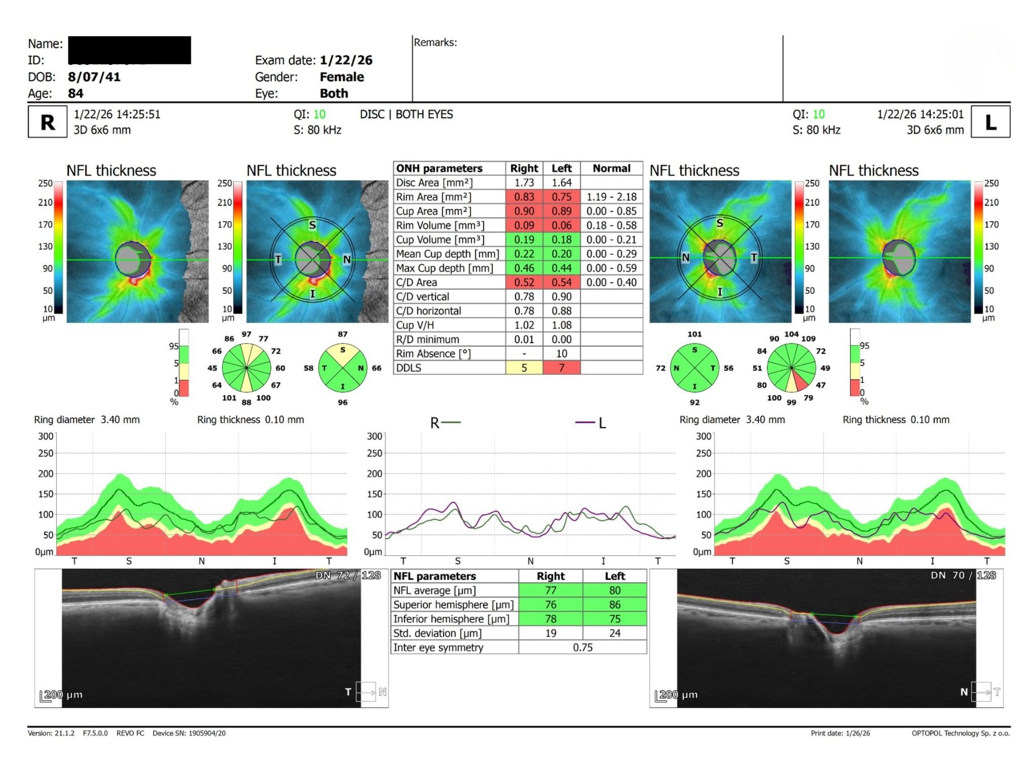
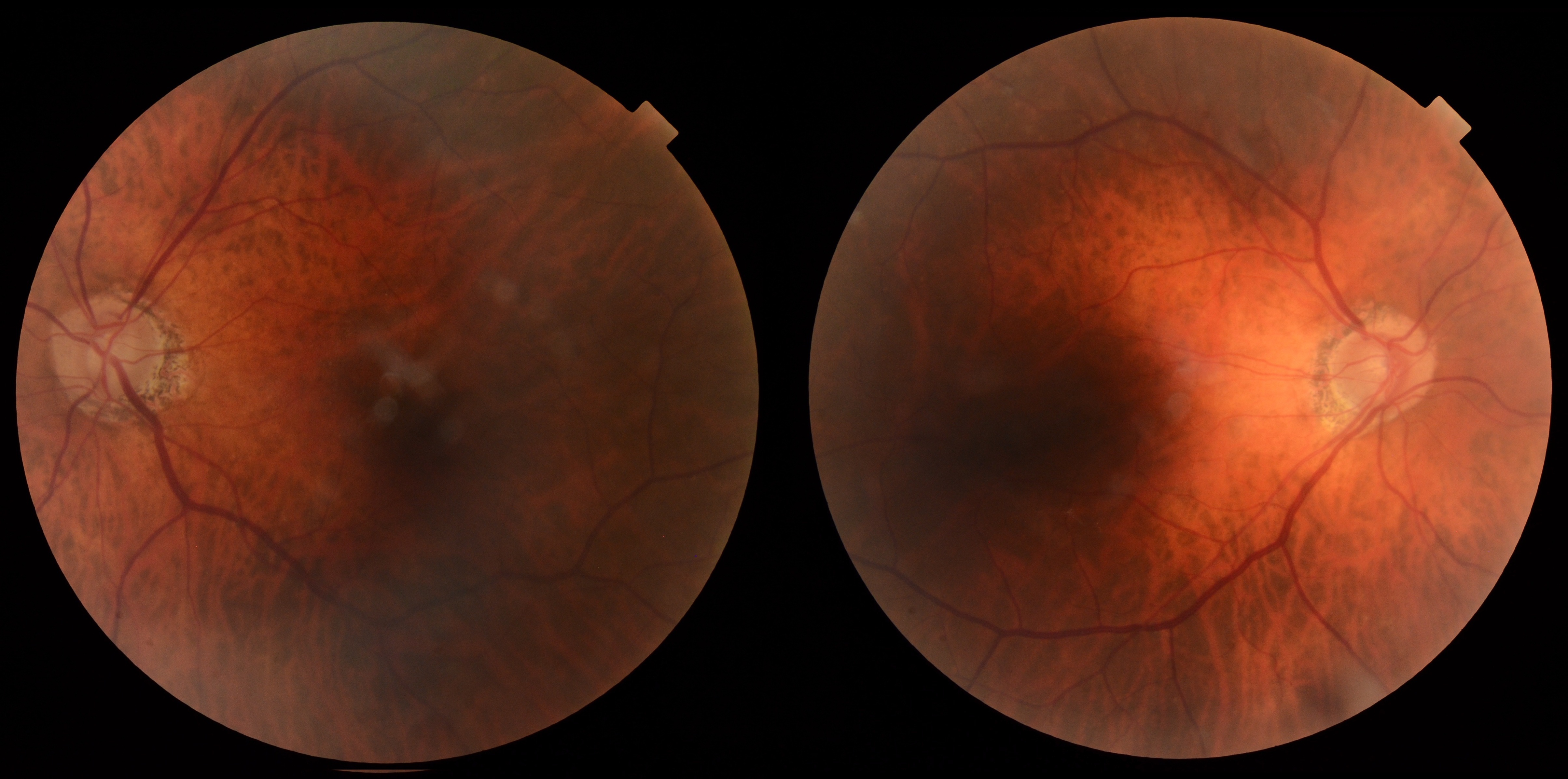
84-year-old female with large cup-to-disc ratios, normal OCT RNFL and visual fields, and stable intraocular pressures under observation.
Case Study: Large Cupping With Normal Testing
An optometrist uploaded this case to Care1.
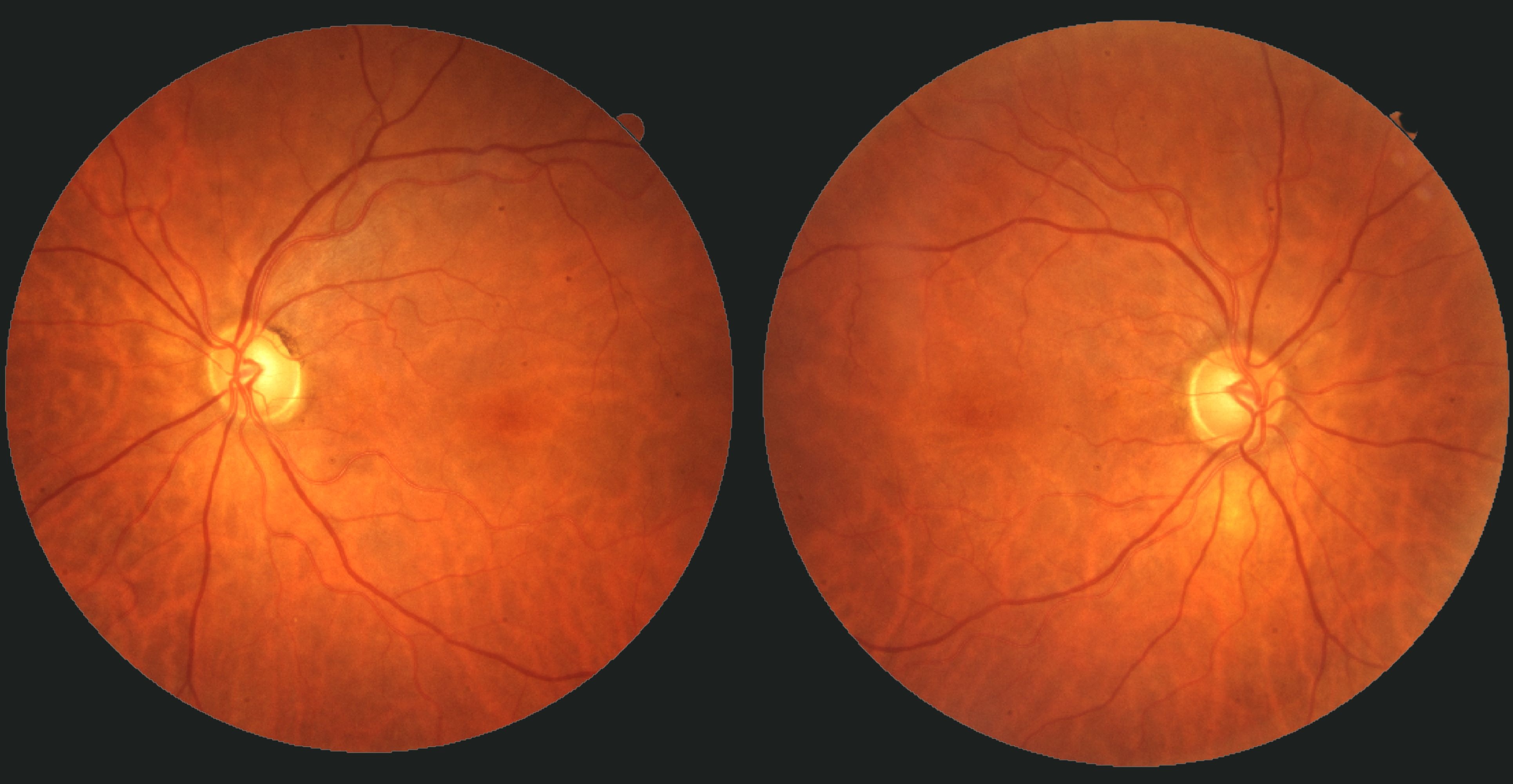
An 84-year-old female was referred as a glaucoma suspect due to large cup-to-disc ratios. BCVA was 20/20 OU with IOPs of 18 mmHg OU and thick corneas (571/572 μm). Anterior segment examination showed pseudophakia, mild meibomian gland dysfunction, conjunctivochalasis, and papillary allergic changes. Posterior segment examination revealed healthy optic nerves with intact neuroretinal rims, cup-to-disc ratios of approximately 0.5 to 0.6, normal OCT RNFL findings, and full visual fields.
In a patient with large cup-to-disc ratios but normal OCT RNFL, visual fields, and IOPs, is observation alone appropriate or should follow-up be intensified?
An ophthalmology subspecialist provided a virtual consult within 1-2 weeks through Care1. Scroll below to see their diagnosis.
Care1 Ophthalmologist's Teleconsult
The optic nerves demonstrated moderate physiologic cupping without evidence of structural or functional progression. OCT RNFL imaging and visual field testing were within normal limits in both eyes, while central corneal thickness measurements were above average, reducing concern for occult pressure-related damage.
Although the cup-to-disc ratios were relatively large, the combination of normal imaging, full visual fields, stable intraocular pressures, and advanced age supported a low-risk glaucoma suspect profile. Continued monitoring with repeat OCT RNFL, visual field testing, and optic nerve assessment was recommended to confirm long-term stability.
Care1 AI's Clinical Insight
"Physiologic Optic Disc Cupping"
Large cup-to-disc ratios can occur as a normal anatomic variation, particularly in patients with larger optic discs. According to the AAO EyeWiki discussion of primary open-angle glaucoma differential diagnosis, physiologic cupping is characterized by large, often symmetric cups without corresponding retinal nerve fiber layer loss or visual field defects. Careful assessment of neuroretinal rim integrity and longitudinal follow-up are essential when differentiating physiologic cupping from glaucomatous optic neuropathy.
Unlock your full revenue potential
✔ Specialist consults within 1-2 weeks ✔ AI-powered clinical support ✔ Greater confidence in complex decision making
In the Ocular Hypertension Treatment Study (OHTS), every 40 μm decrease in central corneal thickness was associated with a substantially higher risk of developing primary open-angle glaucoma, highlighting the powerful protective significance of thick corneas when interpreting intraocular pressure measurements and glaucoma risk.
Reference: Gordon MO, Beiser JA, Brandt JD, et al. The Ocular Hypertension Treatment Study: Baseline factors that predict the onset of primary open-angle glaucoma. Arch Ophthalmol. 2002;120(6):714-720. doi:10.1001/archopht.120.6.714

.jpg)
.jpg)