76-year-old with stable vision and peripheral retinal lesion concern on imaging, asymptomatic, requiring specialist evaluation guidance.
Case Study: Peripheral Retinal Lesion Concern
An optometrist uploaded this case to Care1.
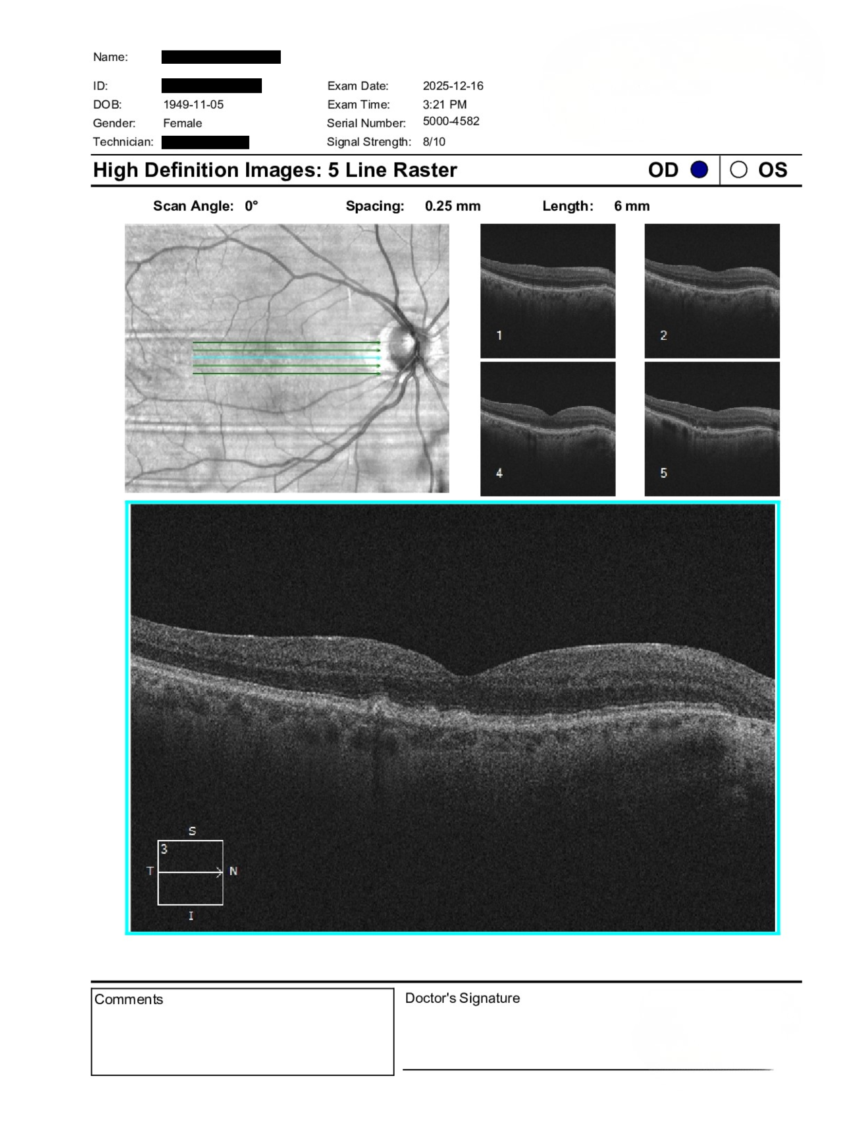
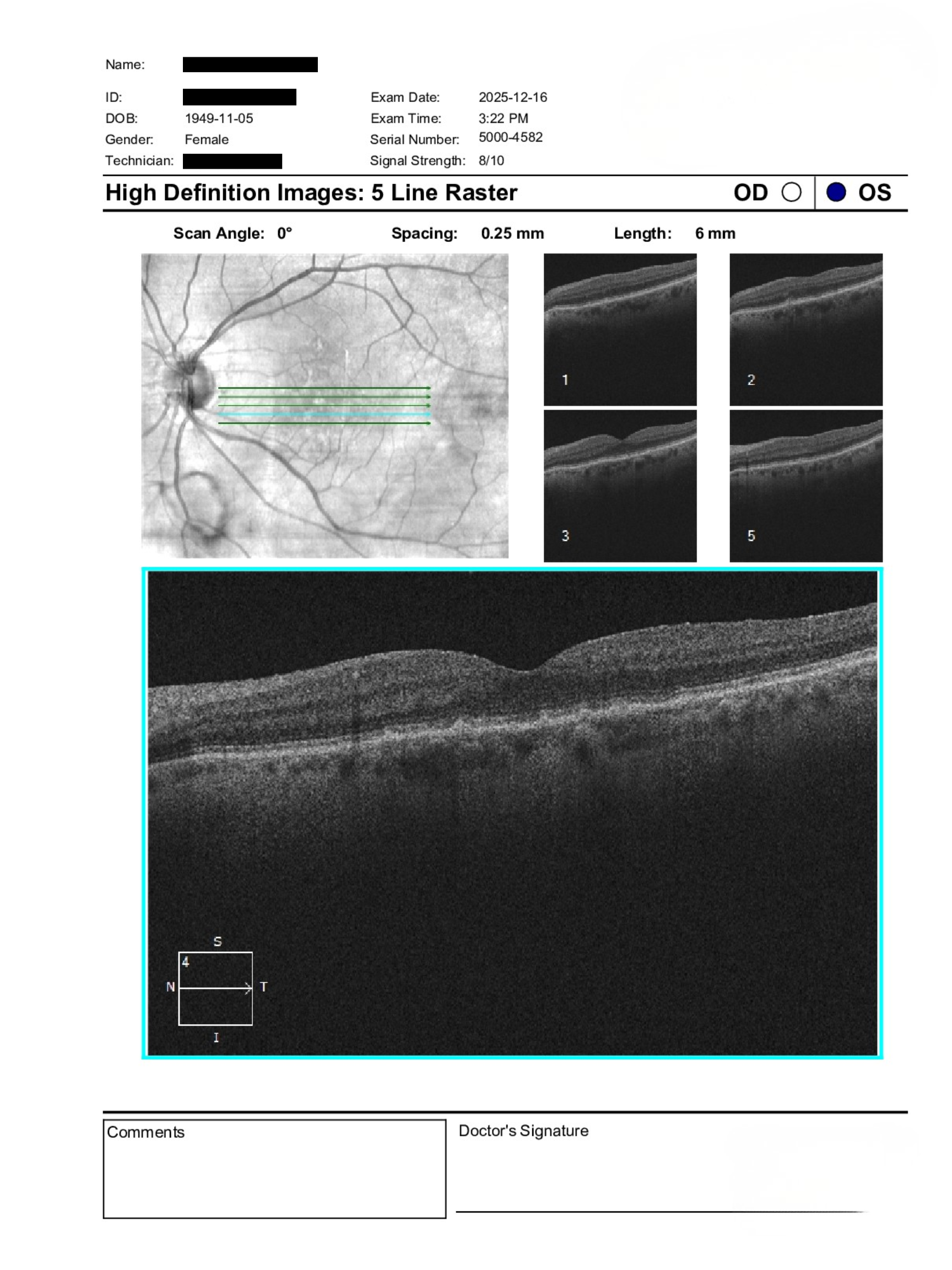
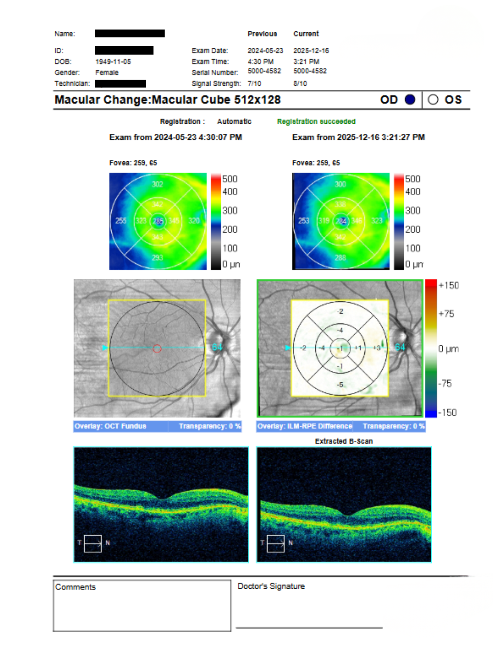
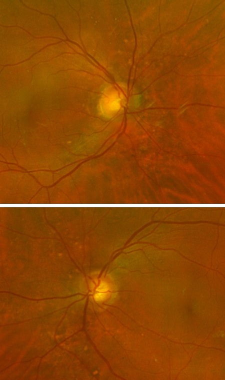
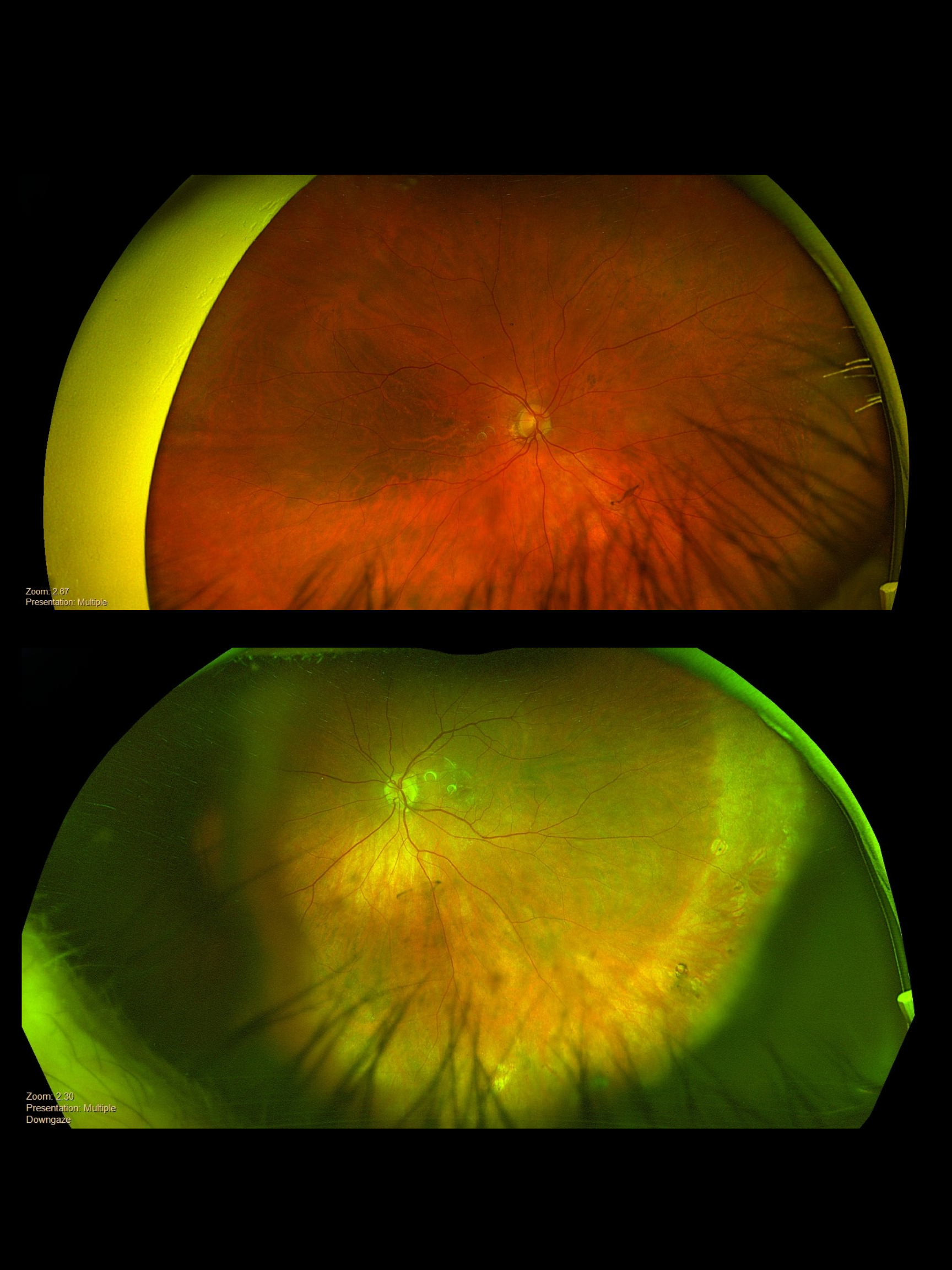
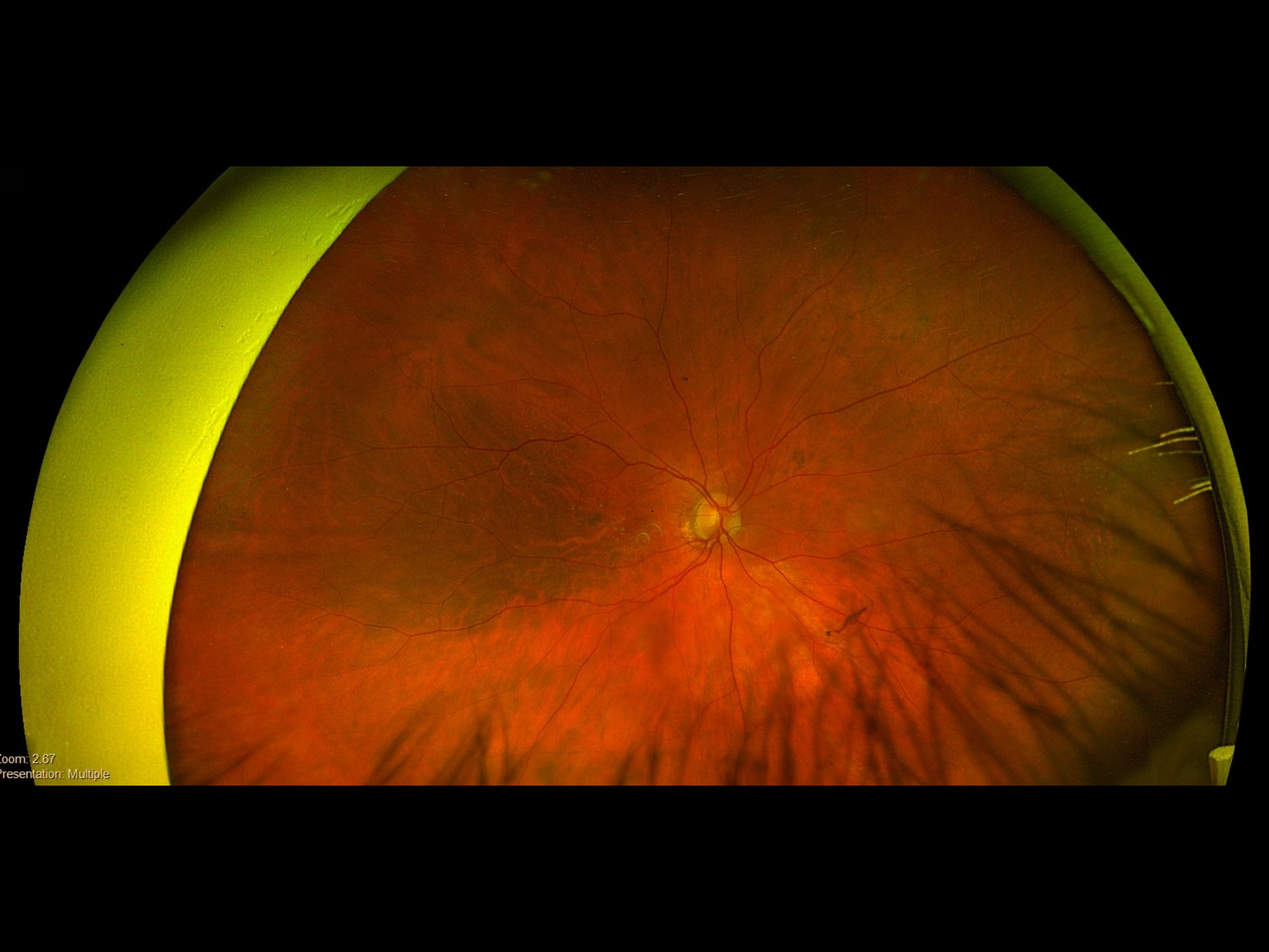
76-year-old female referred for evaluation of a suspected peripheral retinal lesion in the left eye noted on routine exam. BCVA is 20/30 OD and 20/40 OS. IOP measures 19 mmHg OD and 17 mmHg OS. Anterior segment is unremarkable with clear PCIOLs OU. Posterior segment shows stable drusen OU with no macular fluid on OCT. A questionable far peripheral lesion is noted in the left eye at 4–5 o’clock without symptoms of flashes or floaters.
Does the peripheral OS lesion require in-person retinal assessment or can it be safely monitored?
A retina specialist provided a virtual consult within 1-2 weeks through Care1. Scroll below to see their diagnosis.
Care1 Ophthalmologist’s Teleconsult
Imaging review suggests a likely peripheral retinal break in the left eye. Although the patient is currently asymptomatic, peripheral breaks in this location may carry a higher risk profile depending on morphology and vitreoretinal traction status.
There is no evidence of macular involvement or exudative change on OCT, and the posterior pole findings remain stable with dry age-related changes. Given the uncertainty on peripheral visualization and potential risk considerations, in-person retinal assessment is recommended to confirm the diagnosis and discuss management options, including observation versus prophylactic treatment.
Care1 AI’s Clinical Insight
Peripheral retinal breaks often occur at areas of vitreoretinal adhesion and can be asymptomatic, especially when associated with posterior vitreous detachment. Risk of progression depends on lesion type, size, and associated traction. Careful dilated peripheral examination with scleral depression remains the gold standard for detection. Optical imaging may miss subtle far peripheral pathology, making clinical correlation essential.
Practice at the highest level of medicine
✔ Specialist consults within 1-2 weeks ✔ AI-powered clinical support ✔ Greater confidence in complex decision making
Asymptomatic horseshoe-shaped retinal breaks have a low but non-zero risk of progressing to retinal detachment, particularly in the presence of vitreoretinal traction. Long-term observational studies suggest most remain stable without intervention.
Reference: Byer NE. Natural history of asymptomatic retinal breaks. Ophthalmology. 1989;96(9):1428–1434.
Clinical Pearls
Peripheral lesions may need careful scleral evaluation

.png)