36-year-old with headaches, dizziness, and peripheral retinal findings despite normal OCT and visual field testing.
Case Study: Peripheral Exudate and Headaches
An optometrist uploaded this case to Care1.
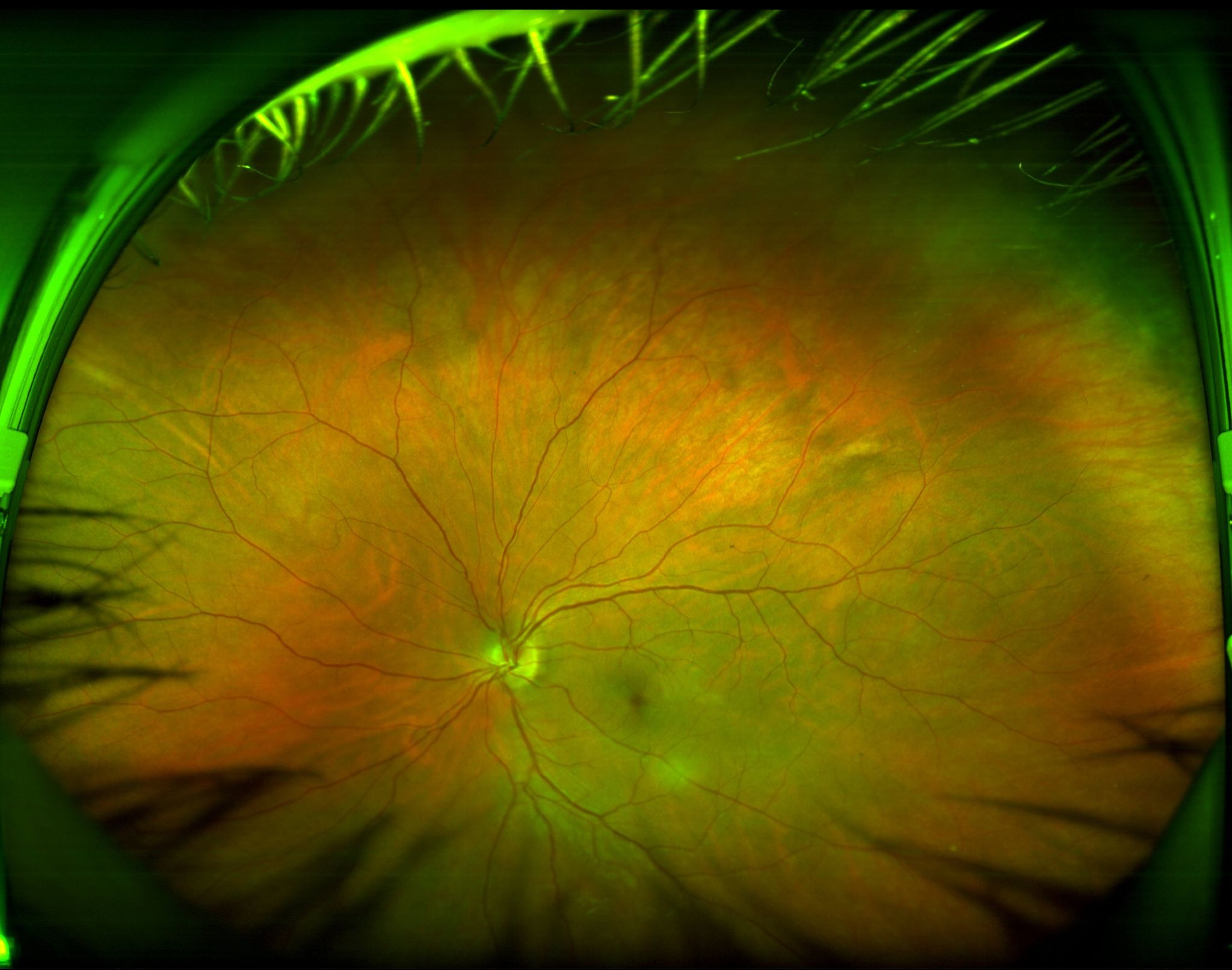
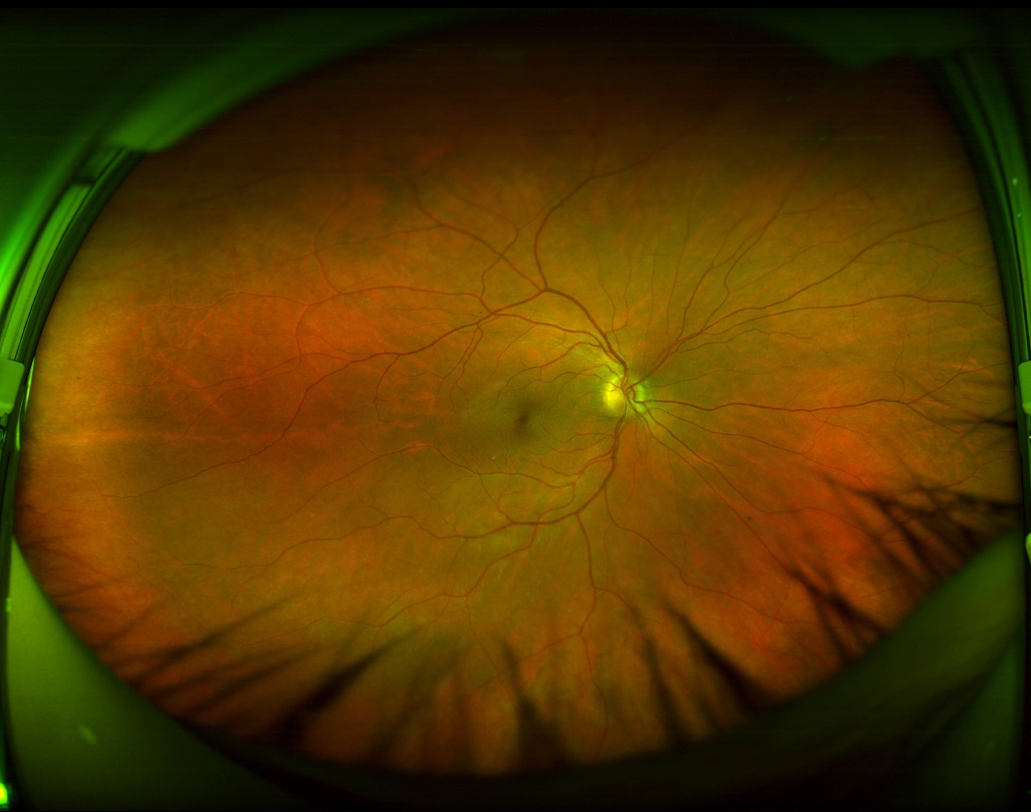
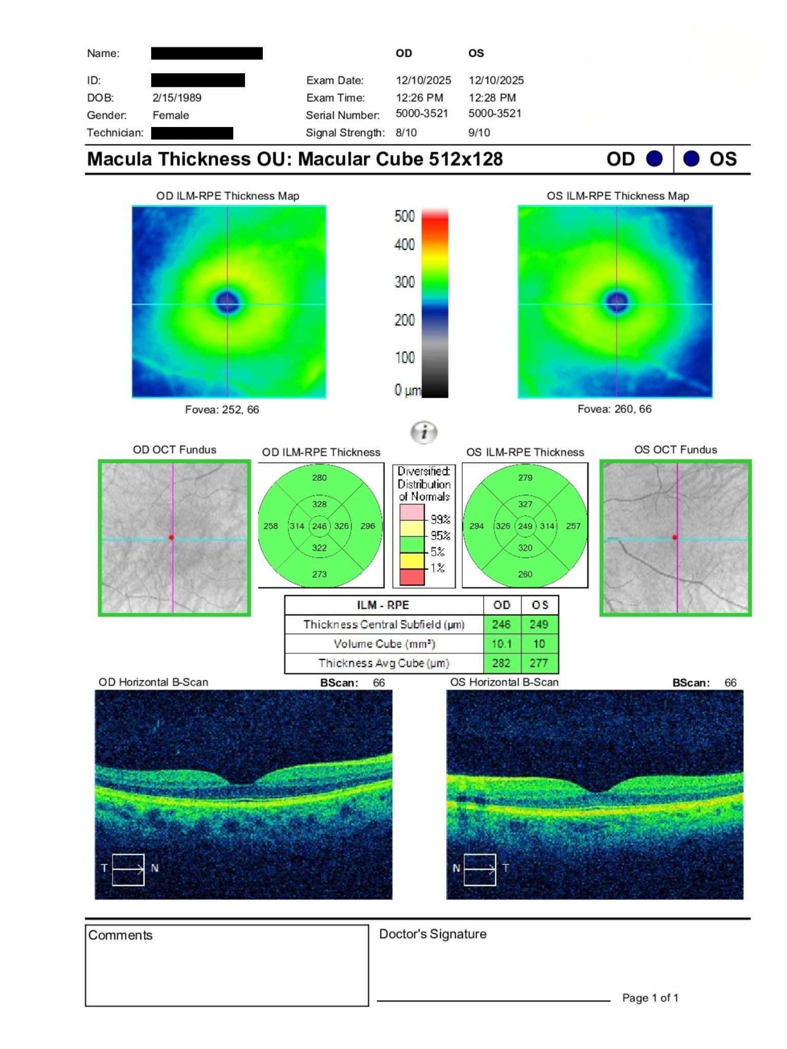
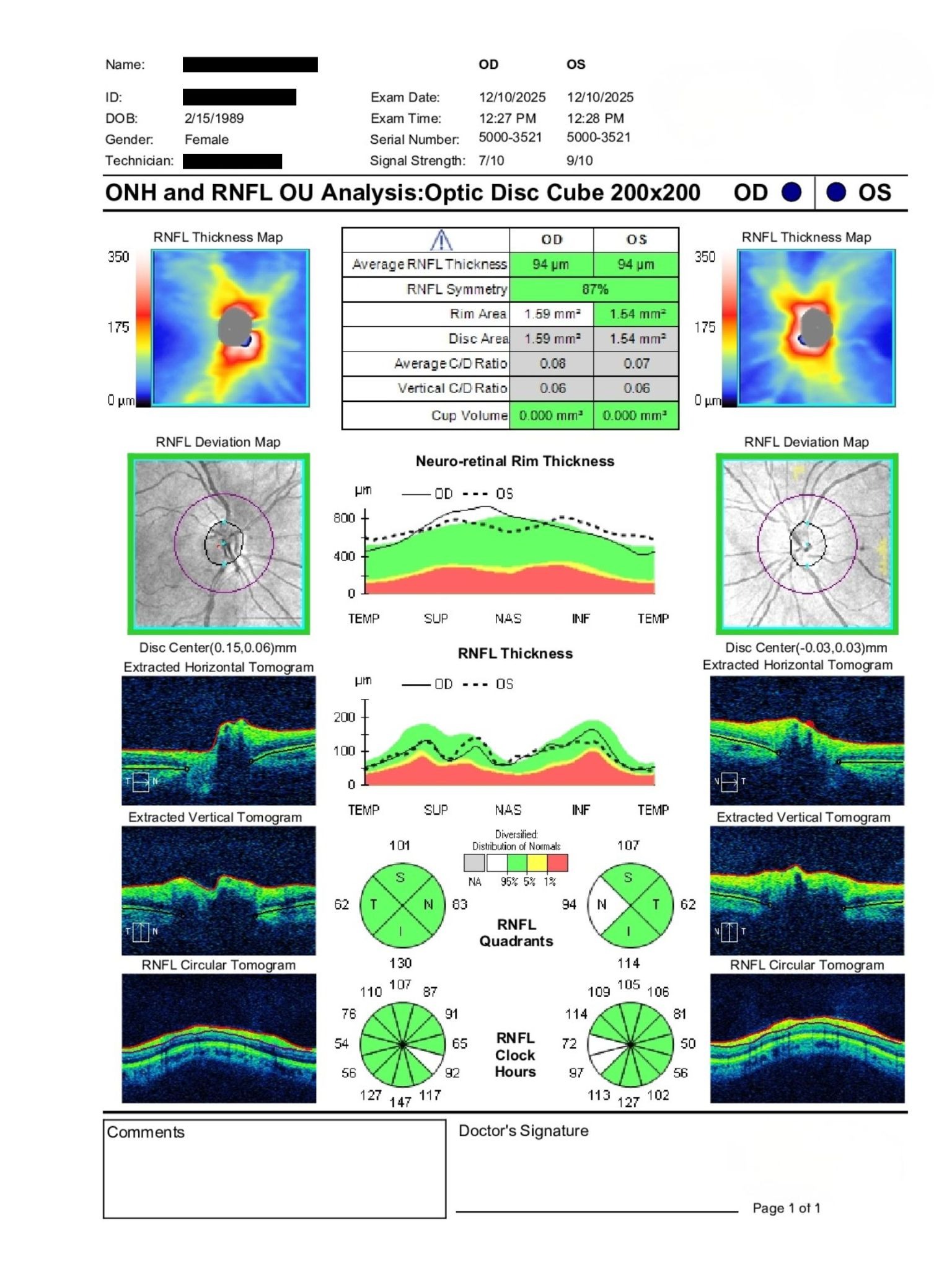
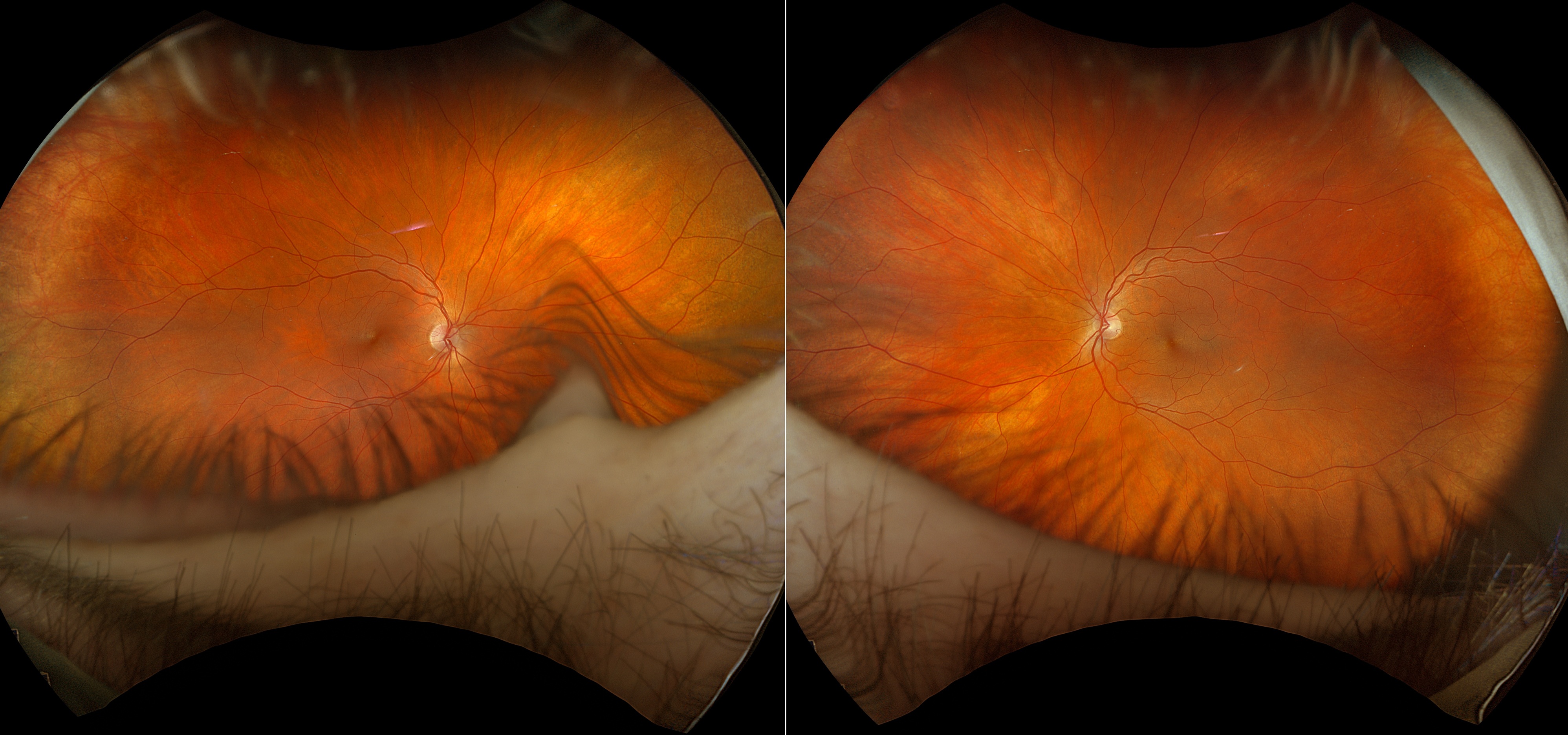
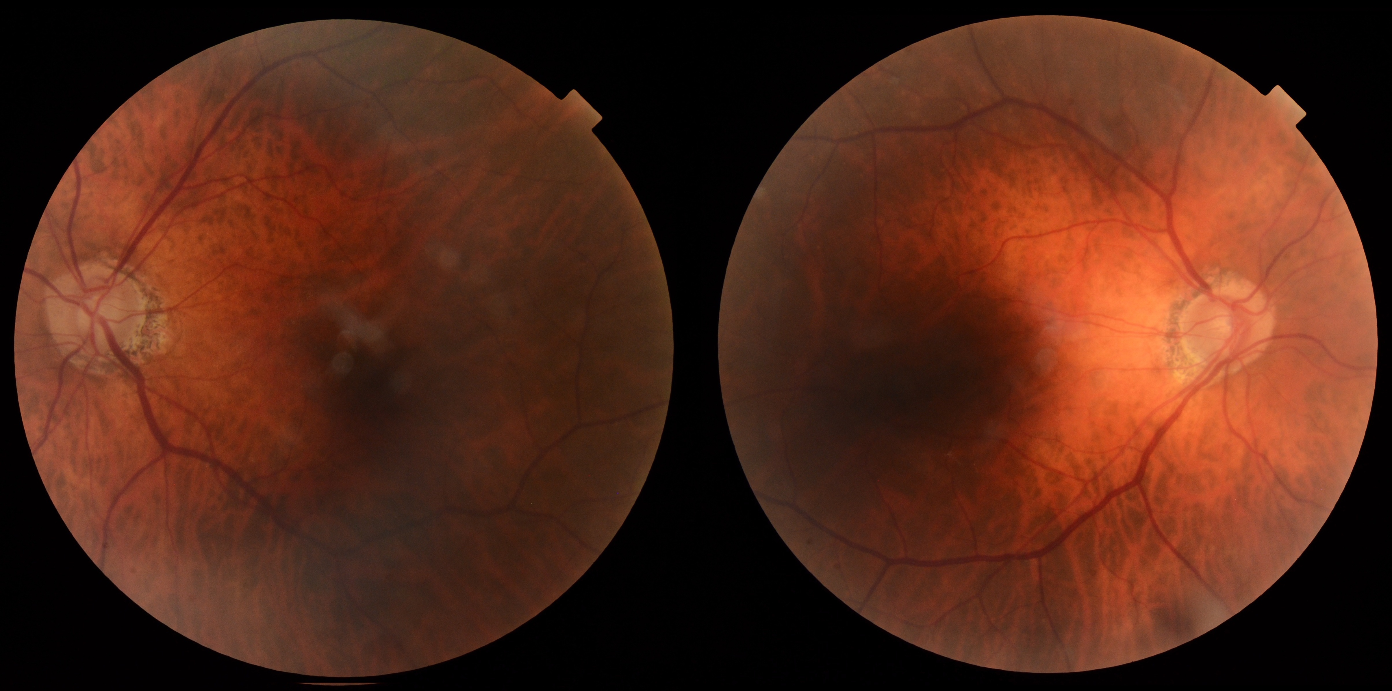
A 36-year-old female was referred for retinal evaluation due to a left peripheral retinal exudate, new-onset headaches, dizziness, postural symptoms, and speech difficulties. BCVA was 20/20 OU with IOPs of 18 mmHg OU. Anterior segment examination was largely unremarkable aside from slight corectopia OD. OCT RNFL and macular imaging were within normal limits, with small optic discs and no edema. Fundus examination revealed an isolated far peripheral exudate OS without retinal tears or detachment.
Do these peripheral retinal findings warrant further retinal or neuro-ophthalmic investigation?
A retina specialist provided a virtual consult within 1-2 weeks through Care1. Scroll below to see their diagnosis.
Care1 Ophthalmologist's Teleconsult
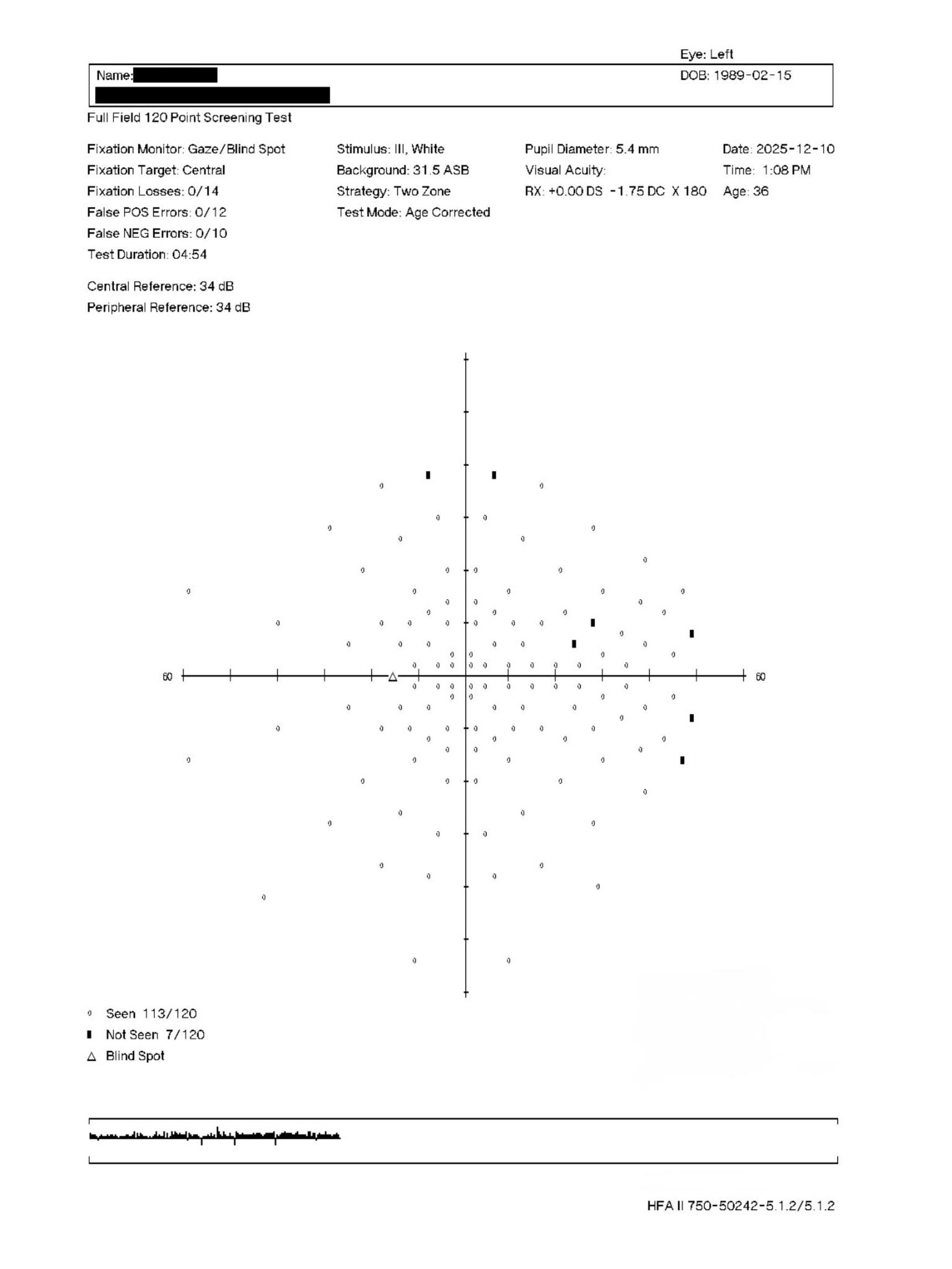
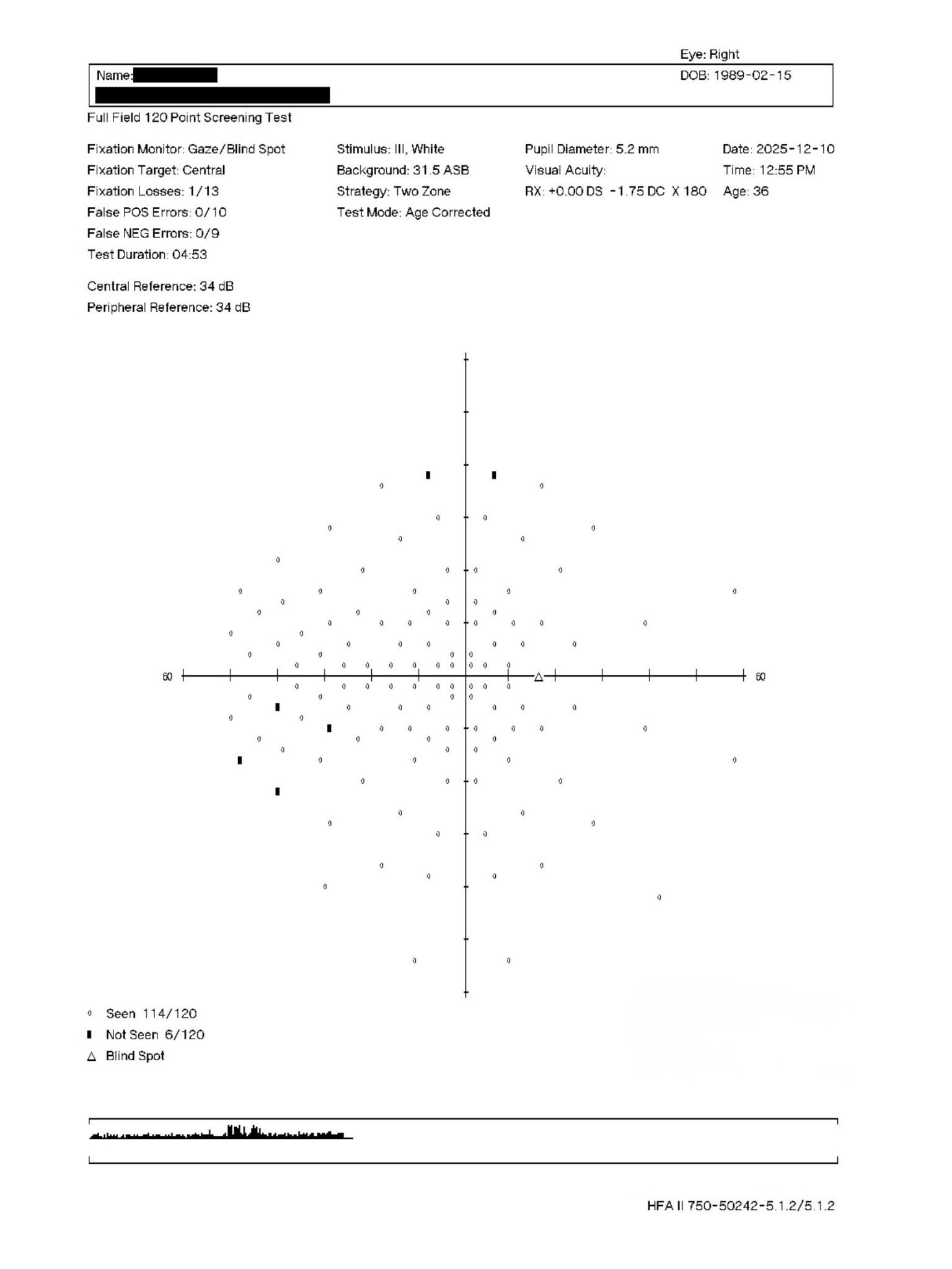
Review of the retinal imaging and diagnostic testing showed small optic nerves that appeared within normal anatomical limits. OCT RNFL, macular imaging, fundus photography, and visual field testing did not demonstrate evidence of optic neuropathy, retinal edema, or other significant retinal pathology.
The peripheral retinal finding appeared isolated, with only minimal retinal involvement related to the patient's systemic history. Continued monitoring, annual follow-up, and ongoing emphasis on blood pressure and glycemic control were recommended. Additional retinal specialist assessment was not considered necessary based on the available findings.
A subsequent corneal review of the topography demonstrated regular with-the-rule astigmatism. While the keratometry readings were elevated, the available topography did not demonstrate definitive evidence of corneal ectasia. If concern remains, advanced corneal tomography may help identify subtle posterior corneal changes not visible on standard topography.
Care1 AI's Clinical Insight
Peripheral retinal exudates can occur secondary to a variety of retinal vascular conditions and may be discovered incidentally during routine examination. Careful evaluation of associated retinal findings, systemic risk factors, and multimodal imaging can help determine whether the lesion represents active pathology or a stable peripheral change. Widefield imaging and longitudinal monitoring are often valuable when assessing isolated peripheral retinal abnormalities.
Unlock your full revenue potential
✔ Specialist consults within 1-2 weeks ✔ AI-powered clinical support ✔ Greater confidence in complex decision making
Ultra-widefield retinal imaging can identify peripheral retinal vascular abnormalities that may be missed on conventional fundus examination, improving detection of clinically significant peripheral pathology in patients with systemic vascular disease.
Reference: Silva PS, Cavallerano JD, Sun JK, Soliman AZ, Aiello LM, Aiello LP. Peripheral lesions identified by mydriatic ultrawide field imaging: distribution and potential impact on diabetic retinopathy severity. Ophthalmology. 2013;120(12):2587-2595. doi:10.1016/j.ophtha.2013.05.004