59-year-old male with excellent vision and mild CSDME OS reviewed via Care1 to confirm appropriate observation and follow-up.
Case Study: Mild Macular Changes
An optometrist uploaded this case to Care1.
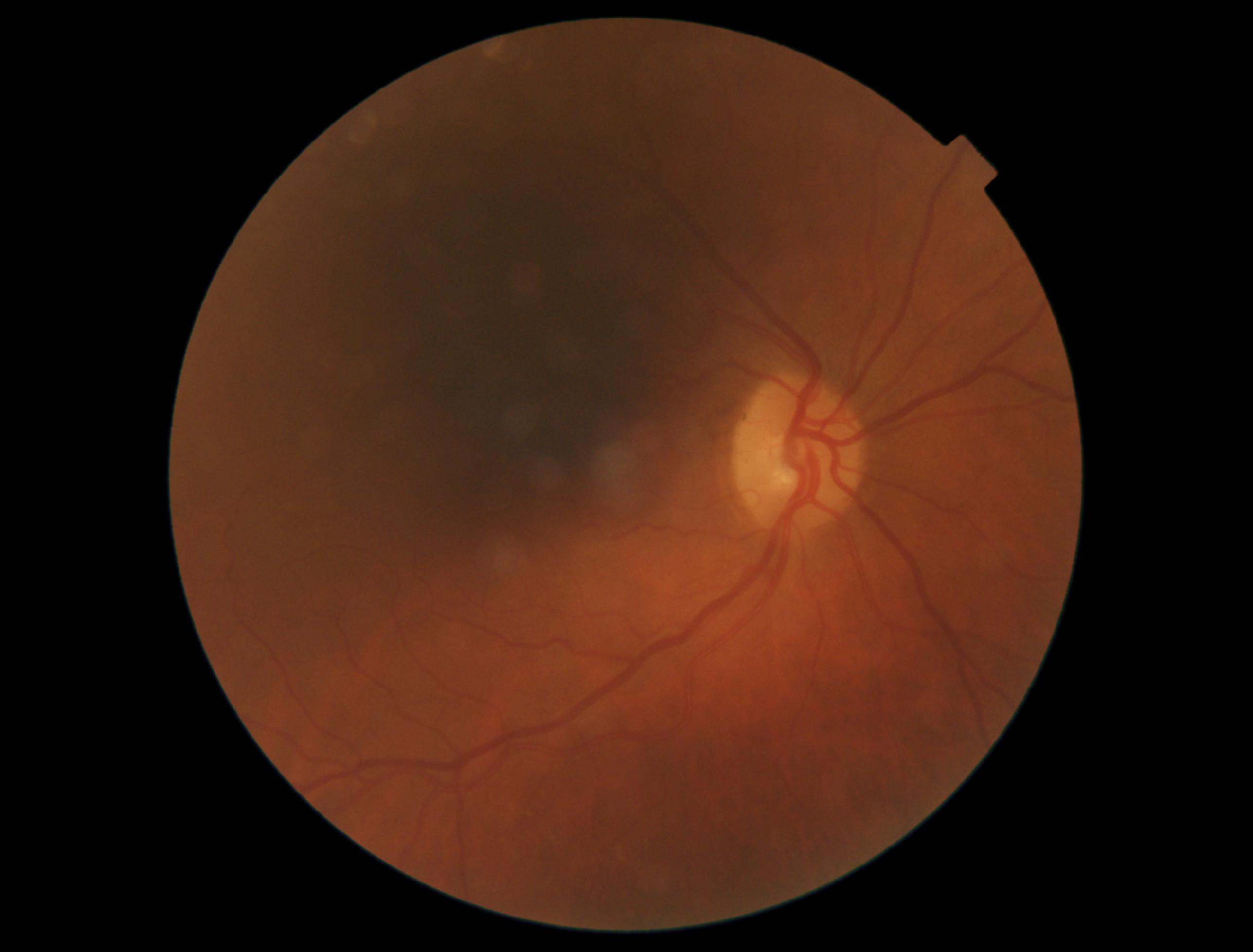
A 59-year-old male patient was referred for ophthalmology review due to concern for clinically significant diabetic macular edema (CSDME) in the left eye, identified on OCT imaging. The patient was asymptomatic at the time of assessment and reported no visual complaints, with excellent visual acuity in both eyes.
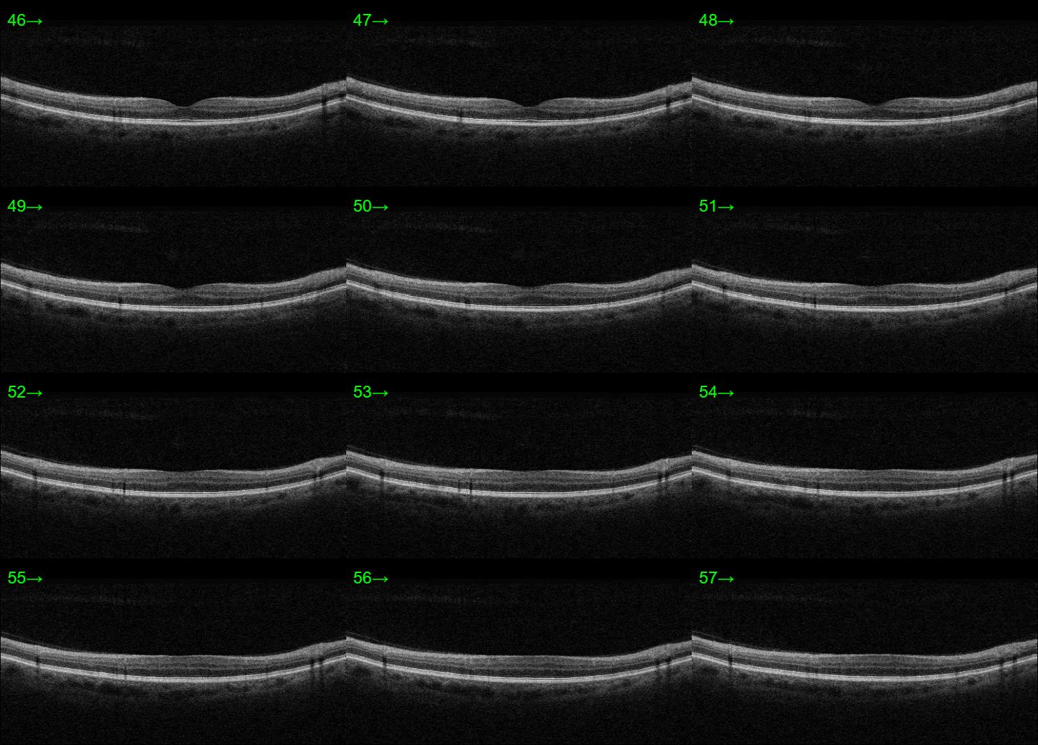
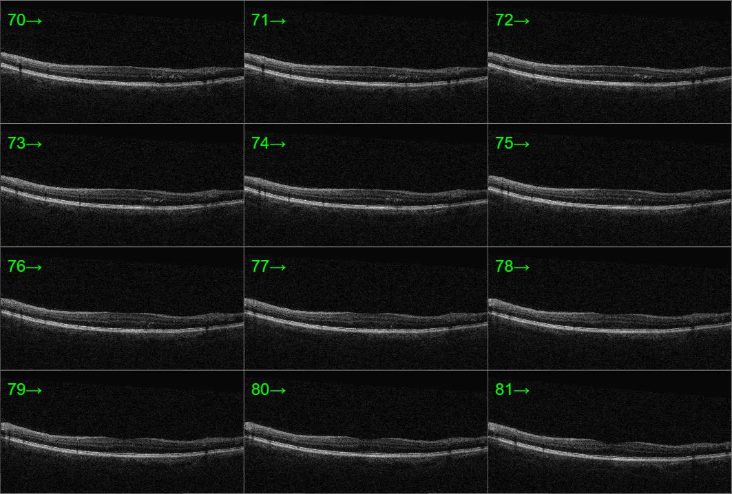
In-office examination showed best-corrected visual acuity of 20/20 in both eyes. Intraocular pressure measurements were not provided. Anterior segment findings were not documented. Posterior segment evaluation with OCT demonstrated minimal cystic changes within the macula of the left eye, raising concern for early or mild macular edema. No abnormal posterior segment findings were noted in the right eye.
Given the OCT findings suggestive of possible CSDME in the setting of preserved visual acuity and no symptoms, would continued observation with routine follow-up be appropriate at this stage, or would you recommend treatment or additional investigation at this time?
An ophthalmology subspecialist provided a virtual consult within 1-2 weeks through Care1. Scroll below to see their diagnosis.
Care1 Subspecialist’s Key Takeaways
This patient has very mild macular changes in the left eye, with excellent visual acuity. OCT shows minimal cystic edema. Clinical evidence suggests observation is appropriate for patients with good vision, as edema may fluctuate without affecting visual outcomes. Follow-up should continue as scheduled, with specialist review to guide any future intervention if vision declines.
Care1 AI’s Clinical Insight
Diabetic macular edema (DME) is defined as the accumulation of excess fluid in the macular region of the retina due to leakage from damaged retinal capillaries, typically associated with diabetic retinopathy. This fluid buildup causes thickening of the retina in the macula, which may occur at any stage of retinopathy and can be detected through clinical examination and imaging such as OCT. DME may be asymptomatic initially but can lead to blurred or distorted vision as retinal swelling involves or threatens the fovea, the area responsible for central vision.
Did You Know?
Diabetic macular edema occurs when high blood sugar damages the blood–retina barrier, causing fluid to leak into the macula and sometimes forming lipid deposits called hard exudates.
Bunt‑Milam A, Saari JC, Klock IB, et al. Zonulae adherentes pore size in the external limiting membrane of the rabbit retina. Invest Ophthalmol Vis Sci. 1985;26(10):1377–1380.