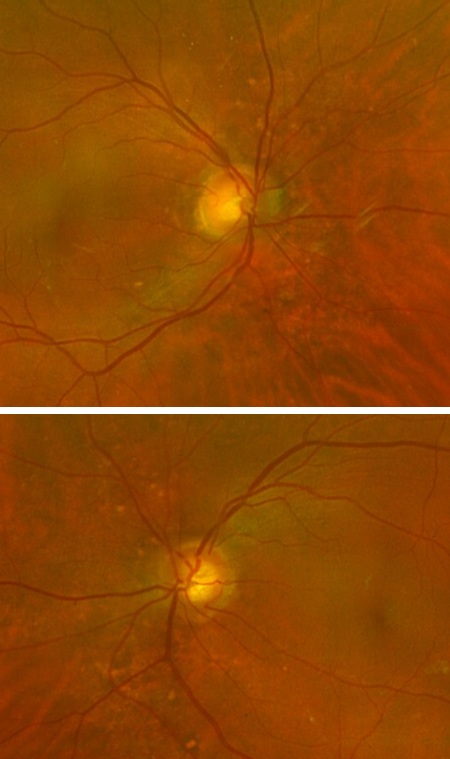
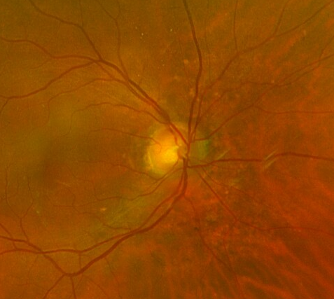
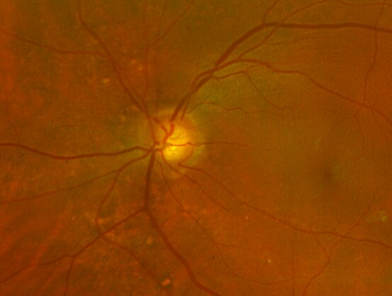
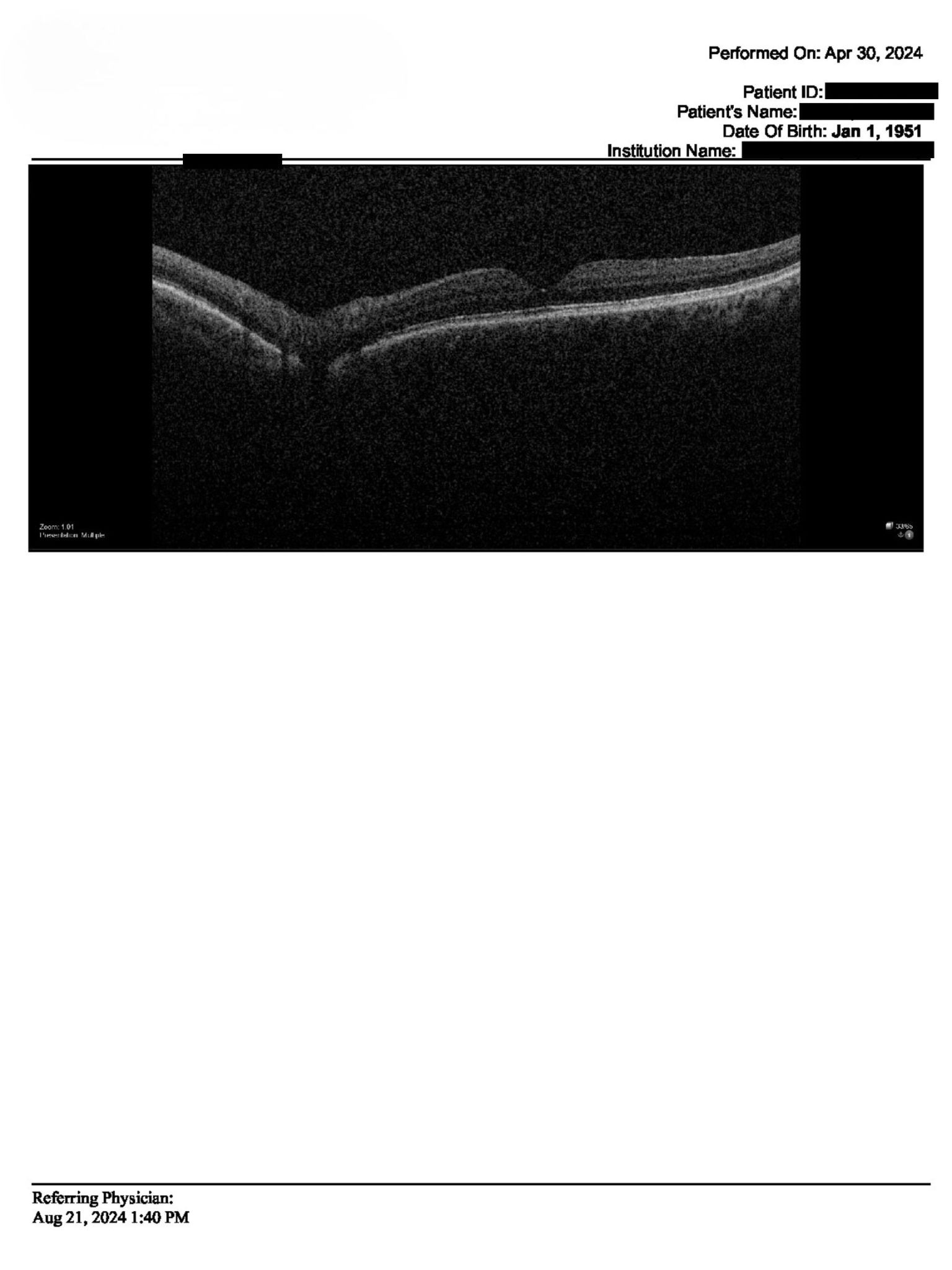
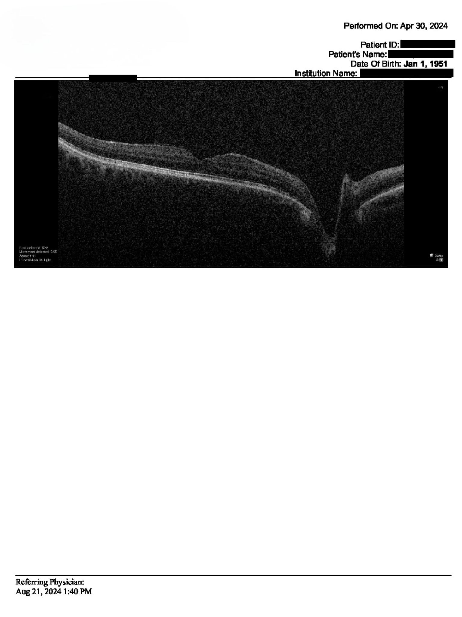
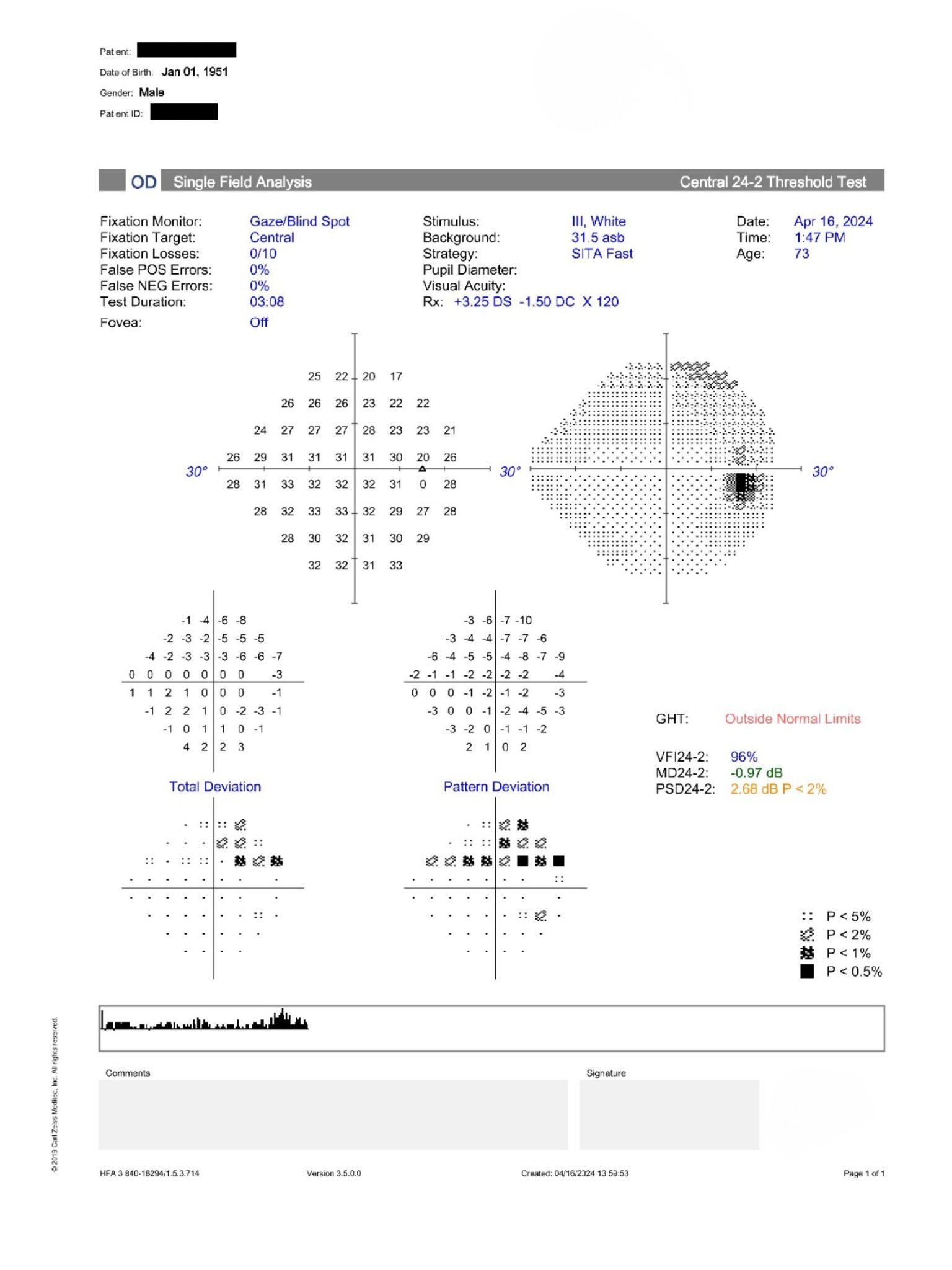
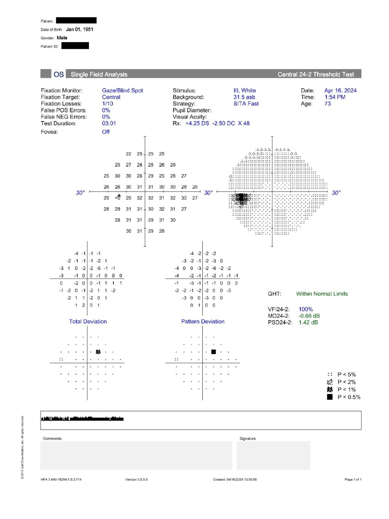
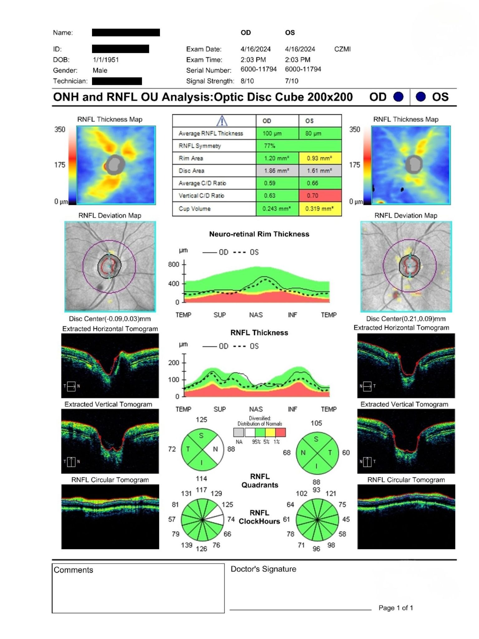
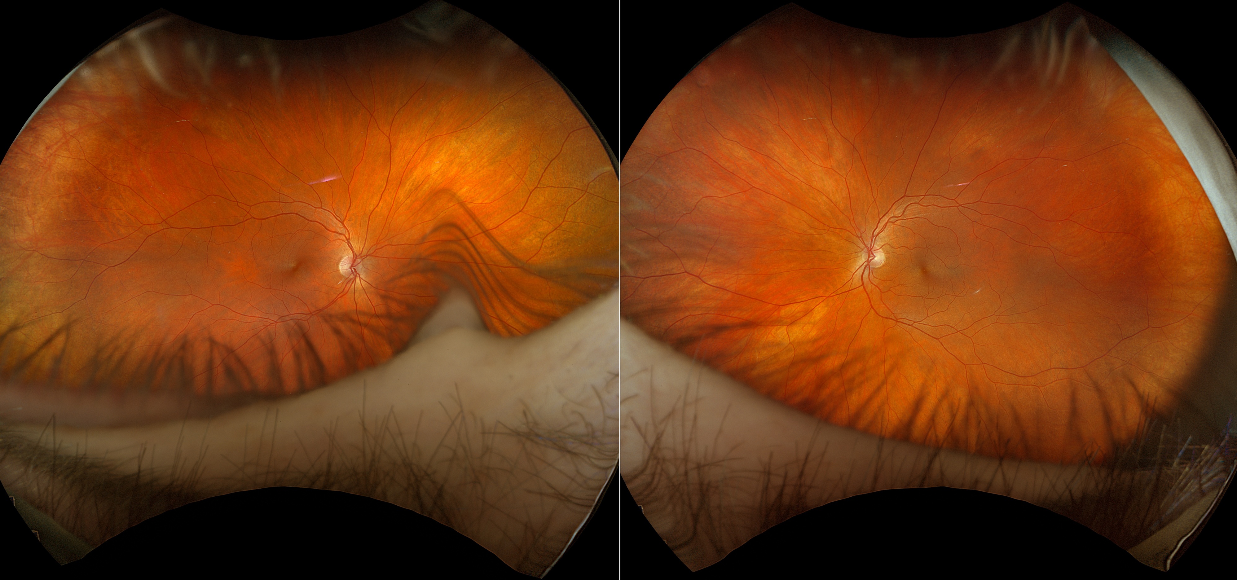
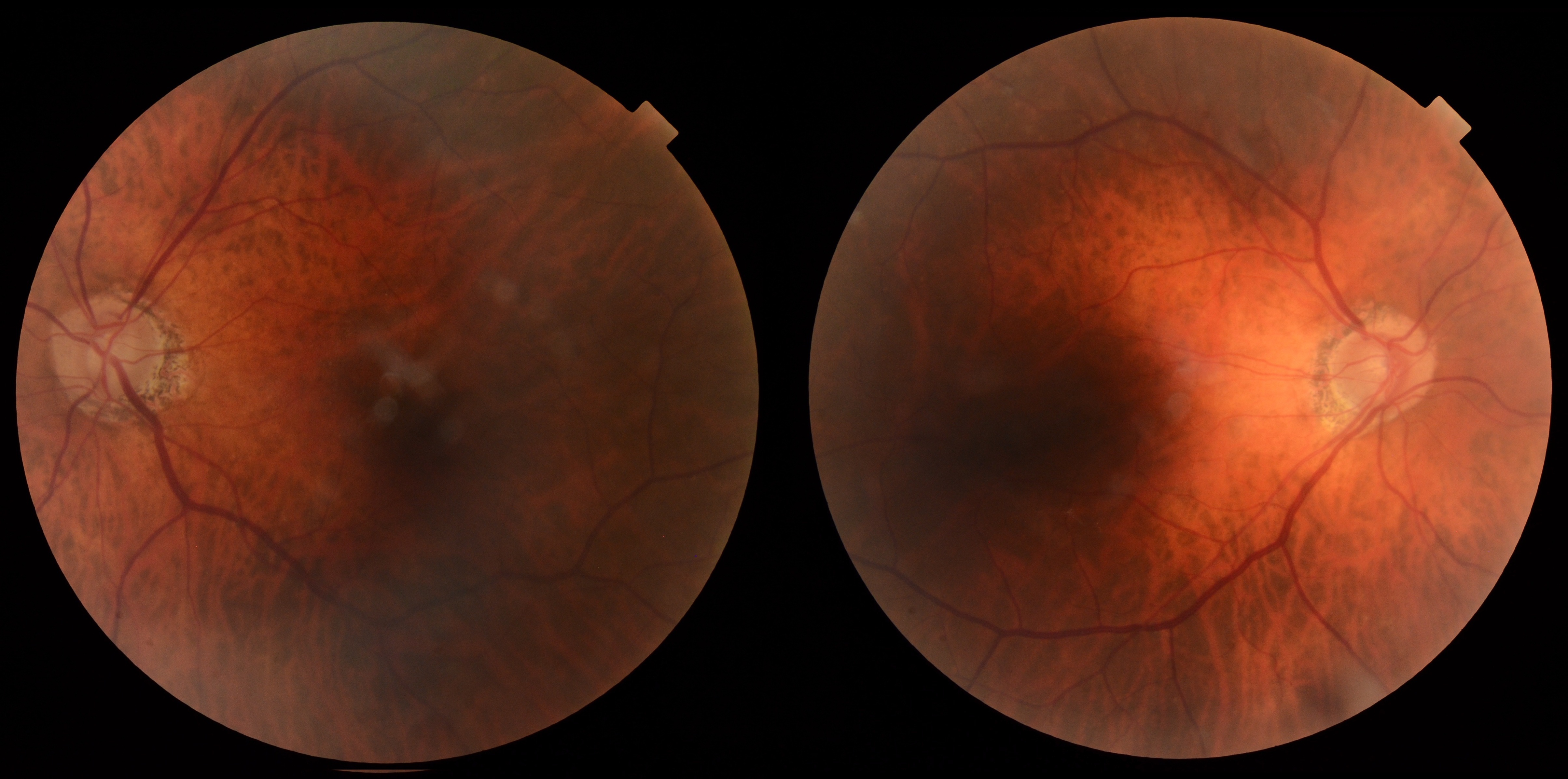
75-year-old with large cup-disc ratios, thin corneas, and stable OCT RNFL findings undergoing glaucoma suspect monitoring.
Case Study: Large Cup Disc Asymmetry
An optometrist uploaded this case to Care1.
A 75-year-old male was referred for ongoing glaucoma suspect monitoring due to a strong family history of glaucoma, large cup-to-disc ratios, and thin corneas. BCVA was 20/20 OD and 20/25 OS. IOP measured 20 mmHg OD and 21 mmHg OS by applanation. Anterior segment examination showed pseudophakia OU with open angles and quiet anterior chambers. Posterior segment findings included large optic nerves with cup-disc asymmetry, stable OCT RNFL measurements, and visual fields without significant glaucomatous defects.
Given the patient's family history, large cup-disc ratios, thin pachymetry, and stable structural and functional testing, what is the most appropriate follow-up strategy?
An ophthalmology subspecialist provided a virtual consult within 1-2 weeks through Care1. Scroll below to see their diagnosis.
Care1 Ophthalmologist's Teleconsult
This case demonstrates the importance of correlating optic nerve appearance with structural and functional testing when assessing glaucoma risk. Despite large cup-disc ratios, intraocular pressure remained within the target range, OCT RNFL findings were stable compared with prior imaging, and visual field testing showed only nonspecific changes without structure-function correlation. Continued annual monitoring is appropriate for this glaucoma suspect given the family history and optic nerve appearance.
Care1 AI's Clinical Insight
Large Physiologic Optic Cups
Large physiologic optic cups can occur in healthy individuals with larger optic nerve heads and may mimic glaucomatous cupping. Evaluation requires careful assessment of neuroretinal rim integrity, retinal nerve fiber layer thickness, visual field status, and longitudinal change over time. Disc size is an important factor when interpreting cup-disc ratios because larger optic discs often have proportionally larger physiologic cups.
Deepen your doctor-patient relationships
✔️ Shorter wait times for specialist input
✔️ More confident, transparent patient conversations
In the Ocular Hypertension Treatment Study, every 40 μm decrease in central corneal thickness was associated with a significantly increased risk of developing primary open-angle glaucoma, highlighting pachymetry as an independent risk factor beyond intraocular pressure alone.
Reference: Gordon MO, Beiser JA, Brandt JD, et al. The Ocular Hypertension Treatment Study: Baseline factors that predict the onset of primary open-angle glaucoma. Arch Ophthalmol. 2002;120(6):714-720. doi:10.1001/archopht.120.6.714