Patient follow-up shows large optic cups and pressure changes; therapy adjusted. Is close monitoring enough or are further changes needed?
Case Study: Large Cupping with Visual Defects
An optometrist uploaded this case to Care1.
A 75-year-old male patient presented for a follow-up, reporting no new visual concerns. He has a history of arthritis and previously variable intraocular pressure control. Ocular history is notable for glaucoma, currently managed with Lumigan nightly OU.
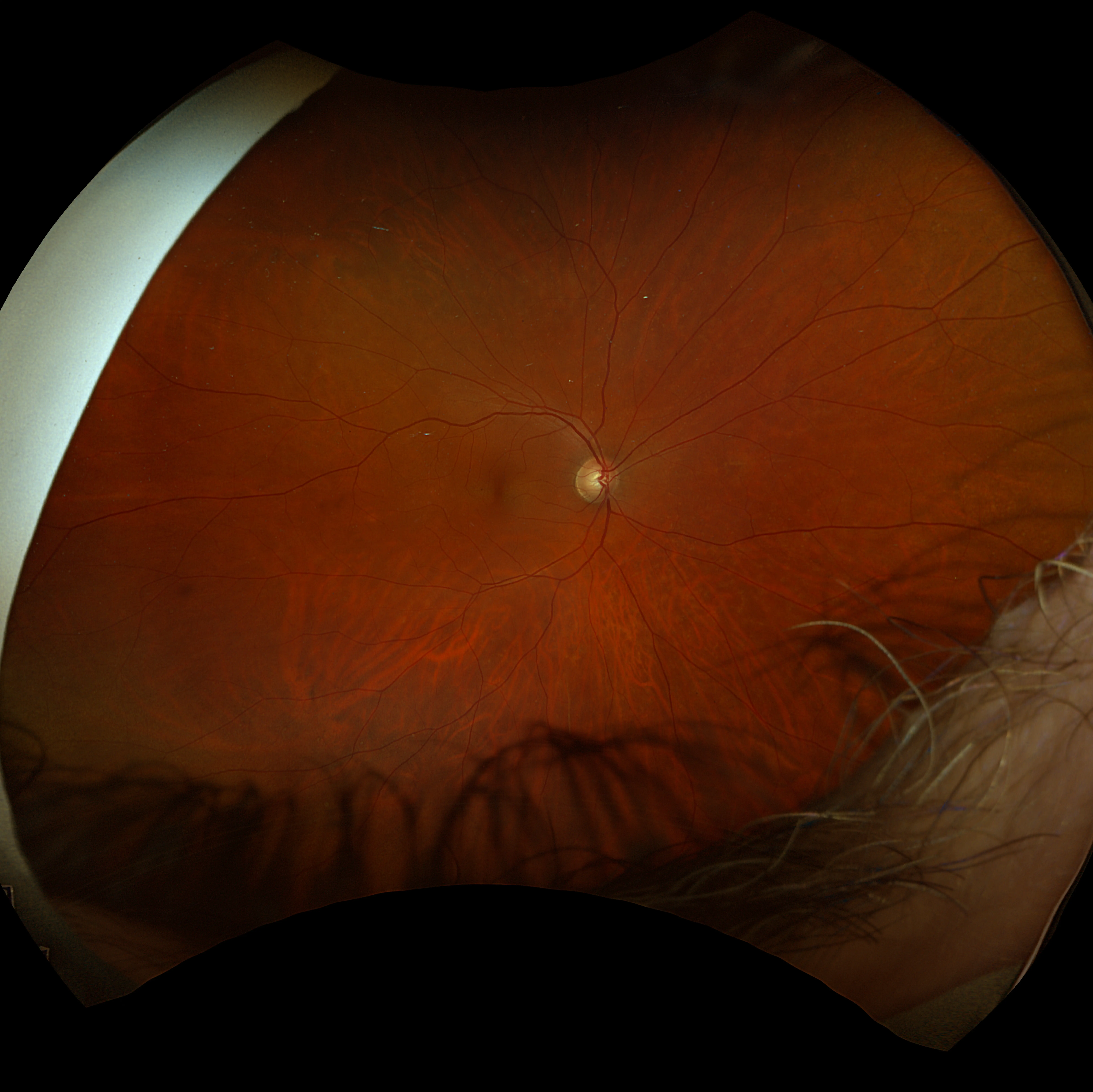
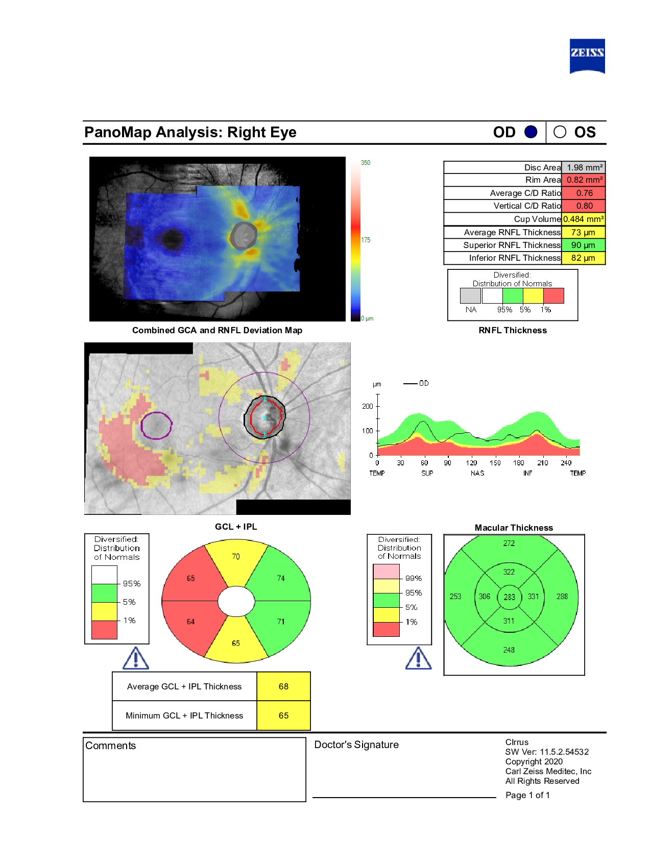
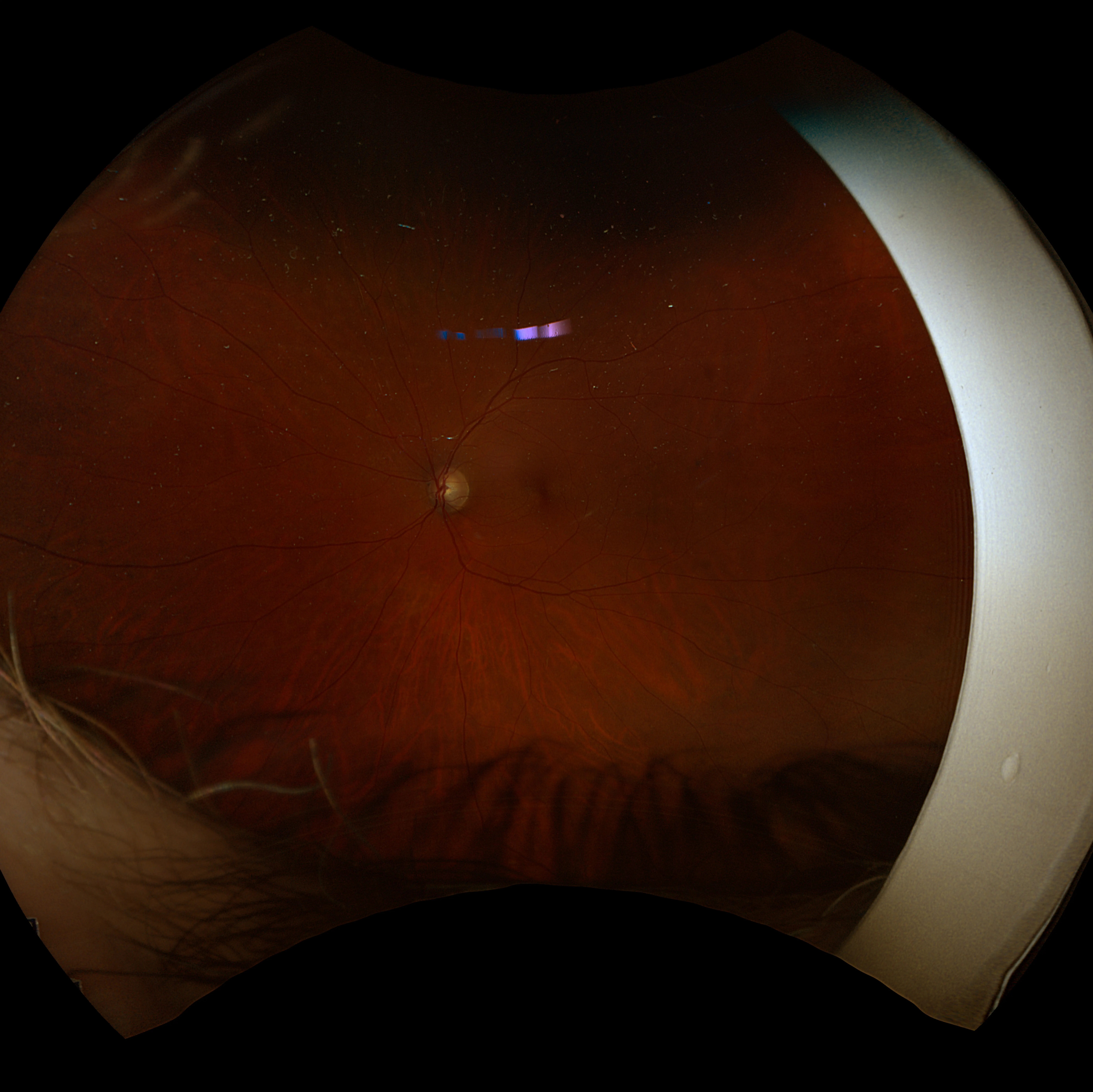
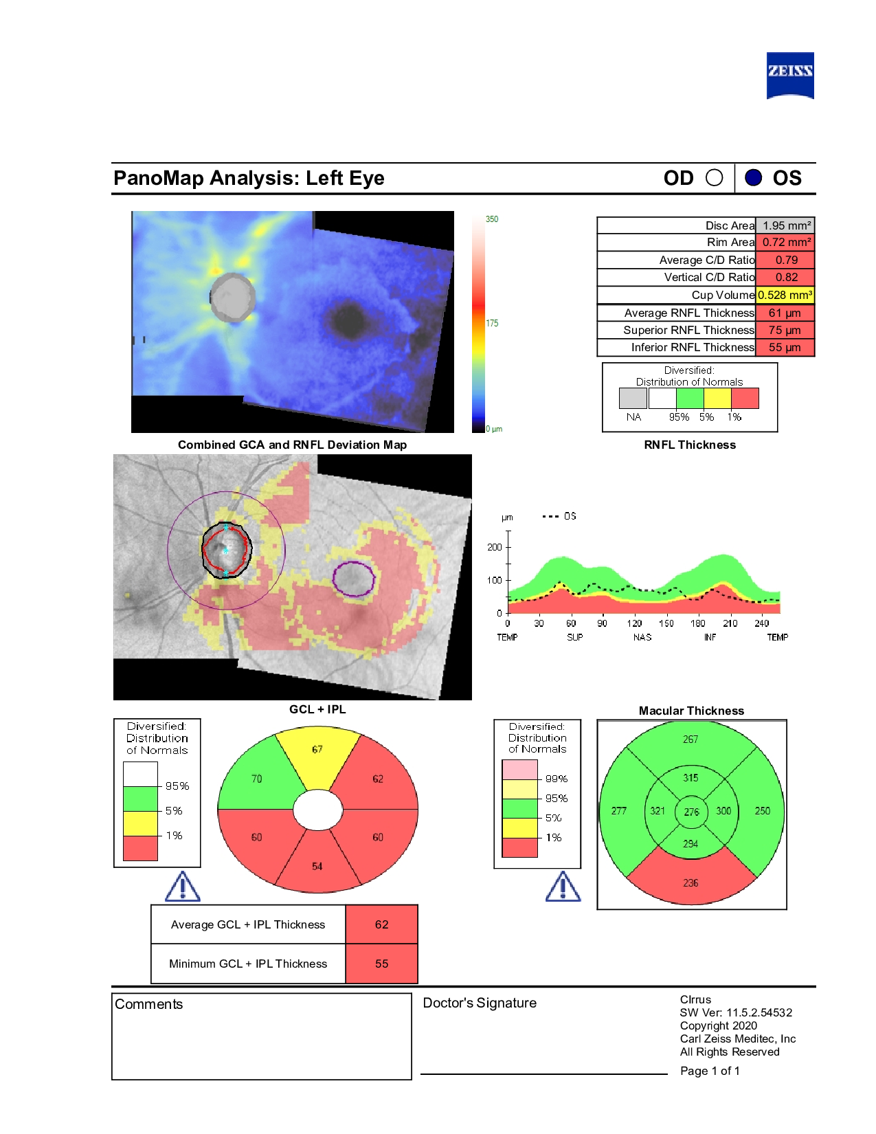
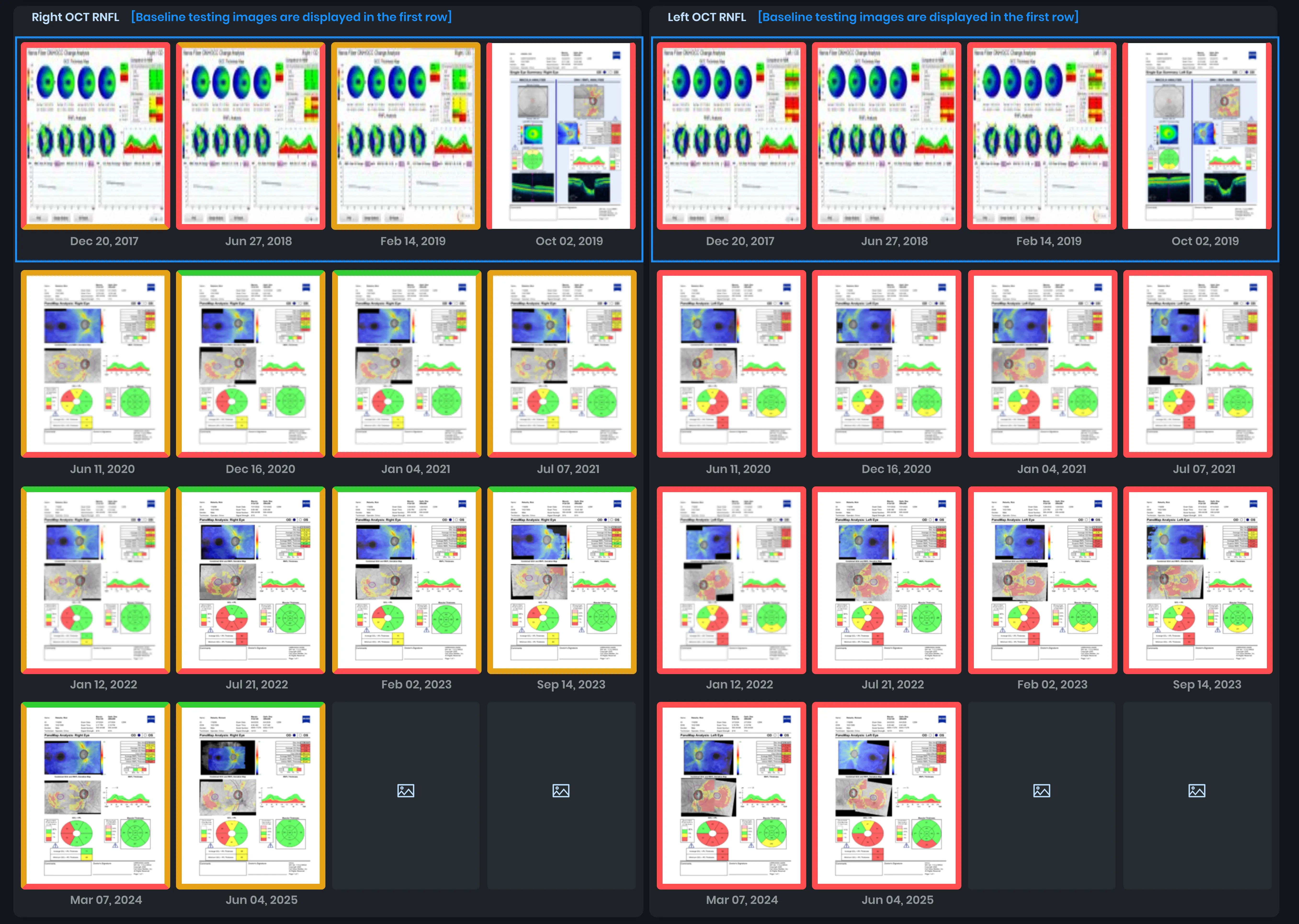
In-office examination showed best-corrected visual acuity of 20/20 in both eyes. Intraocular pressure was IOP OD 21 mmHg, OS 18 mmHg. Anterior segment examination revealed mild ocular surface findings and early lens changes bilaterally. Optic nerve evaluation demonstrated large cups in both eyes, with greater cupping in the left eye and concern for possible structural change on imaging. The corneas were noted to be thin bilaterally.
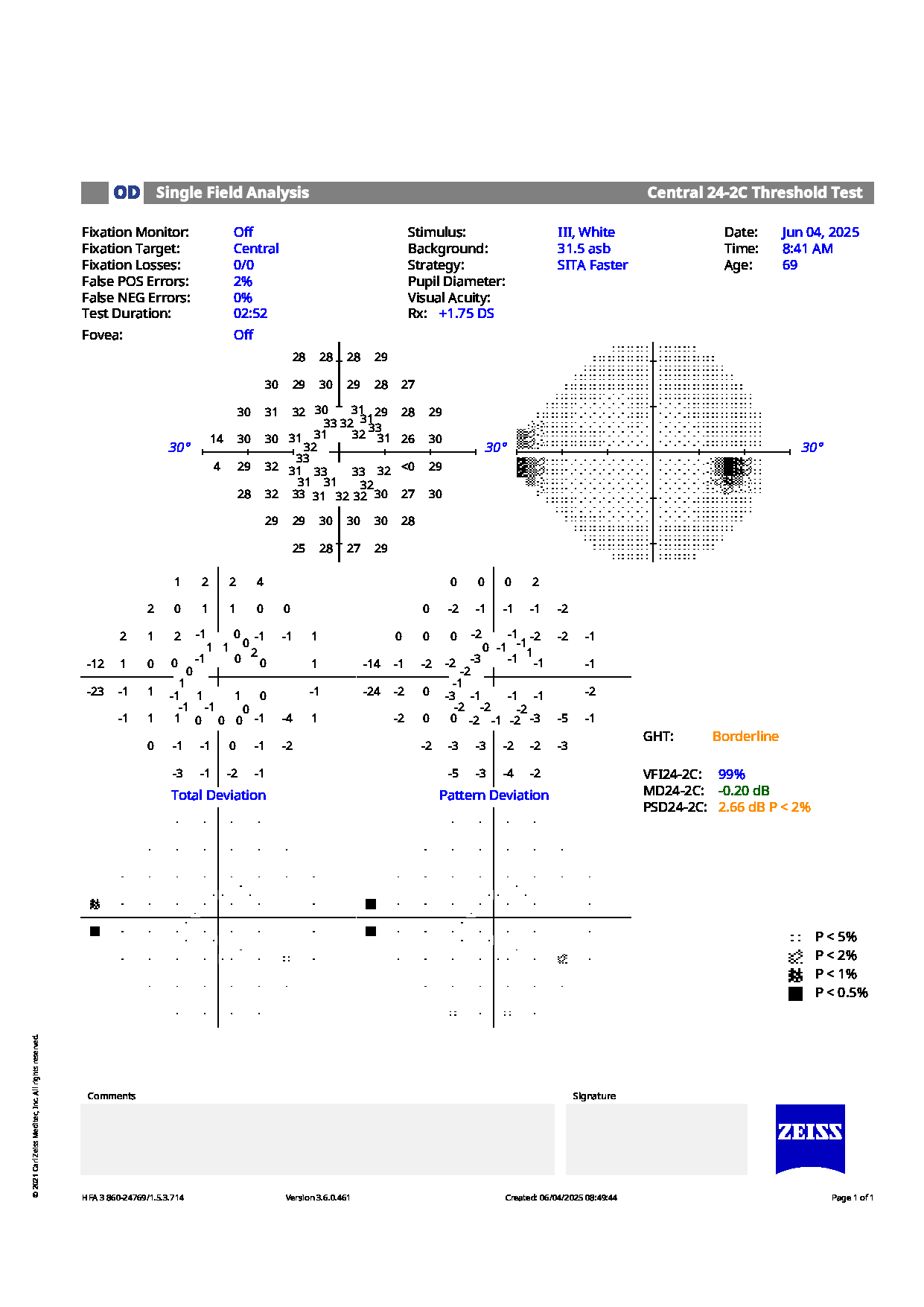
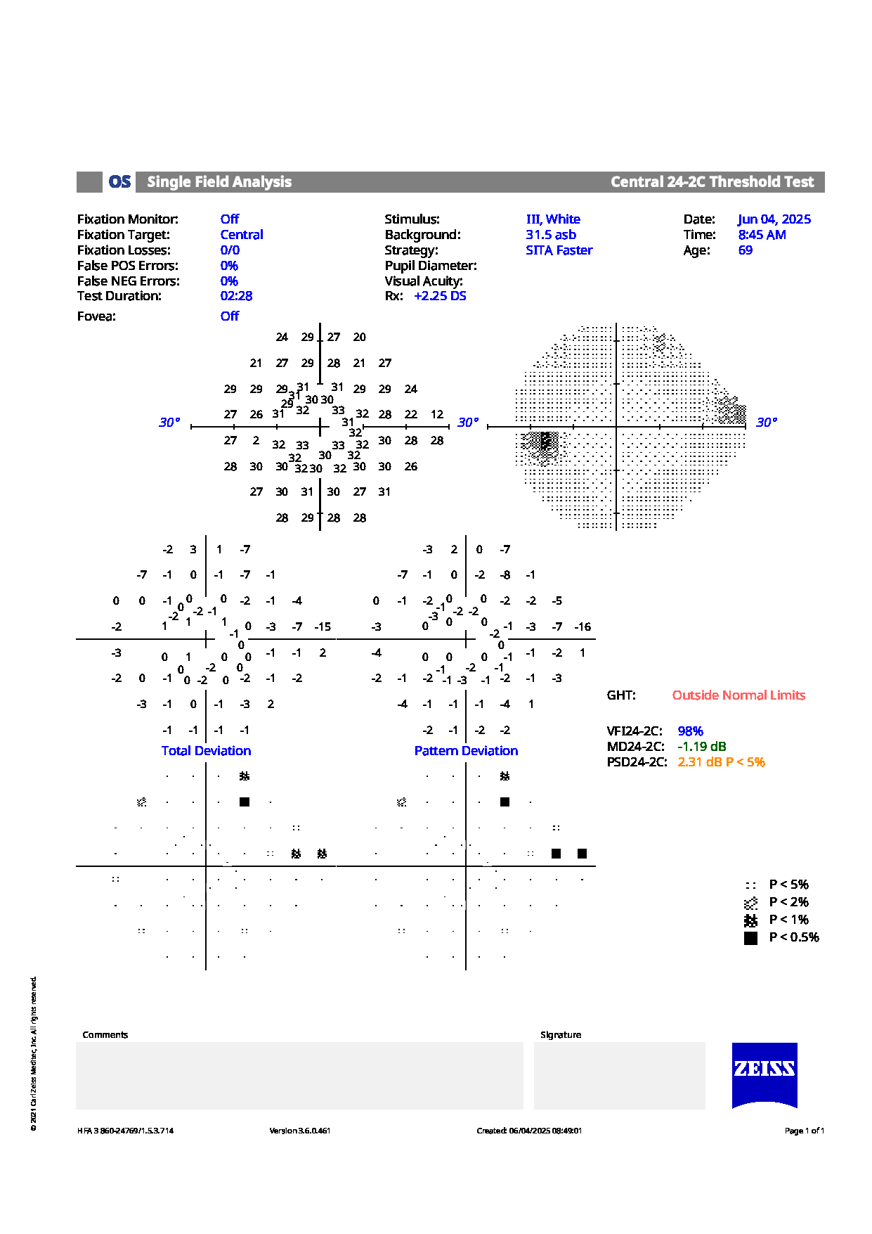
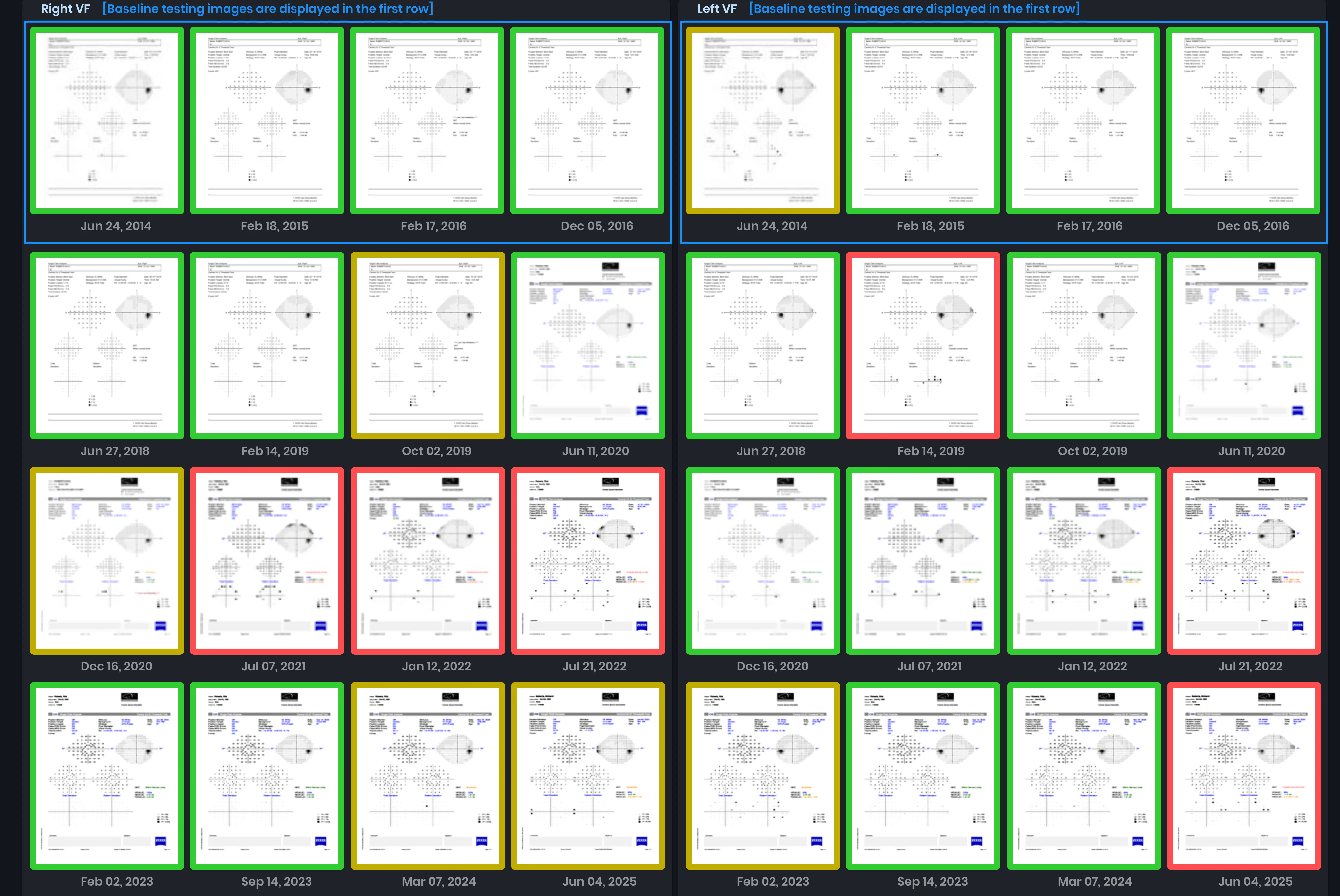
Visual field testing showed a nasal step in the right eye and superior depressions in the left eye. Based on these findings, the patient was not meeting previously established pressure targets on their current regimen. XALATAN nightly OU was initiated to replace Lumigan, with plans for a short-term pressure reassessment and escalation to combination treatment if targets remain unmet.
Given the current pressures and structural/functional findings, would you recommend any additional changes to management at this stage, or is the current plan of medication adjustment with close follow-up appropriate?
An ophthalmology subspecialist provided a virtual consult within 1-2 weeks through Care1. Scroll below to see their diagnosis.
Care1 Subspecialist’s Key Takeaways
This patient’s eye pressures are currently above the comfort zone on Lumigan, though OCT RNFL and visual fields remain stable. The patient has been switched to Xalatan, with a 1-month IOP check planned. If pressures remain elevated, escalation to Xalacom may be considered.
Care1 AI’s Clinical Insight
Normal tension glaucoma (NTG) is a form of chronic optic neuropathy similar to primary open‑angle glaucoma but occurring despite intraocular pressures that remain within the normal range. It results in characteristic optic nerve head cupping and retinal nerve fiber layer thinning with corresponding visual field loss, and diagnosis requires careful exclusion of other optic neuropathies and confirmation of open angles with normal pressures. Although the exact cause is unclear, factors independent of eye pressure such as vascular dysregulation and structural susceptibility are believed to contribute, and treatment generally still aims to lower pressure to slow progression.
Did You Know?
Optic disc hemorrhages are closely associated with adjacent retinal nerve fiber layer defects and peripapillary atrophy in NTG eyes, indicating a structural link between hemorrhage location and glaucomatous damage.
Sugiyama K, Tomita G, Kitazawa Y, et al. The associations of optic disc hemorrhage with retinal nerve fiber layer defect and peripapillary atrophy in normal‑tension glaucoma. Ophthalmology. 1997;104(11):1926–1933. doi:10.1016/S0161‑6420(97)30005‑0