Case Study: Chronic Retinal Changes in Diabetic Patient
An optometrist uploaded this case to Care1.
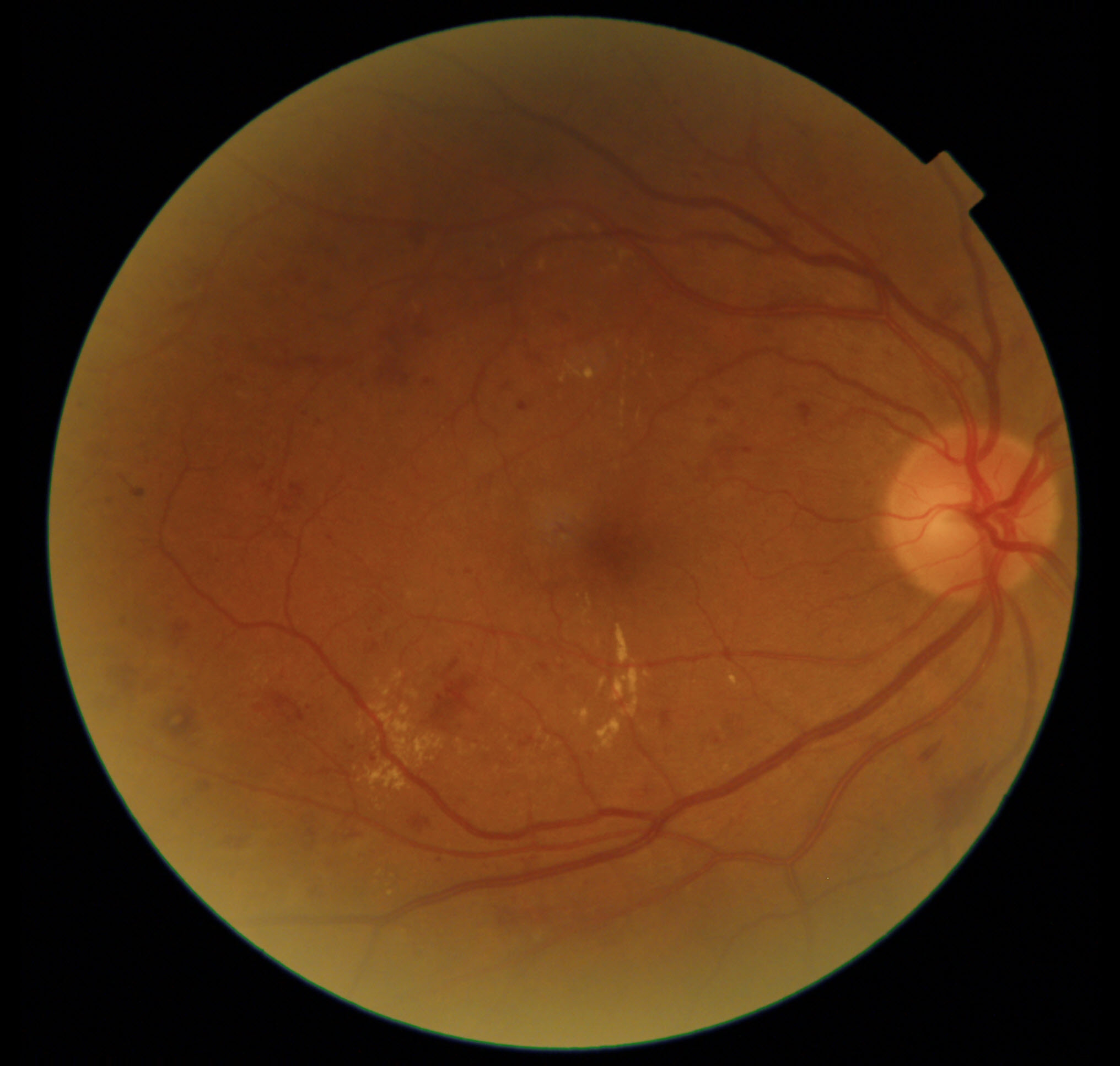
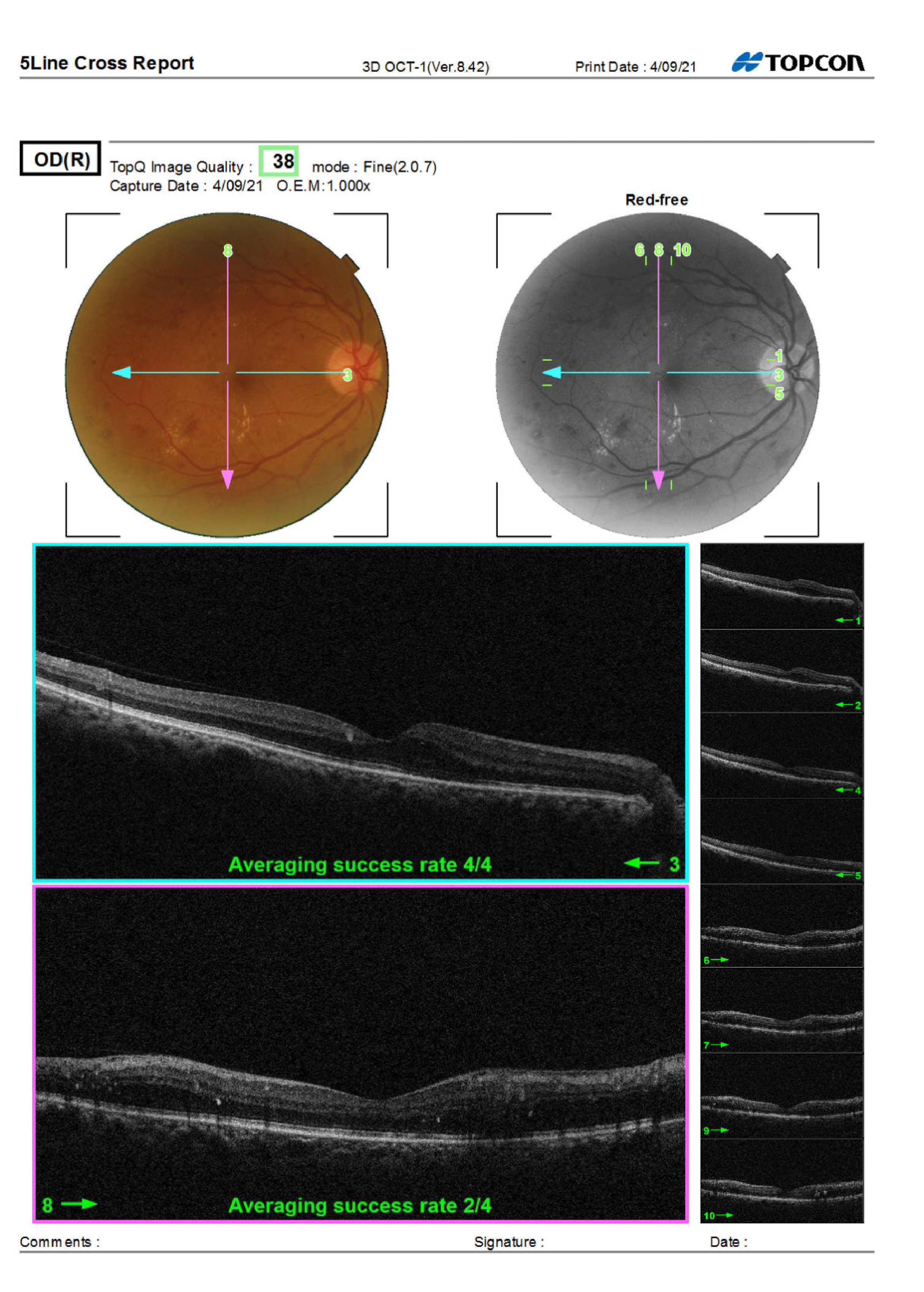
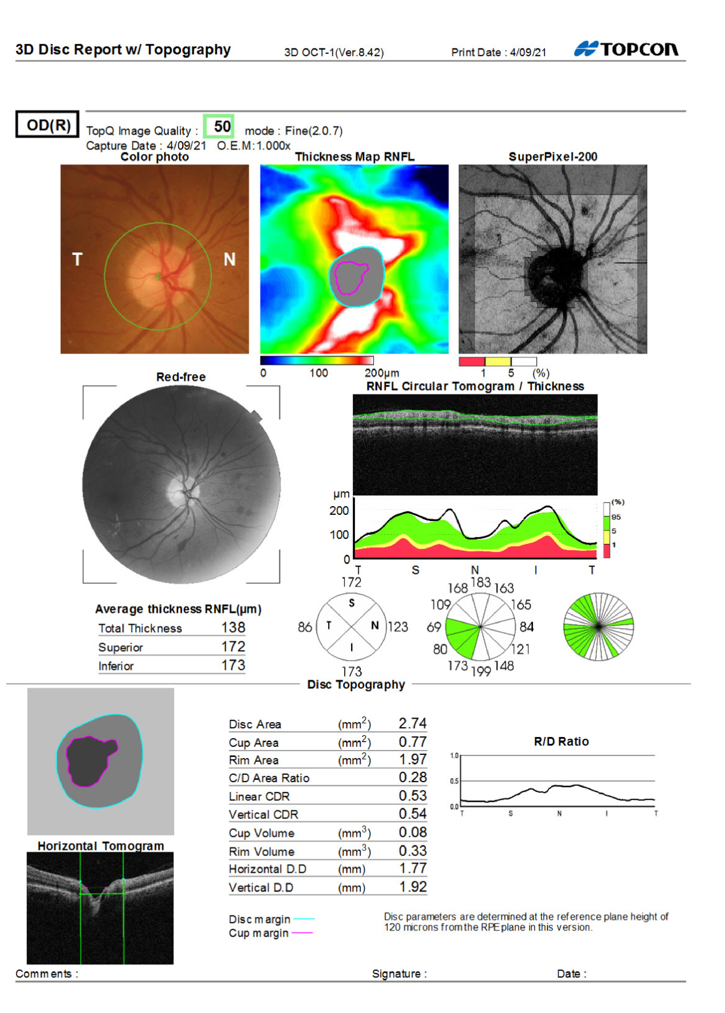
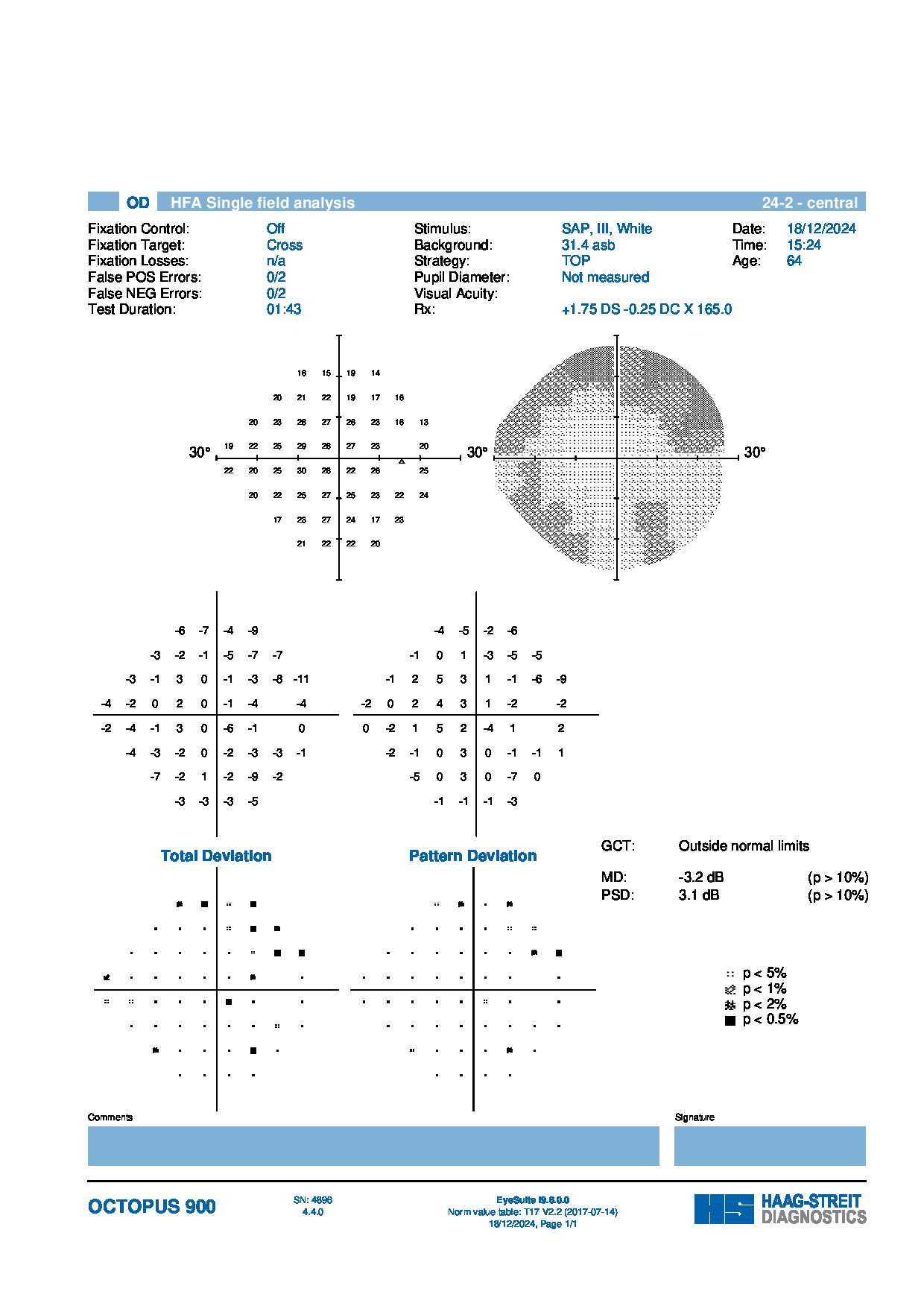
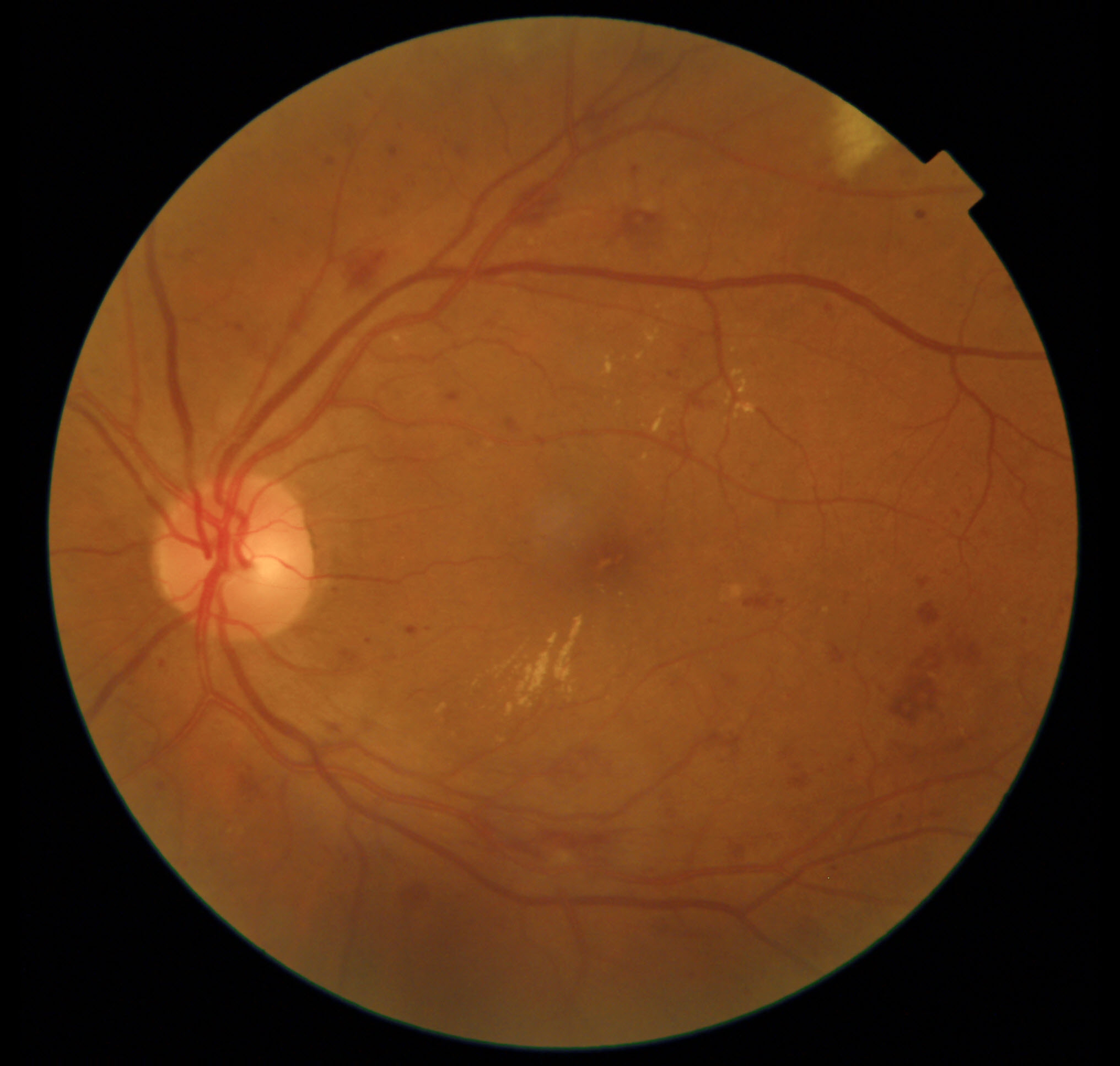
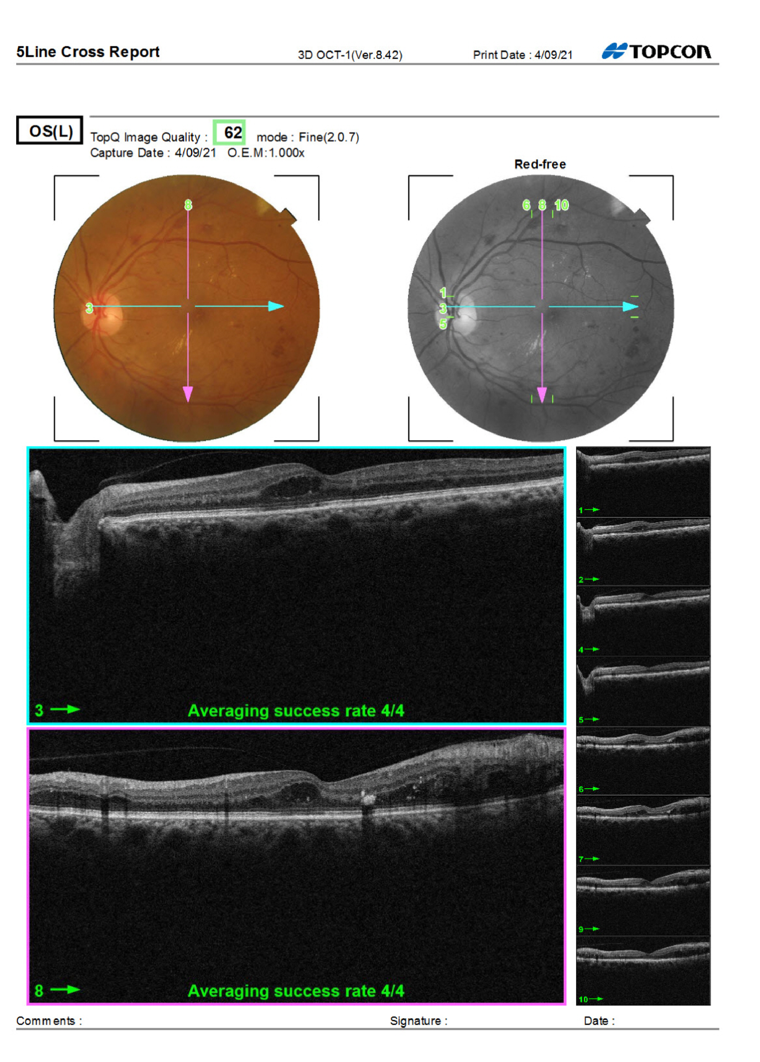
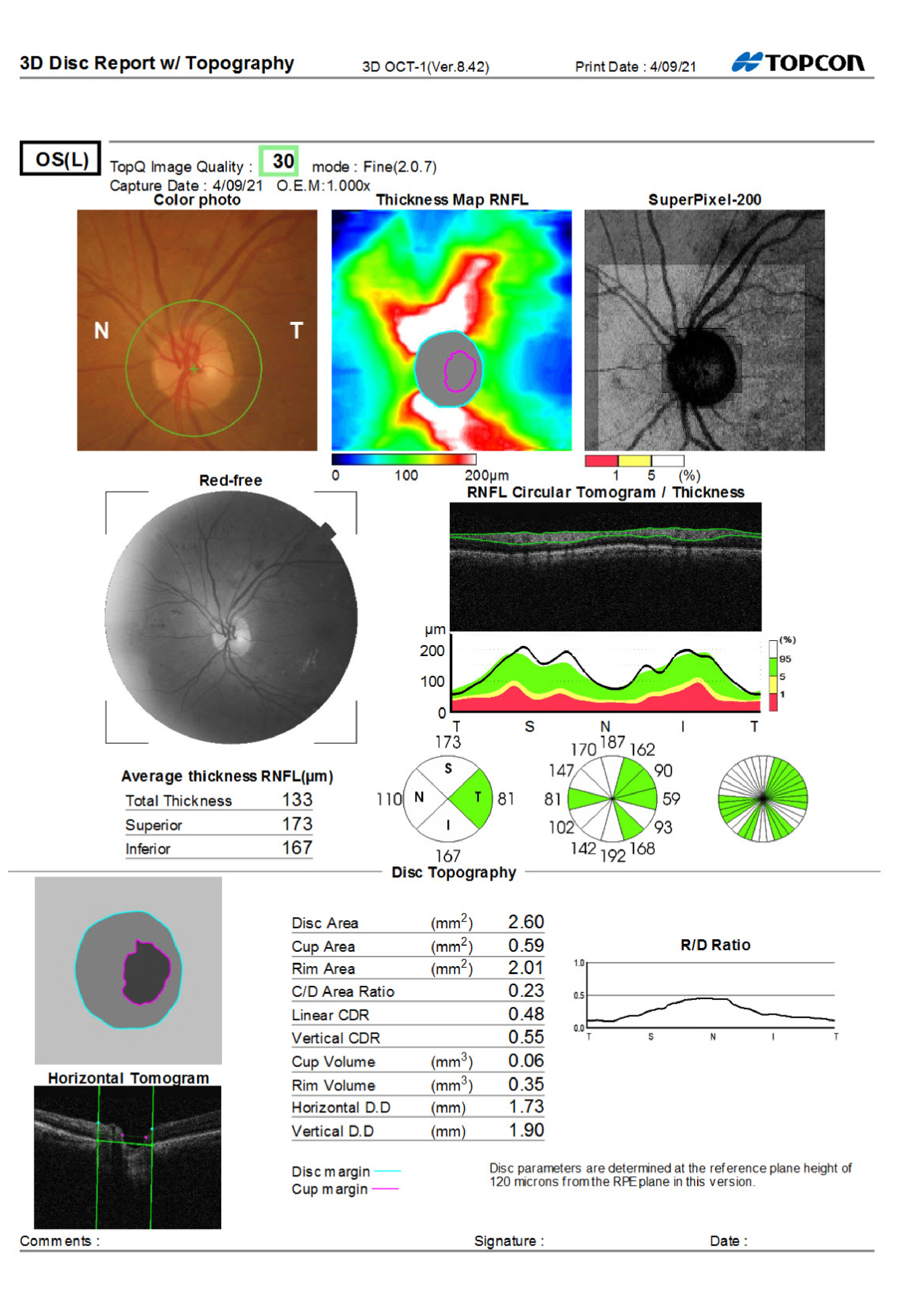
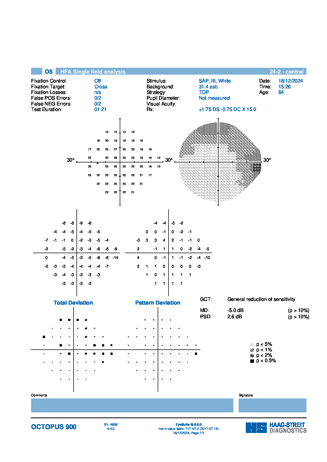
A 60-year-old female with a long history of diabetes and hypertension presented for a follow-up visit. Eye pressures are stable, and visual acuity remains excellent. Retinal examination revealed scattered macular exudates and peripheral changes, consistent with chronic retinal disease.
The optometrist noted that ongoing monitoring is essential and emphasized tight blood sugar control. Follow-up with the ophthalmologist is planned.
What is the recommended follow-up interval for monitoring this patient’s retinal status, and should additional imaging or intervention be considered at this time?
An ophthalmology subspecialist provided a virtual consult within 1-2 weeks through Care1. Scroll below to see their diagnosis.
Care1 Subspecialist’s Key Takeaways
Referral was reviewed by the specialist, who agreed with the concerns raised during the exam. Imaging showed notable retinal changes that require close monitoring, and the specialist advised that the patient is at higher risk of needing treatment soon. Follow-up with the specialist in four months is recommended, while the optometrist continues routine observation and care.
Care1 AI’s Clinical Insight
Diabetic retinopathy (DR) is a progressive microvascular complication of diabetes that affects the retinal blood vessels and can lead to vision loss. Early stages, known as nonproliferative diabetic retinopathy (NPDR), are characterized by microaneurysms, hemorrhages, and capillary leakage, while advanced stages may progress to proliferative diabetic retinopathy (PDR) with abnormal new vessel growth. Risk factors include poor glycemic control, hypertension, and duration of diabetes. Pathophysiologically, ischemic retina releases VEGF, causing increased vascular permeability, retinal swelling, and angiogenesis.
Did You Know?
The microaneurysms and retinal hemorrhages seen in diabetic retinopathy often contain high levels of vascular endothelial growth factor (VEGF), a protein that promotes new blood vessel formation and increased vascular permeability. This suggests that abnormal signaling in response to retinal ischemia plays a central role in disease progression.
Anderson DH, Mullins RF, Hageman GS, Johnson LV. A role for local inflammation in the formation of drusen in the aging eye. American Journal of Ophthalmology. 2002;134(3):411–431. doi:10.1016/s0002-9394(02)01357-7