37-year-old male with incidental vitreous traction OS and normal exam findings, raising management and referral considerations.
Case Study: Large Temporal Vitreous Tuft
An optometrist uploaded this case to Care1.
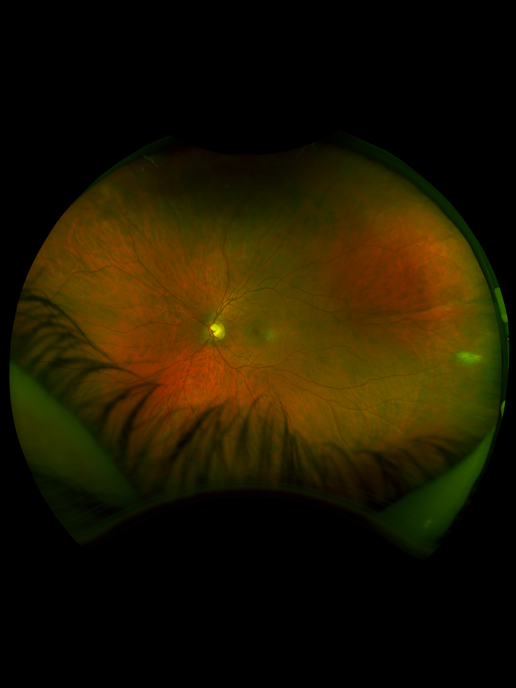
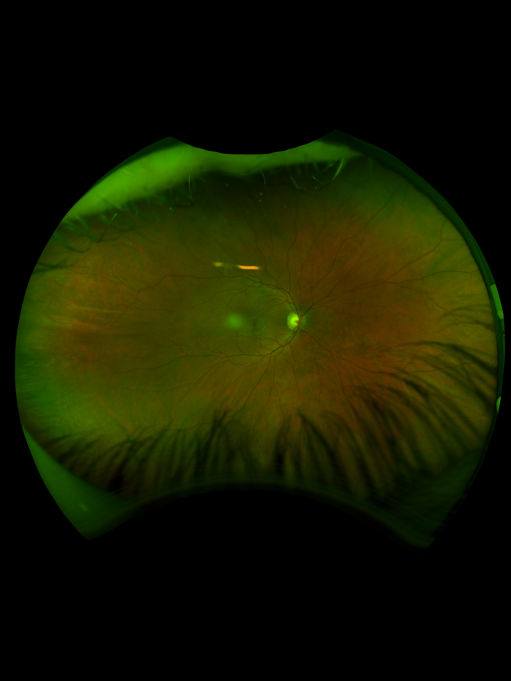
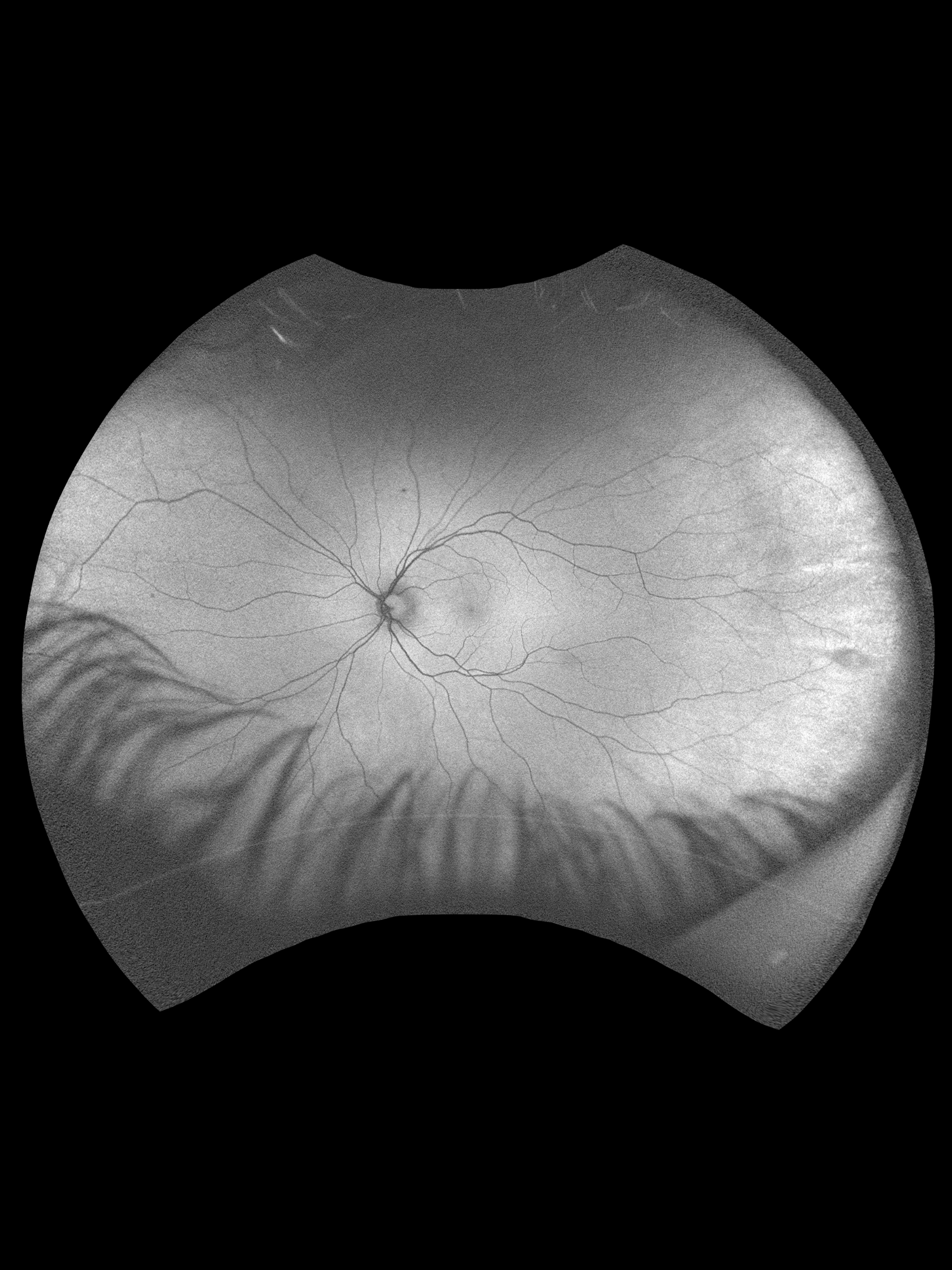
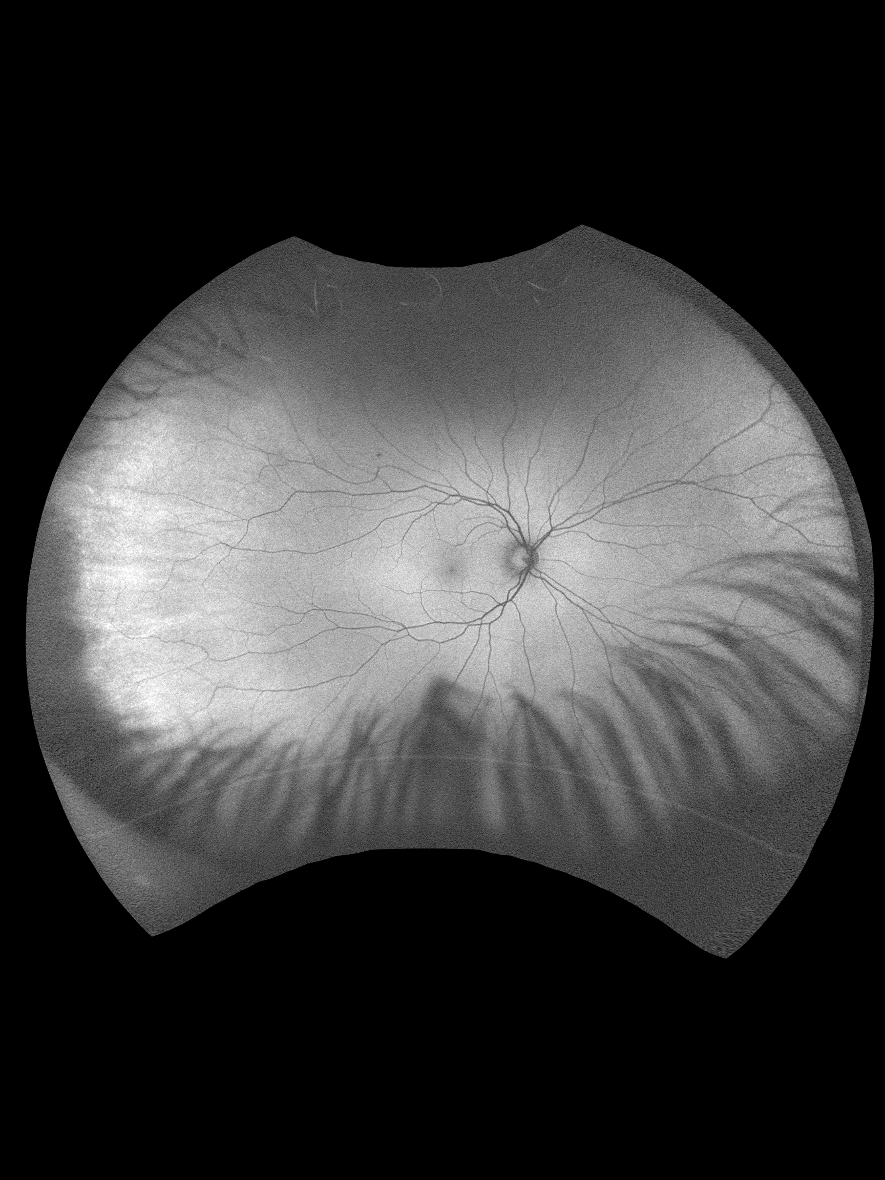
A 37-year-old male was referred for evaluation of a large temporal vitreous tuft OS found during a routine exam. He is asymptomatic with no prior imaging for comparison and a history of LASIK OU. BCVA was not documented. IOPs were 12 mmHg OD and 13 mmHg OS. Anterior segment was largely unremarkable aside from trace blepharitis and mild pigment findings OD. Posterior segment showed healthy discs with C/D ratios of 0.65 OD and 0.60 OS. Peripheral exam revealed a large vitreous tuft with traction at 3 o’clock OS.
Would you recommend observation or non-urgent retinal consultation for this vitreous traction finding?
A retina specialist provided a virtual consult within 1-2 weeks through Care1. Scroll below to see their diagnosis.
Care1 Ophthalmologist's Teleconsult
The findings are consistent with a vitreous tuft rather than a retinal tear, and immediate treatment is not required. These lesions are typically benign but may warrant monitoring due to their association with vitreoretinal traction. Referral for a non-urgent in-person retinal evaluation is reasonable to confirm the diagnosis and counsel the patient. Continued observation with routine follow-up is appropriate, with education on symptoms of retinal complications.
Further in-person evaluation and management is recommended for this patient.
Care1 AI’s Clinical Insight
Vitreoretinal tufts are peripheral retinal abnormalities characterized by focal vitreous adhesion and retinal gliosis. They are often discovered incidentally during dilated fundus examination and may be associated with increased vitreoretinal traction. While generally benign, they can occasionally predispose to retinal breaks under tractional forces.
Practice at the highest level of medicine
✔ Specialist consults within 1-2 weeks ✔ AI-powered clinical support ✔ Greater confidence in complex decision making
Vitreoretinal tufts are present in up to 5 percent of the population but account for a disproportionately higher percentage of retinal tears due to focal vitreous traction.
Reference: Byer NE. Lattice degeneration of the retina. Surv Ophthalmol. 1979;23(4):213–248.
Clinical Pearls
Large vitreous tufts are usually benign but require monitoring
Peripheral traction may increase risk of retinal tears
Educate patients on symptoms of retinal detachment