43-year-old with bilateral drusen and normal vision raises concern for early macular changes and genetic risk.
Case Study: Bilateral Drusen in Young Adult
An optometrist uploaded this case to Care1.
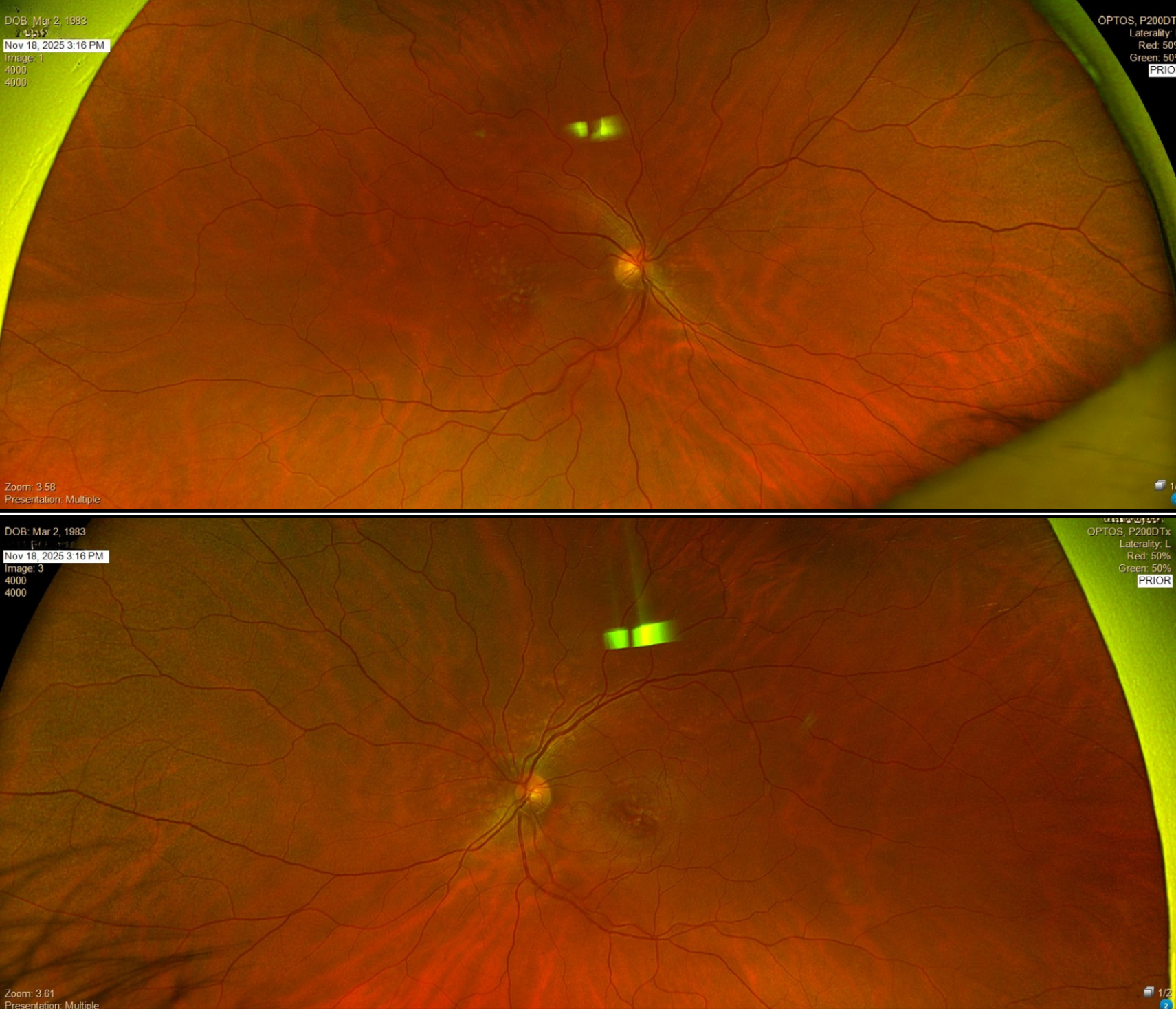
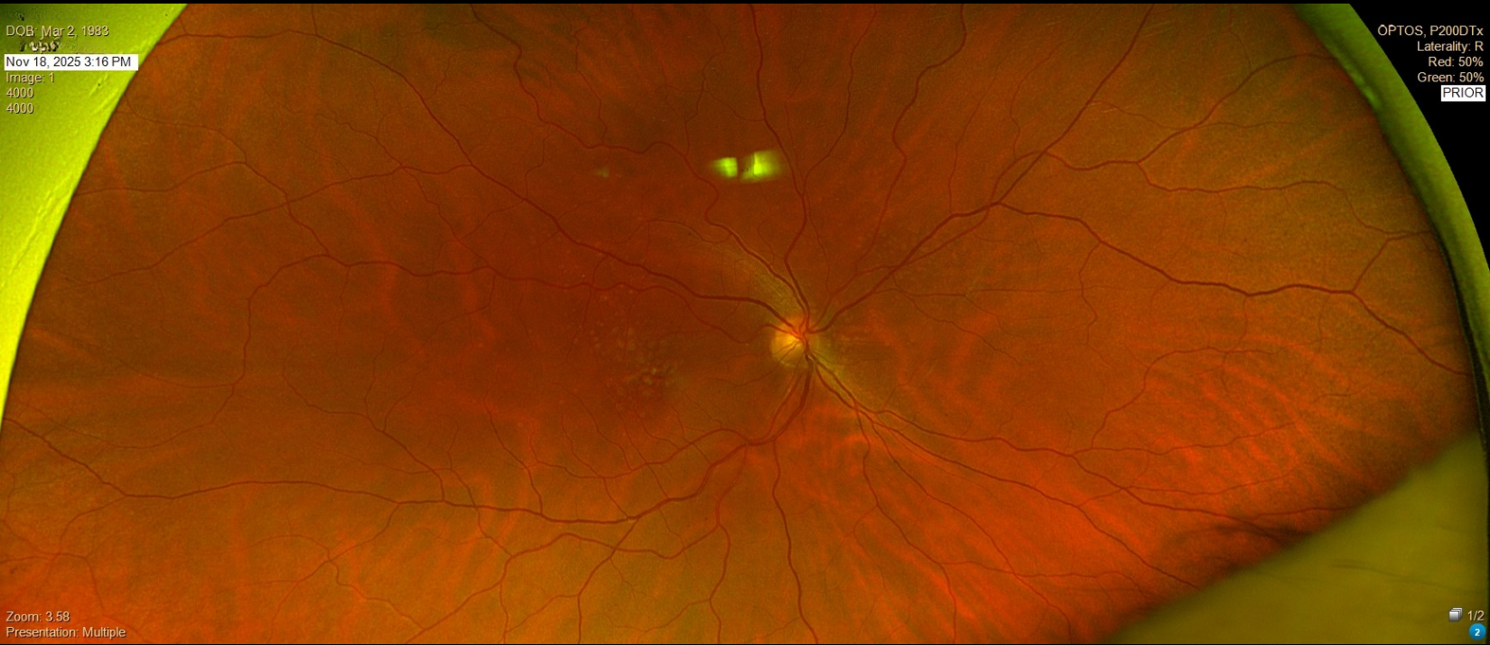
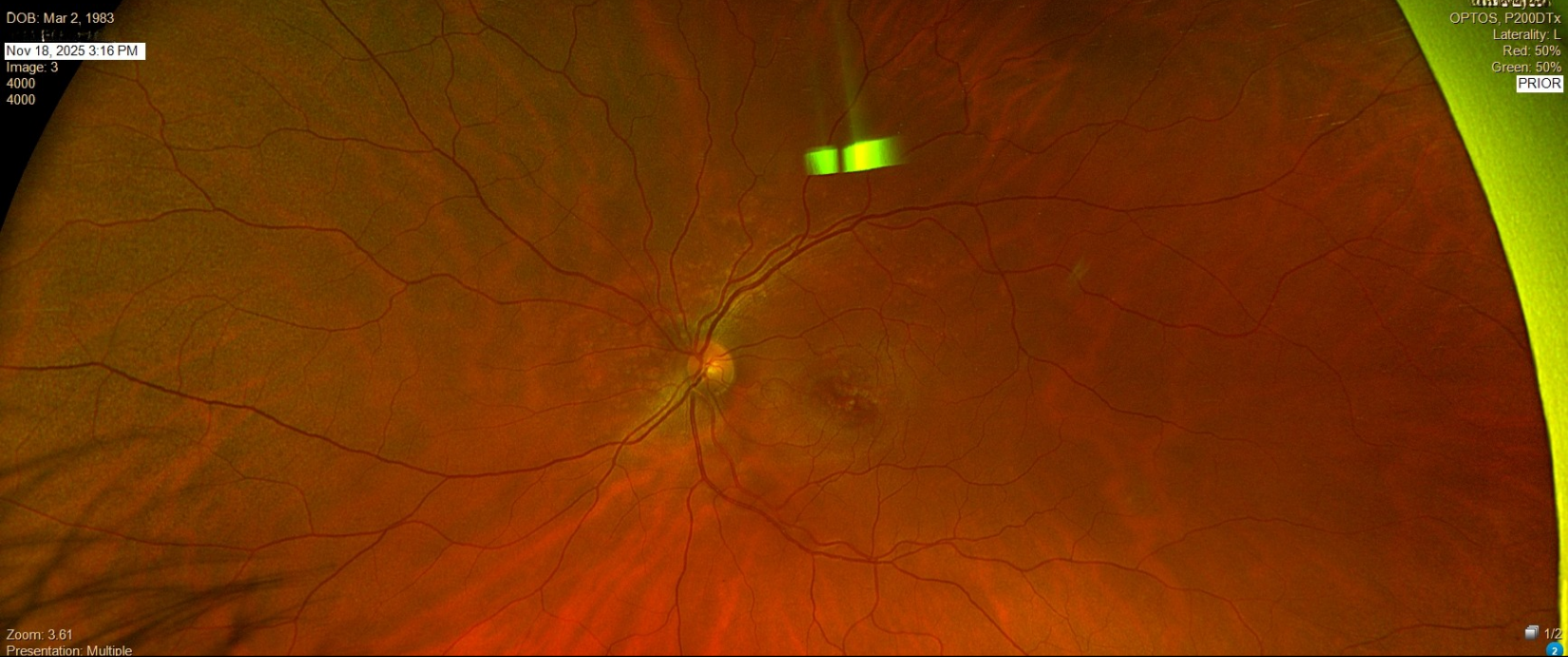
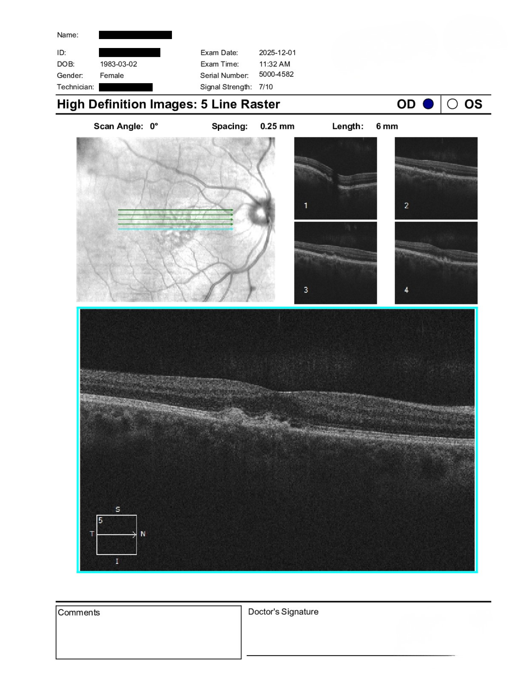
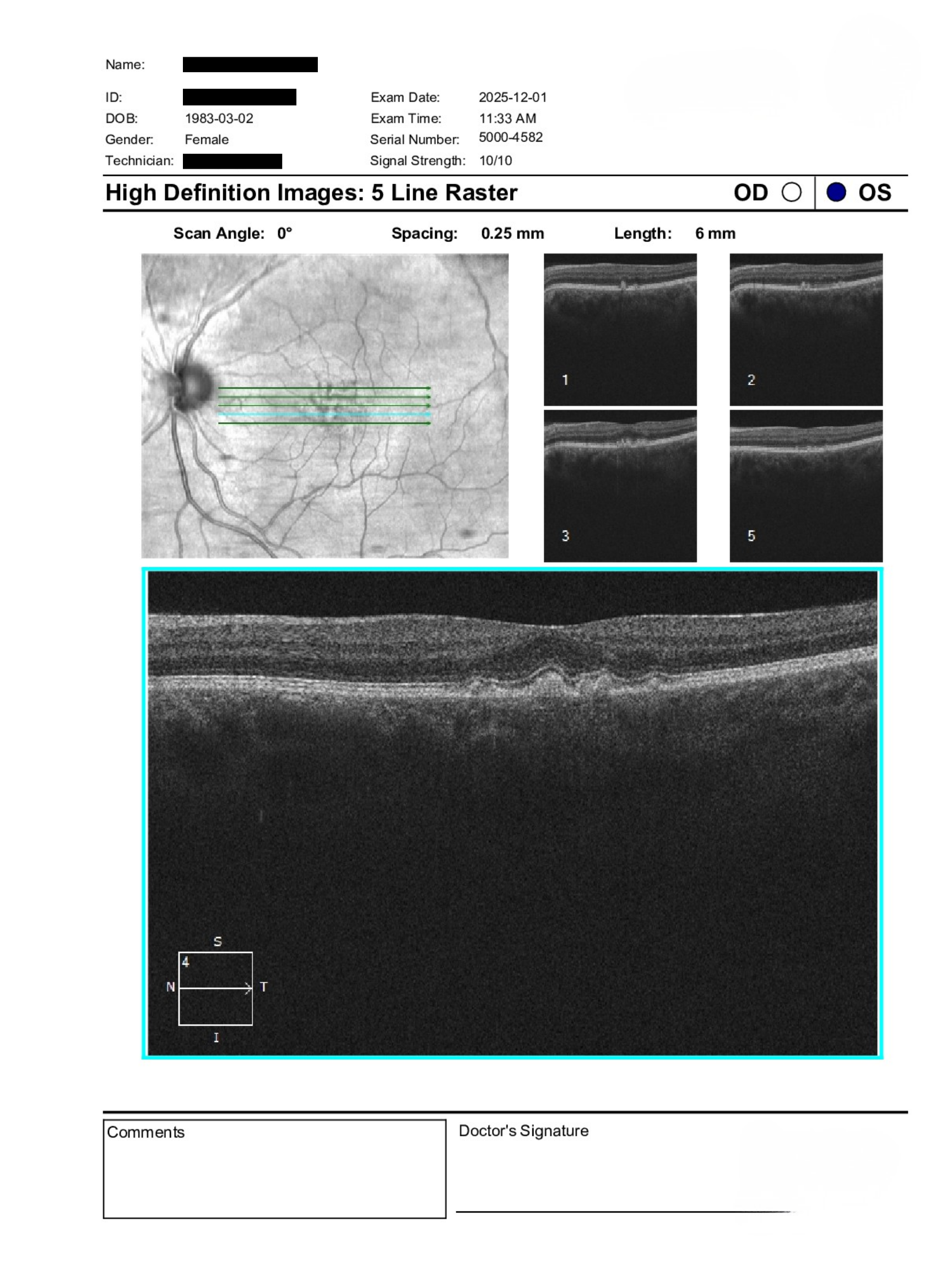
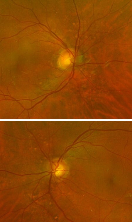
A 43-year-old patient was referred for evaluation of suspected macular dystrophy with a history of drusen and prior genetic testing suggesting possible retinal associations. BCVA is 20/20 in both eyes with IOPs of 14 mmHg OD and 16 mmHg OS. Anterior segment examination is unremarkable aside from trace nuclear sclerosis. Posterior segment shows bilateral drusen with otherwise healthy discs, vasculature, and peripheral retina. OCT imaging was performed to further assess macular structure.
What is the role of genetic testing and referral in a patient with suspected inherited macular changes but preserved vision?
A retina specialist provided a virtual consult within 1-2 weeks through Care1. Scroll below to see their diagnosis.
Care1 Ophthalmologist's Teleconsult
Findings are most consistent with a pattern dystrophy given the patient’s age and imaging features. These conditions may have a genetic component, though routine genetic testing is not typically required unless there are systemic associations, progressive vision loss, or clear signs of inherited retinal disease such as photoreceptor disruption.
Pattern dystrophies can overlap with other inherited or metabolic syndromes, but many cases remain stable with good visual function. Referral for genetic evaluation is optional and should be guided by patient interest and clinical progression. Ongoing monitoring is appropriate, with consideration for in-person ophthalmology assessment if changes develop.
Care1 AI’s Clinical Insight
Pattern dystrophies are a group of inherited macular disorders characterized by pigment deposition at the level of the retinal pigment epithelium. They often present in mid-adulthood and may mimic age-related macular degeneration on clinical exam. Visual prognosis is generally good, though slow progression can occur over time.
Practice at the highest level of medicine
✔ Specialist consults within 1-2 weeks ✔ AI-powered clinical support ✔ Greater confidence in complex decision making