90-year-old male follow-up shows mild epiretinal changes, optic nerve cupping, and slight RNFL thinning; monitoring interval under review.
Case Study: Elevated Eye Pressures and Optic Changes
An optometrist uploaded this case to Care1.
A 90-year-old male patient presented for a routine follow-up, reporting no new visual concerns. He takes multiple systemic medications and has a history of posterior vitreous detachment in the left eye. Ocular history is notable for open-angle glaucoma, previously treated with Travatan Z, and currently managed with Vyzulta (latanoprostene bunod 0.024%) once nightly OU.
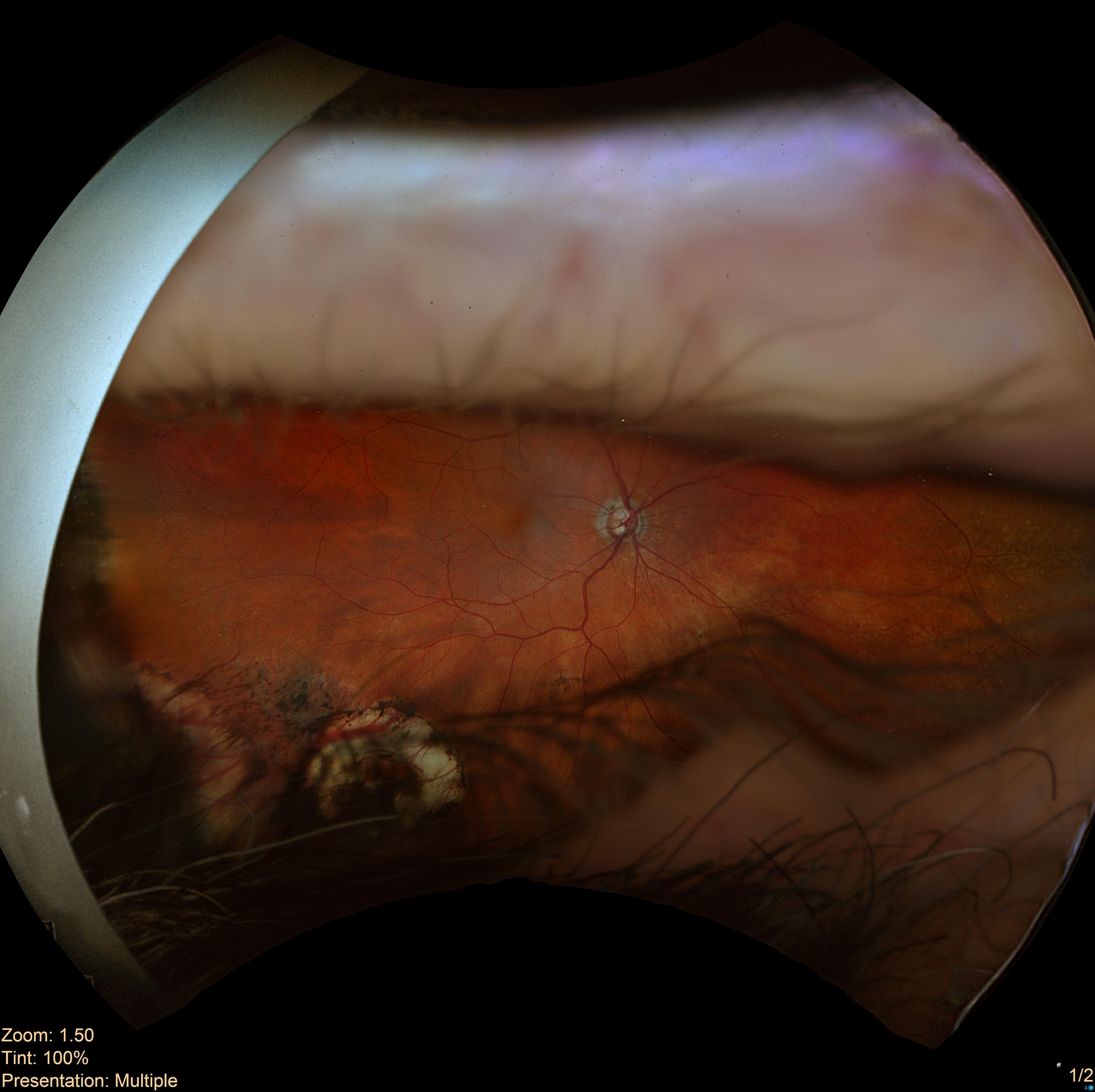
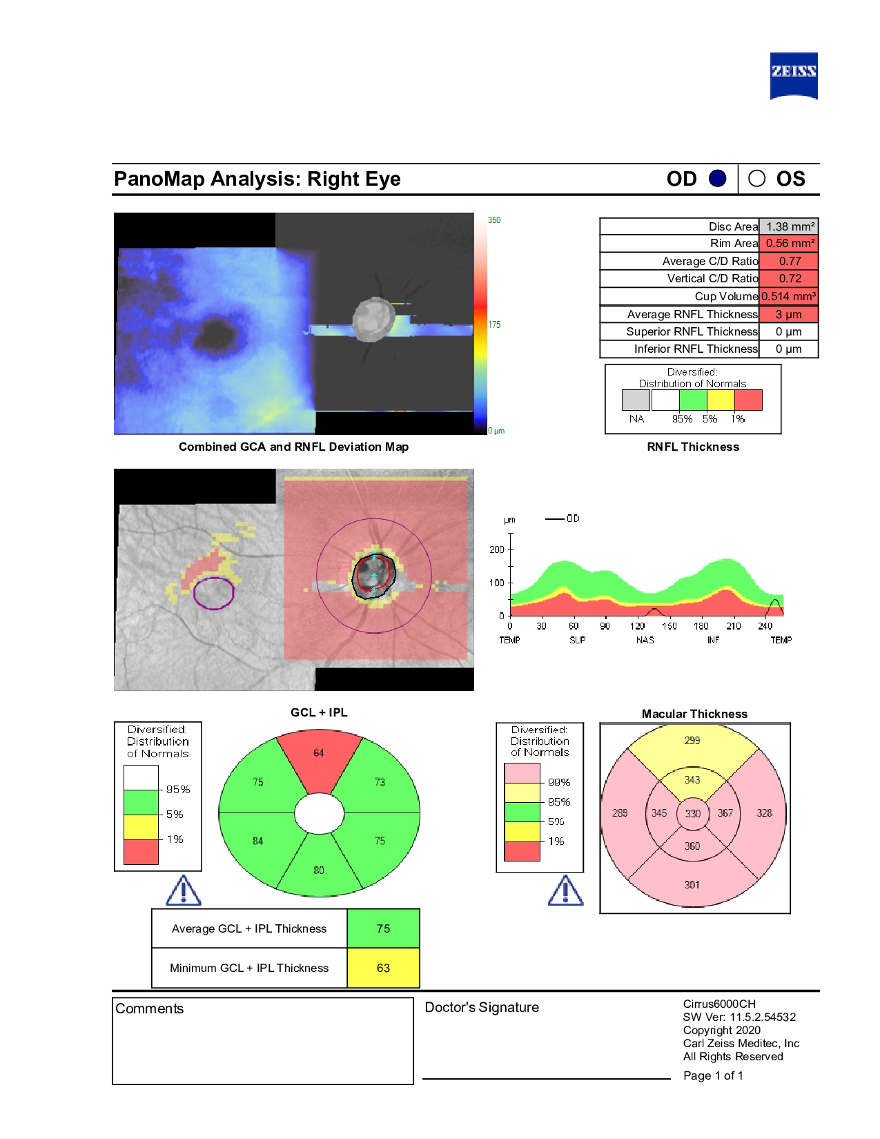
In-office examination showed intraocular pressure of 21 mmHg OD and 22 mmHg OS. Anterior segment evaluation was unremarkable. Posterior segment examination demonstrated mild epiretinal membranes in both eyes, peripheral cryo scarring in the right eye, and a posterior vitreous detachment in the left eye. Optic nerve assessment revealed large cups (C/D 0.80 H/0.80 V in both eyes), with a thinner superior rim in the right eye and a shallower cup in the left eye. OCT indicated slight thinning of the retinal nerve fiber layer in the right eye.
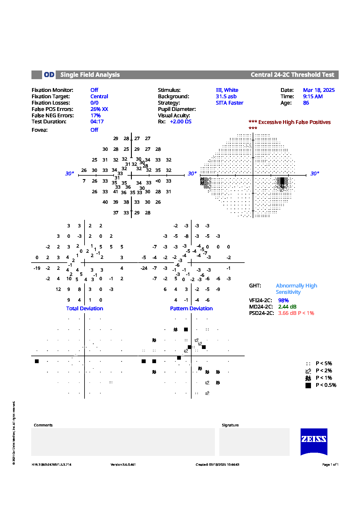
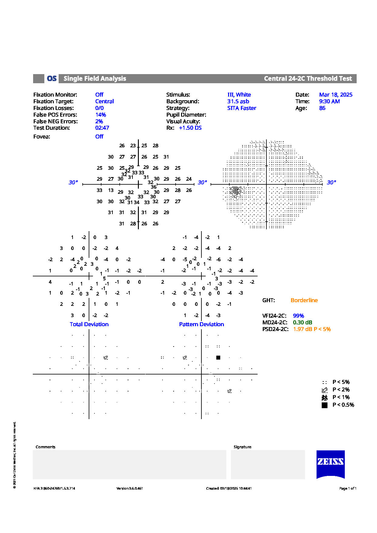
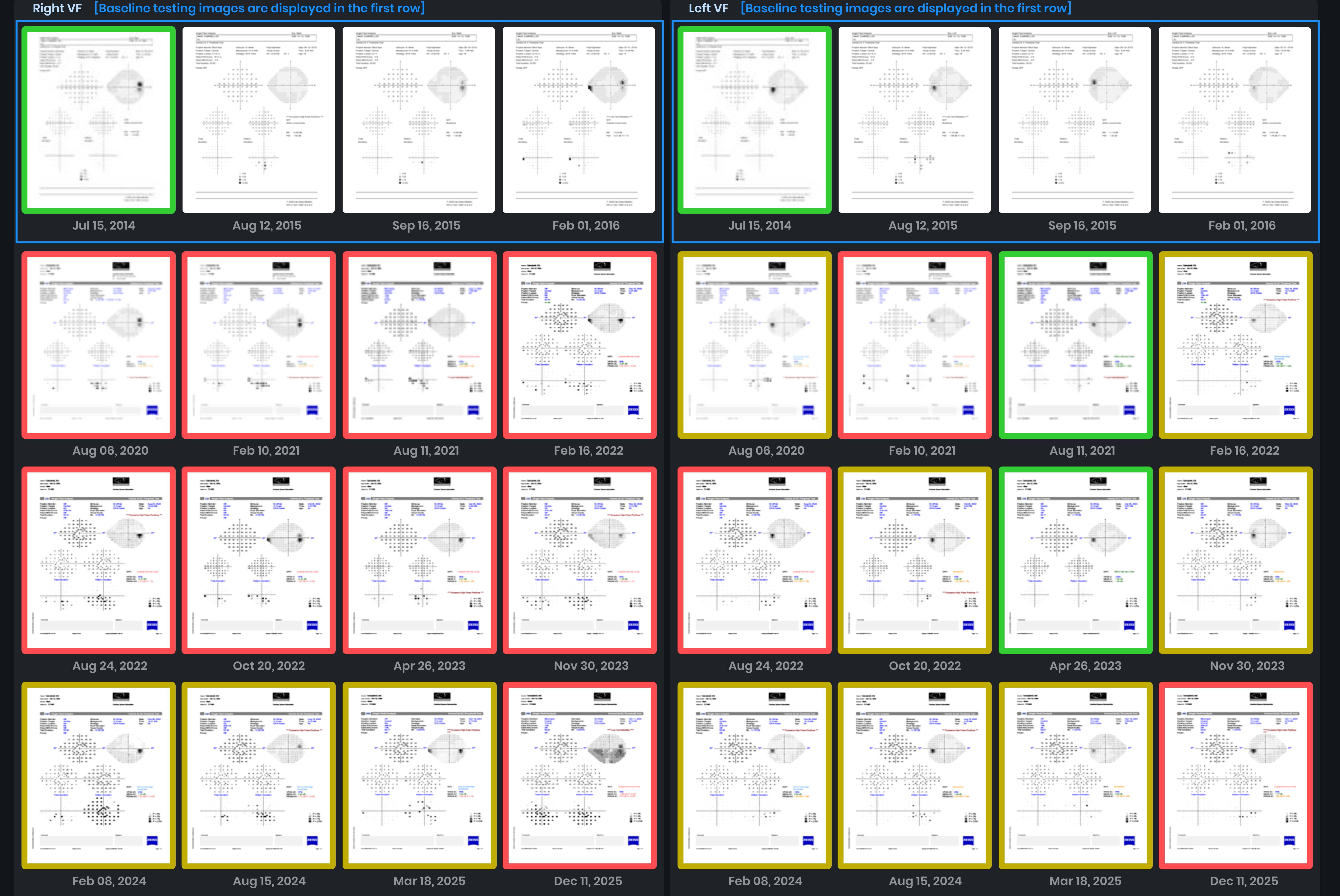
Visual field testing showed high false positives OD, with no definitive defects OS.
Given the optic nerve appearance, peripheral retinal changes, and subtle structural thinning, would you recommend continuing the current monitoring interval, or is a shorter follow-up or additional intervention indicated at this stage?
An ophthalmology subspecialist provided a virtual consult within 1-2 weeks through Care1. Scroll below to see their diagnosis.
Care1 Subspecialist’s Key Takeaways
The patient is on Vyzulta OU with intraocular pressure above the comfort zone. OCT and visual field testing were unreliable, though ganglion cell analysis remains stable.
Given the difficulty obtaining consistent measurements and the advanced stage of the disease, the specialist recommends escalating treatment. This could include Selective Laser Trabeculoplasty (SLT) or switching to a dual-agent therapy to better achieve target pressures.
Care1 AI’s Clinical Insight
Standard Automated Perimetry (SAP) is a computerized method to measure the visual field by presenting light stimuli at set locations and recording the dimmest visible stimulus at each point. It provides standardized, reproducible visual field data that help assess the extent and progression of visual field loss in conditions like glaucoma. SAP typically uses white‑on‑white testing with algorithms such as SITA to efficiently estimate thresholds across the field. The test also includes measures to monitor fixation and reliability of results.
Did You Know?
One interesting fact from the Standard Automated Perimetry article is that a significant number of retinal ganglion cells may be lost before a visual field defect is detectable on standard automated perimetry. Histological studies have shown that many ganglion cells can be damaged before loss is apparent on SAP, which has motivated development of alternative perimetric tests that target specific ganglion cell pathways for earlier detection.
Kerrigan‑Baumrind LA, Quigley HA, Pease ME, et al. Number of ganglion cells in glaucoma eyes compared with threshold visual field tests. Invest Ophthalmol Vis Sci. 2000;41(3):741‑748.