66-year-old with high eye pressures, myopic discs, mild RNFL thinning, and normal visual fields; monitoring and follow-up decisions under consideration.
Case Study: Elevated Pressures with Myopic Discs
An optometrist uploaded this case to Care1.
A 66-year-old female patient presented for a routine follow-up, reporting no new visual concerns. She mainly wears monthly disposable contact lenses, though tolerance has decreased over time, and occasionally uses glasses. Systemic history is notable for high blood pressure. She is not currently using any topical pressure-lowering drops.
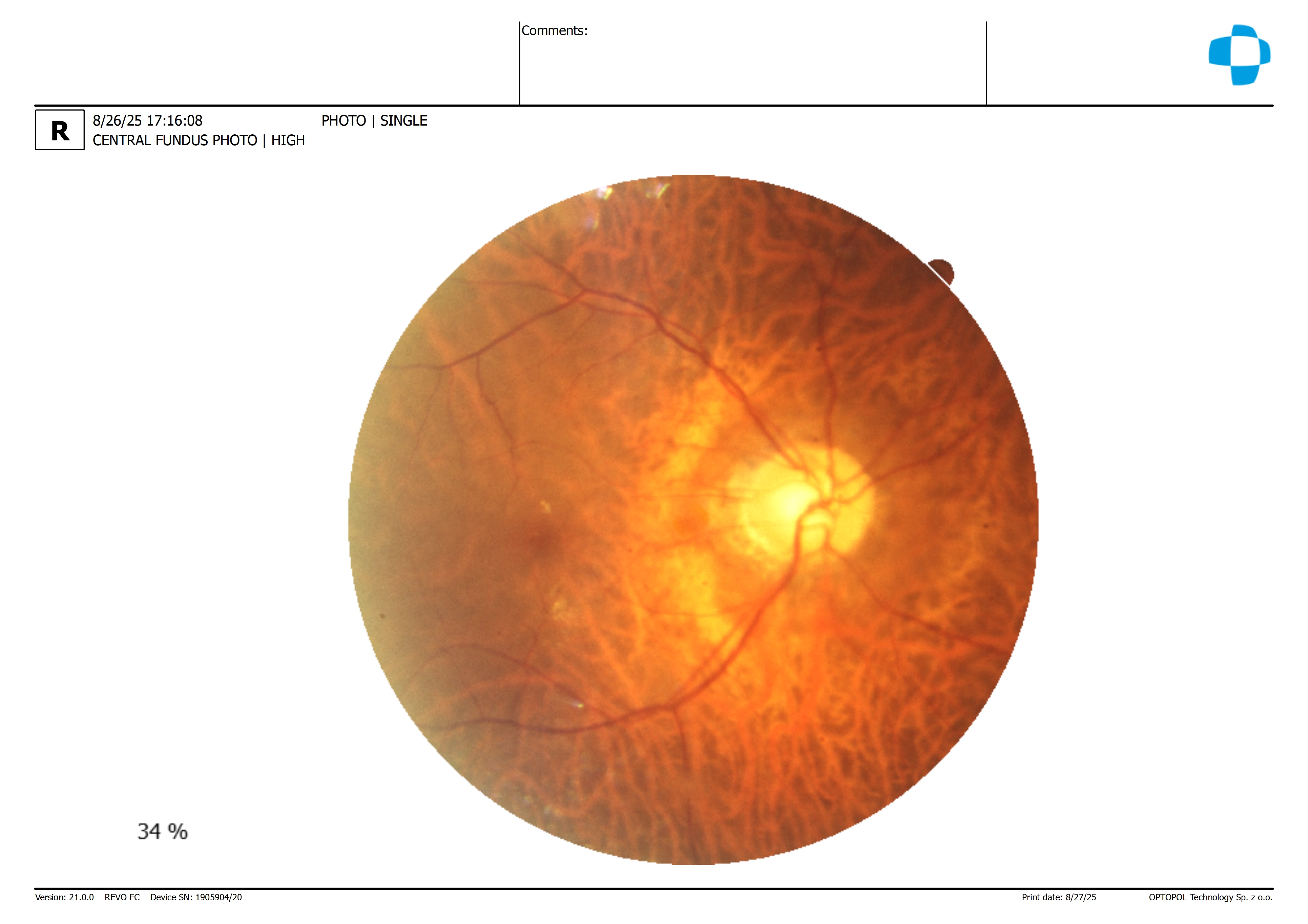
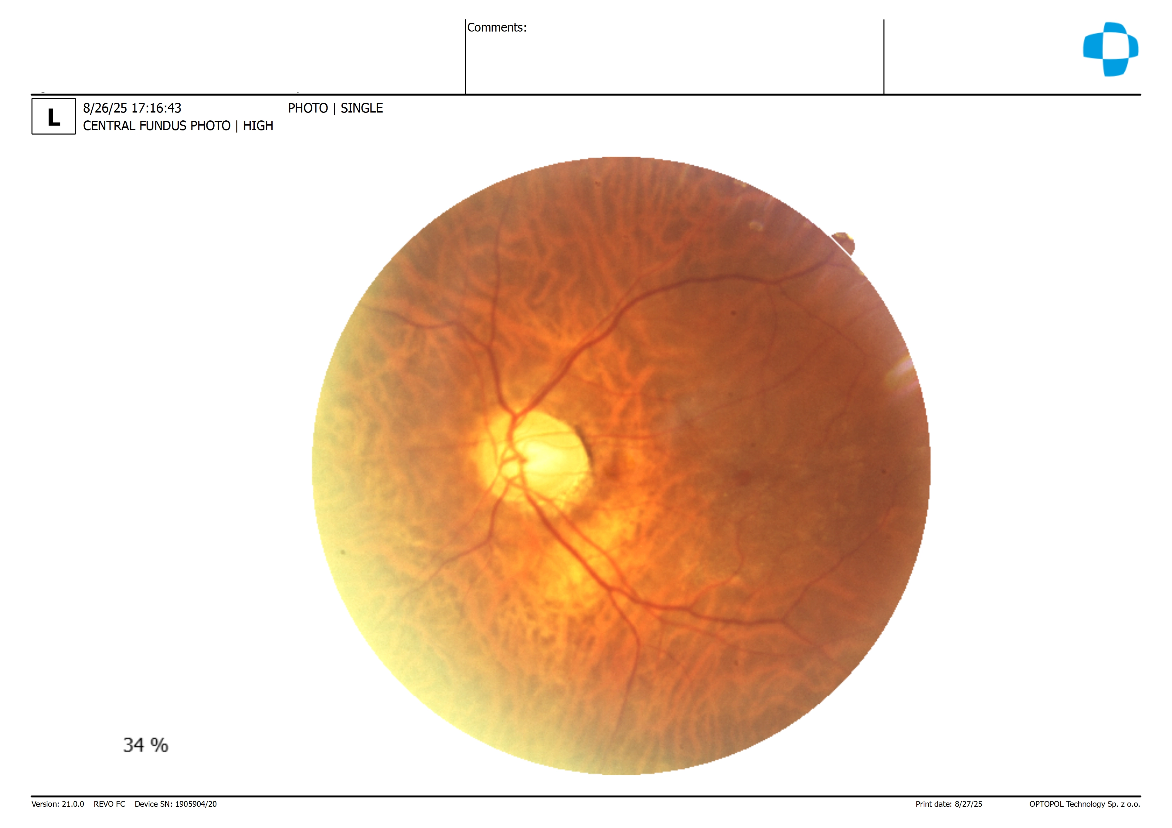
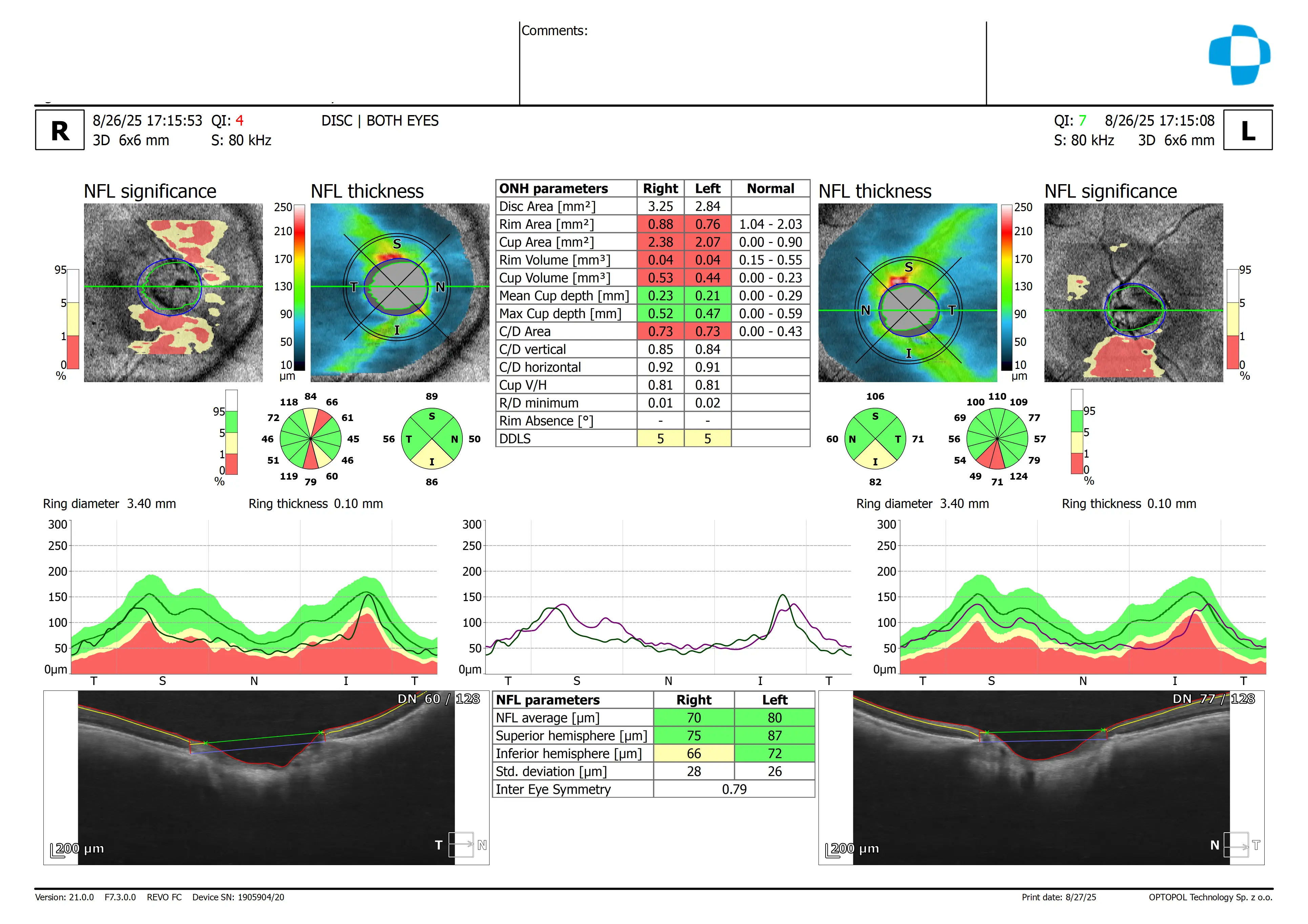
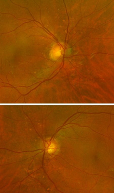
In-office examination showed best-corrected visual acuity of 20/20 in both eyes. Intraocular pressure was IOP OD 23 mmHg, OS 24 mmHg. Anterior segment examination revealed clear lids, lashes, conjunctiva, corneas, and deep quiet anterior chambers. Lens changes were minimal. Optic nerve evaluation demonstrated large discs with intact neural rims and myopic crescents, with mild inferior retinal nerve fiber layer thinning. Posterior segment evaluation showed a mild epiretinal membrane in the left eye.
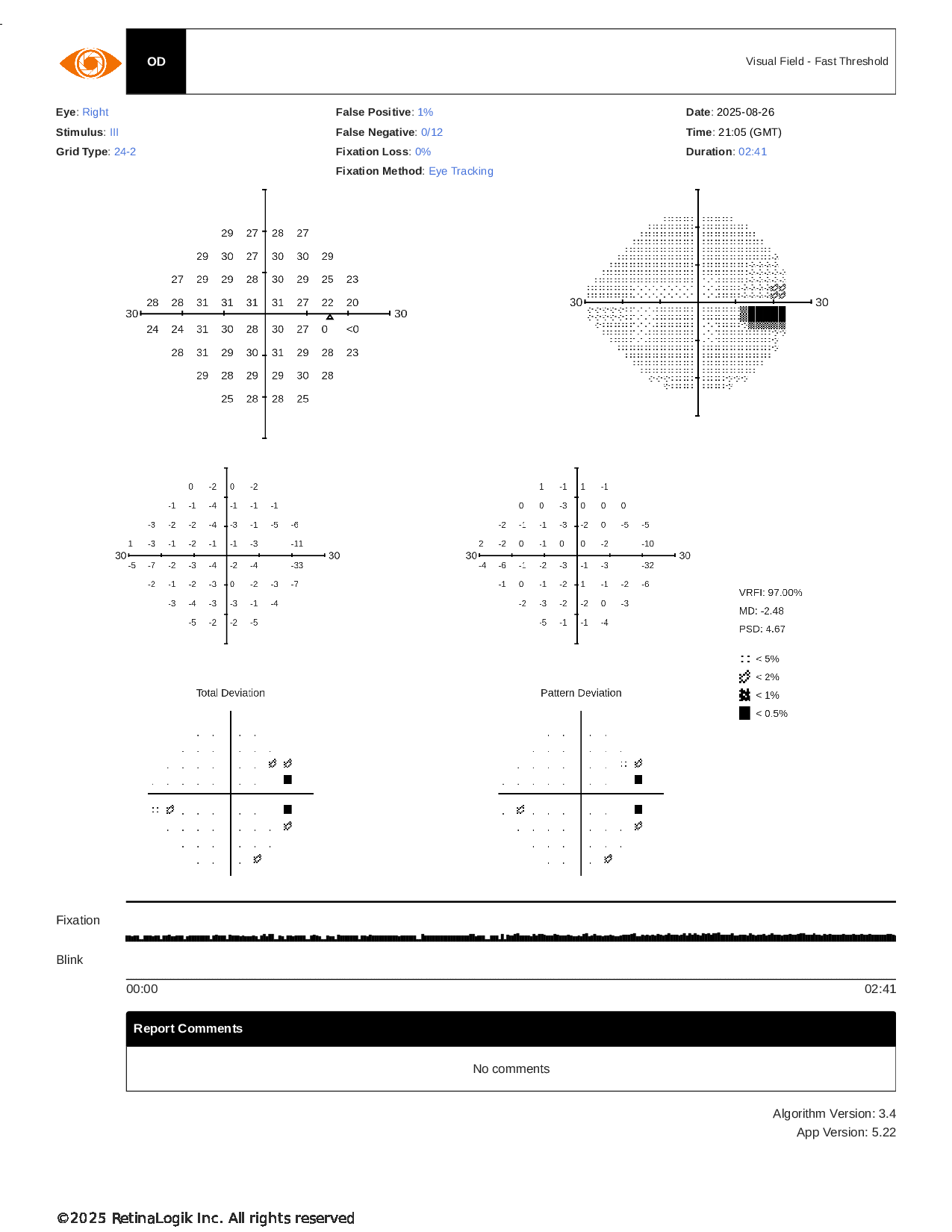
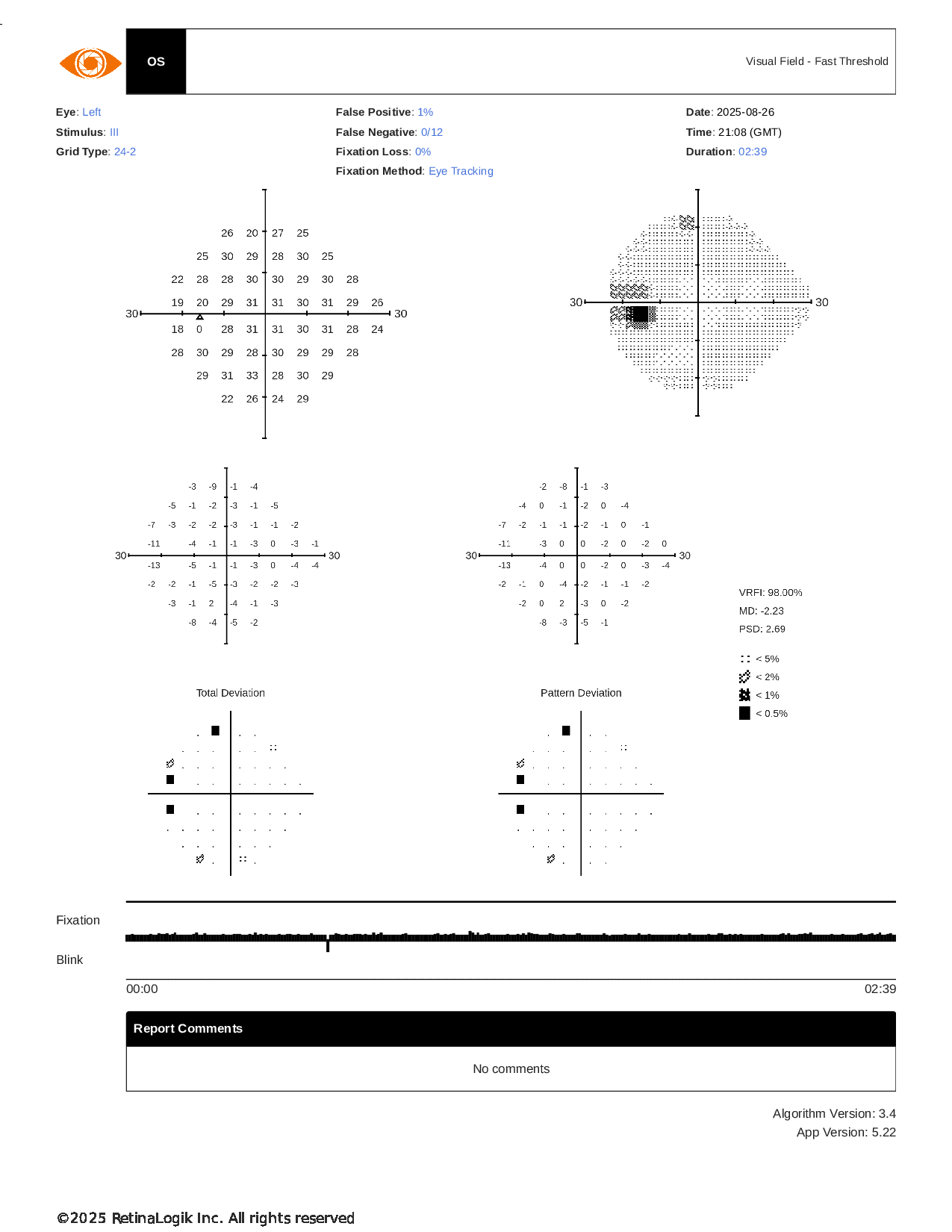
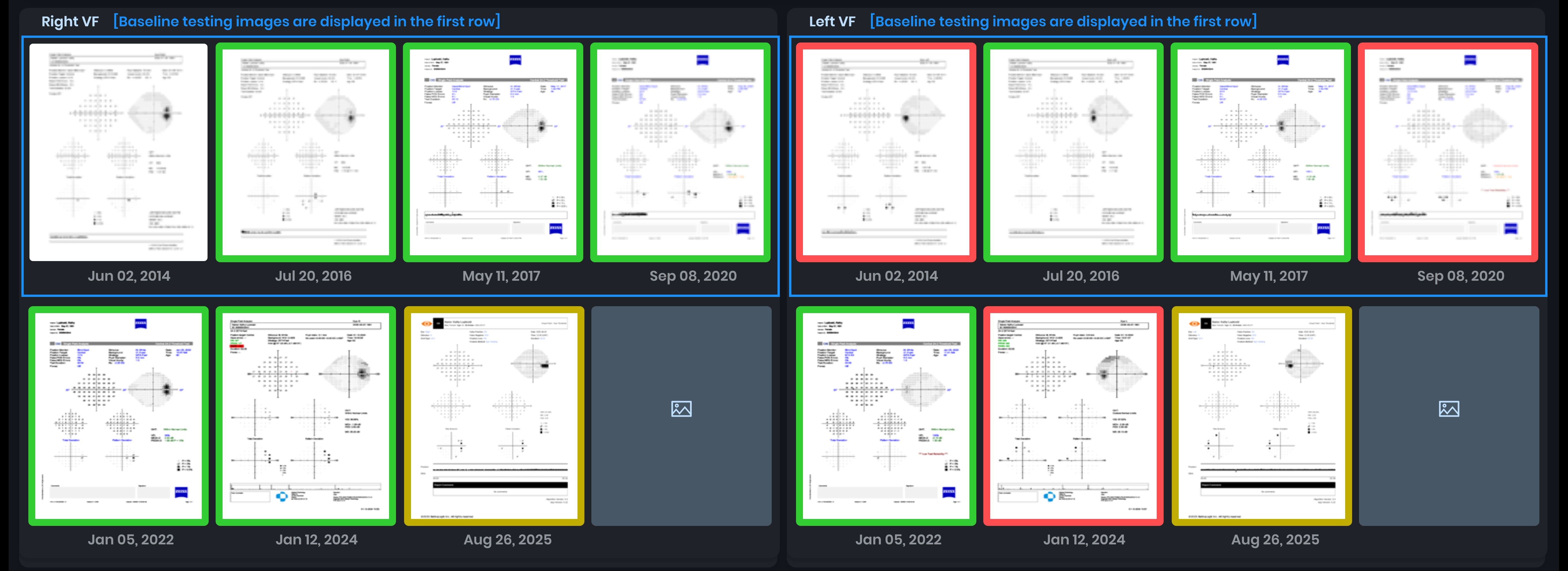
Visual field testing was normal in both eyes. Based on these findings, the patient’s pressures are elevated but she is not at surgical threshold, and yearly monitoring was recommended.
Given the elevated eye pressures and subtle structural changes, would you recommend continuing yearly follow-up, or would a shorter interval or intervention be more appropriate at this stage?
An ophthalmology subspecialist provided a virtual consult within 1-2 weeks through Care1. Scroll below to see their diagnosis.
Care1 Subspecialist’s Key Takeaways
The patient has classic myopic optic nerves with large cups and enlarged blind spots. Given this history, the specialist recommends careful management of eye pressures around 23–25 mmHg.
OCT imaging revealed thinning of the retinal nerve fiber layer (RNFL) inferiorly in both eyes, without corresponding visual field defects.
As a first-line intervention, Selective Laser Trabeculoplasty (SLT) is recommended, with referral to an ophthalmologist. A short-term follow-up in 1-2 months is advised to closely monitor pressures and structural changes.
Care1 AI’s Clinical Insight
Spectral-domain optical coherence tomography (SD-OCT) is a noninvasive imaging technology that provides high-resolution visualization of retinal and optic nerve head structures, including the retinal nerve fiber layer and ganglion cell layers. It allows for objective measurement and comparison to normative databases to detect structural changes. SD-OCT can track changes over time through repeatable scans, helping distinguish true anatomical loss from normal variation. Interpretation requires attention to factors like signal quality and individual anatomical differences.
Did You Know?
Studies have shown that retinal nerve fiber layer (RNFL) thickness measurements obtained with spectral‑domain OCT (SD‑OCT), especially when analyzed by quadrant or clock‑hour sectors, can help distinguish glaucomatous eyes from normal eyes with high diagnostic accuracy.
Mwanza JC, Oakley JD, Budenz DL, Anderson DR. Ability of Cirrus HD‑OCT optic nerve head parameters to discriminate normal from glaucomatous eyes. Ophthalmology. 2011;118:241‑248.