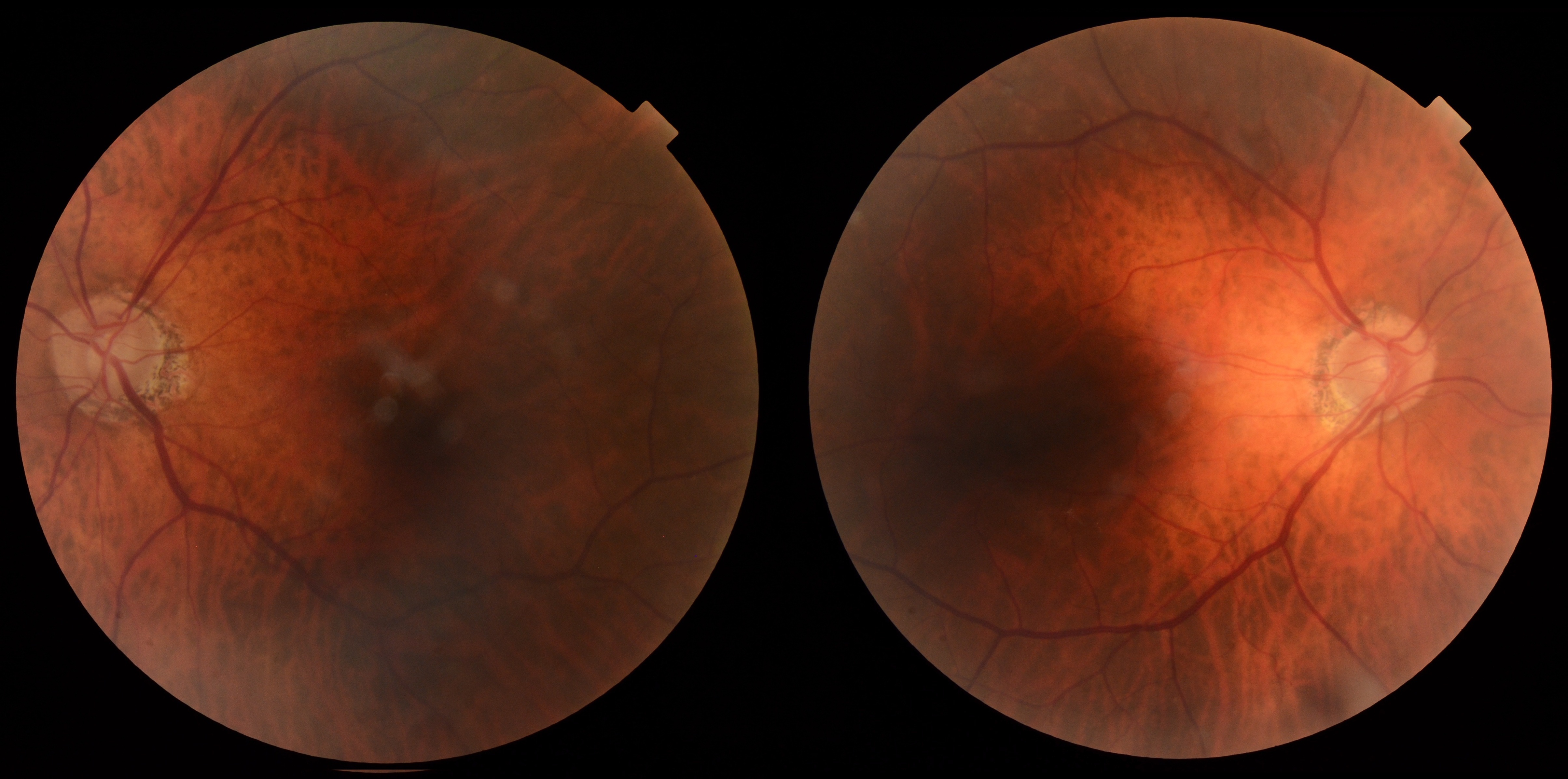
68-year-old female with new left-eye floaters, vitreous traction, and retinal hemorrhage requiring retinal management review.
Case Study: New Floaters With Retinal Hemorrhage
An optometrist uploaded this case to Care1.
A 68-year-old female was referred for evaluation of new floaters in the left eye. BCVA was 20/20 OU, and IOP measured 12 mmHg OD and 11 mmHg OS. Anterior segment findings were unremarkable. Posterior examination revealed a hemorrhagic posterior vitreous detachment with superotemporal vitreous traction and an operculated retinal lesion in the left eye. Follow-up imaging showed enlargement of the tractional area despite improved symptoms, with no obvious retinal detachment identified on dilated examination.
Would this symptomatic hemorrhagic peripheral retinal lesion benefit from retinal treatment or continued observation?
A retina specialist provided a virtual consult within 1-2 weeks through Care1. Scroll below to see their diagnosis.
Care1 Ophthalmologist's Teleconsult
Review of the Optos imaging demonstrated an unremarkable right eye and a superotemporal peripheral retinal hole in the left eye associated with adjacent retinal hemorrhages. The lesion appeared flat without evidence of subretinal fluid.
In the setting of an acute posterior vitreous detachment, careful peripheral retinal examination remains essential for identifying retinal breaks and assessing detachment risk. While observation may be appropriate for some asymptomatic peripheral retinal holes, the presence of acute symptoms and associated retinal hemorrhage increases concern for progression. Given these findings, in-person retinal evaluation and treatment were recommended.
Care1 AI's Clinical Insight
Posterior vitreous detachment occurs when the vitreous separates from the retinal surface and becomes increasingly common with age. Acute symptomatic cases warrant careful peripheral retinal examination because retinal tears and holes may develop during vitreoretinal separation. Associated vitreous or retinal hemorrhage can increase the likelihood of finding a clinically significant retinal break.
Unlock your full revenue potential
✔ Specialist consults within 1-2 weeks ✔ AI-powered clinical support ✔ Greater confidence in complex decision making
Retinal breaks identified in eyes with acute symptomatic posterior vitreous detachment are significantly more likely when vitreous or retinal hemorrhage is present, making hemorrhage one of the most important predictive clinical findings during evaluation.
Reference: Coffee RE, Westfall AC, Davis GH, Mieler WF, Holz ER. Symptomatic posterior vitreous detachment and the incidence of delayed retinal breaks: case series and meta-analysis. Am J Ophthalmol. 2007;144(3):409-413. doi:10.1016/j.ajo.2007.05.002
Clinical Pearls
New floaters require careful peripheral retinal assessment
Retinal hemorrhage increases concern for retinal breaks