75-year-old female with liver disease and alcohol history. Vision stable, possible intervention needed. What follow-up interval is recommended?
Case Study: Vision Changes with Subretinal Atrophy
An optometrist uploaded this case to Care1.
A 75-year-old female presented for a follow-up visit. She has a history of significant alcohol use (approximately 8 oz gin and 8 oz tonic daily), advanced liver disease (cirrhosis, not a transplant candidate), and smokes occasionally (about 1/5 pack per day). Despite these systemic risks, her vision remains surprisingly good.
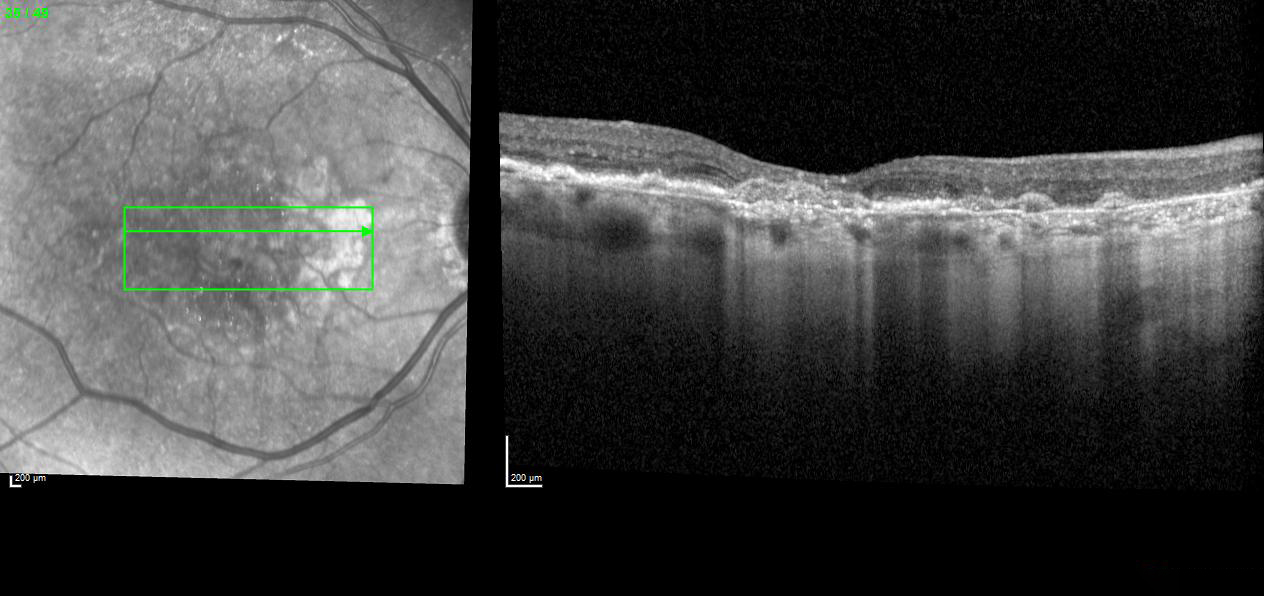
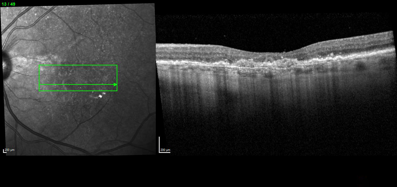
In-office testing and imaging showed findings that may require intervention, though the exact timing and urgency are unclear. The optometrist noted that treatment may be needed, but wanted guidance on follow-up.
If intervention is required now, what is the recommended follow-up interval for this patient?
An ophthalmology subspecialist provided a virtual consult within 1-2 weeks through Care1. Scroll below to see their diagnosis.
Care1 Retina Surgeon’s Key Takeaways
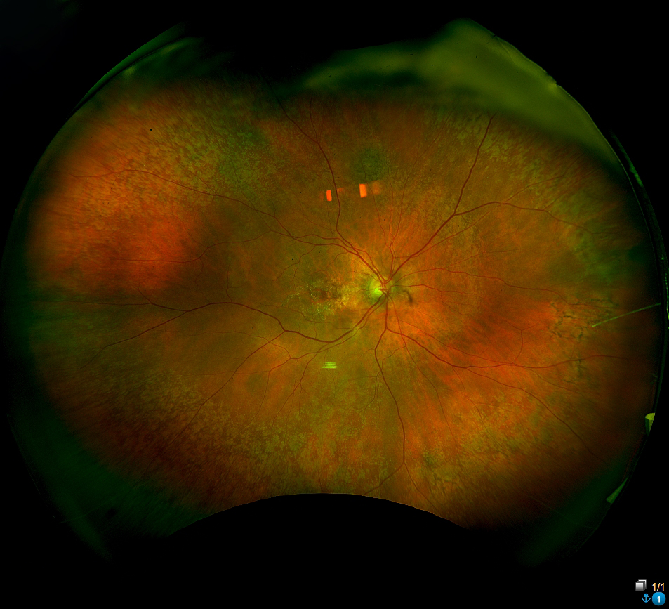
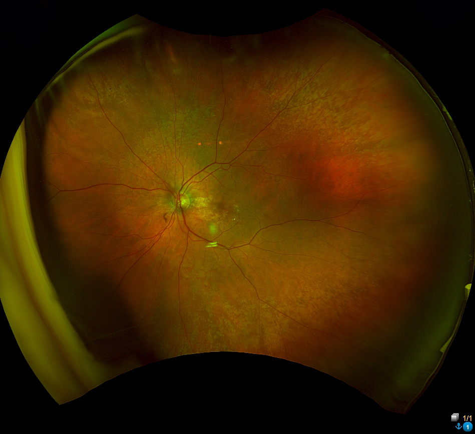
Referral sent to the specialist. The reviewing physician agrees with the initial assessment. Imaging revealed changes in the left eye, including deposits and areas of atrophy. Advanced scans confirmed abnormal blood vessel growth, and treatment with anti-VEGF therapy has been recommended.
Care1 AI’s Clinical Insight
Age-related macular degeneration (AMD) is a progressive retinal disease affecting the central macula, leading to gradual central vision loss. Early stages are marked by drusen and pigment changes in the retinal pigment epithelium, while late stages may involve either geographic atrophy or abnormal blood vessel growth beneath the retina. Genetic factors, age, smoking, and cardiovascular health contribute to risk. Pathophysiologically, AMD involves oxidative stress, complement-mediated inflammation, and disruption of the retinal pigment epithelium–Bruch’s membrane–choriocapillaris complex.
Did You Know?
The deposits under the retina seen in AMD, called drusen, often contain proteins from the body’s immune complement system, including complement proteins C3, C5, and the membrane-attack complex. This suggests AMD may be driven in part by chronic, localized inflammation rather than just aging.
Anderson DH, Mullins RF, Hageman GS, Johnson LV. A role for local inflammation in the formation of drusen in the aging eye. American Journal of Ophthalmology. 2002;134(3):411–431. doi:10.1016/s0002-9394(02)01357-7