85-year-old with macular changes shows stable vision. When is in-person referral needed versus continued monitoring?
Case Study: Monitoring Advanced Macular Changes in an 85-Year-Old Patient
An optometrist uploaded this case to Care1.
An 85-year-old patient with a history of previous intravitreal treatments in one eye was seen for routine follow-up. The patient reported long-standing poor vision in that eye, while the other eye remained functional with mild visual reduction. Both eyes had intraocular lenses from past cataract surgery, and the ocular surfaces were healthy and quiet.
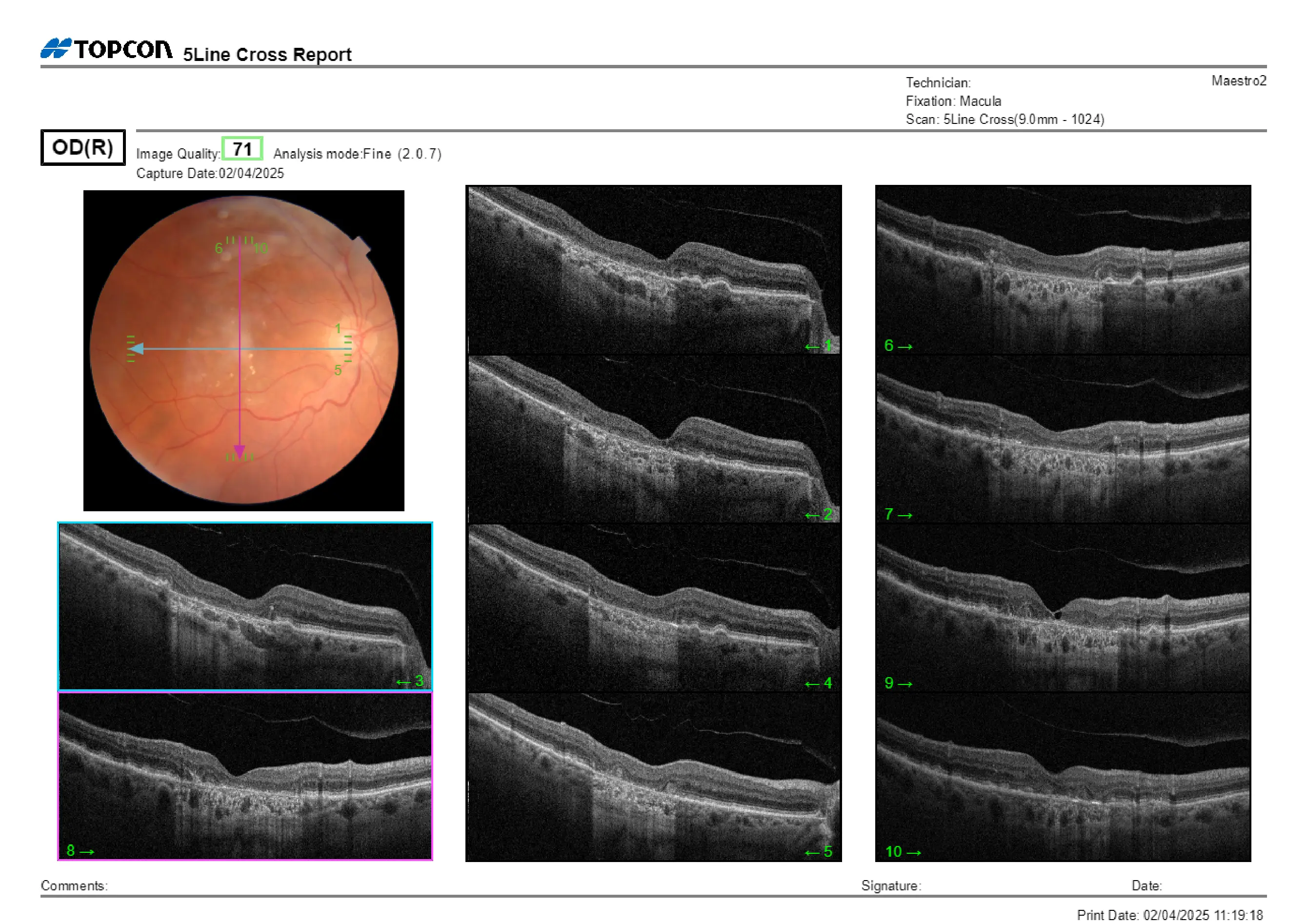
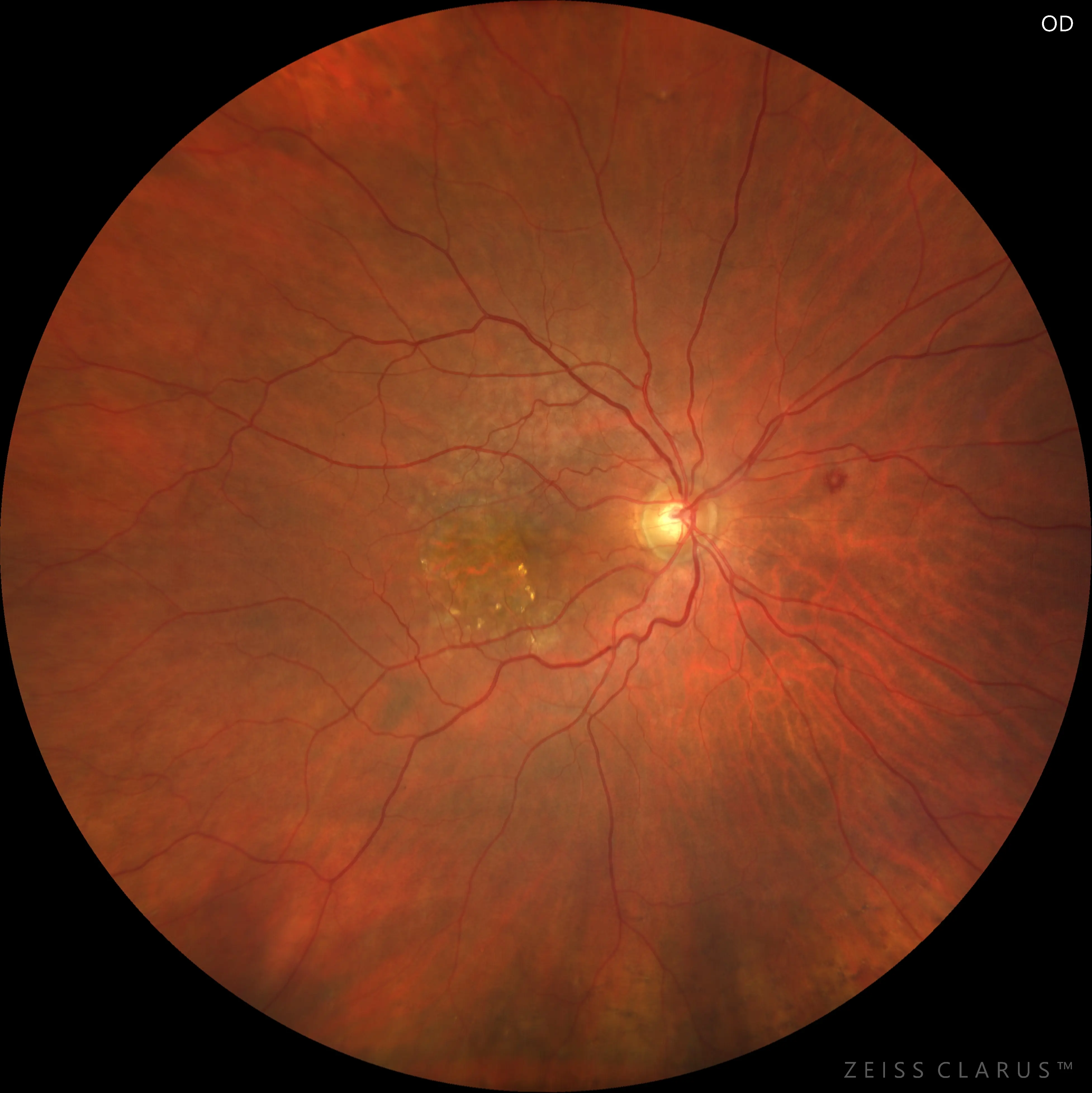
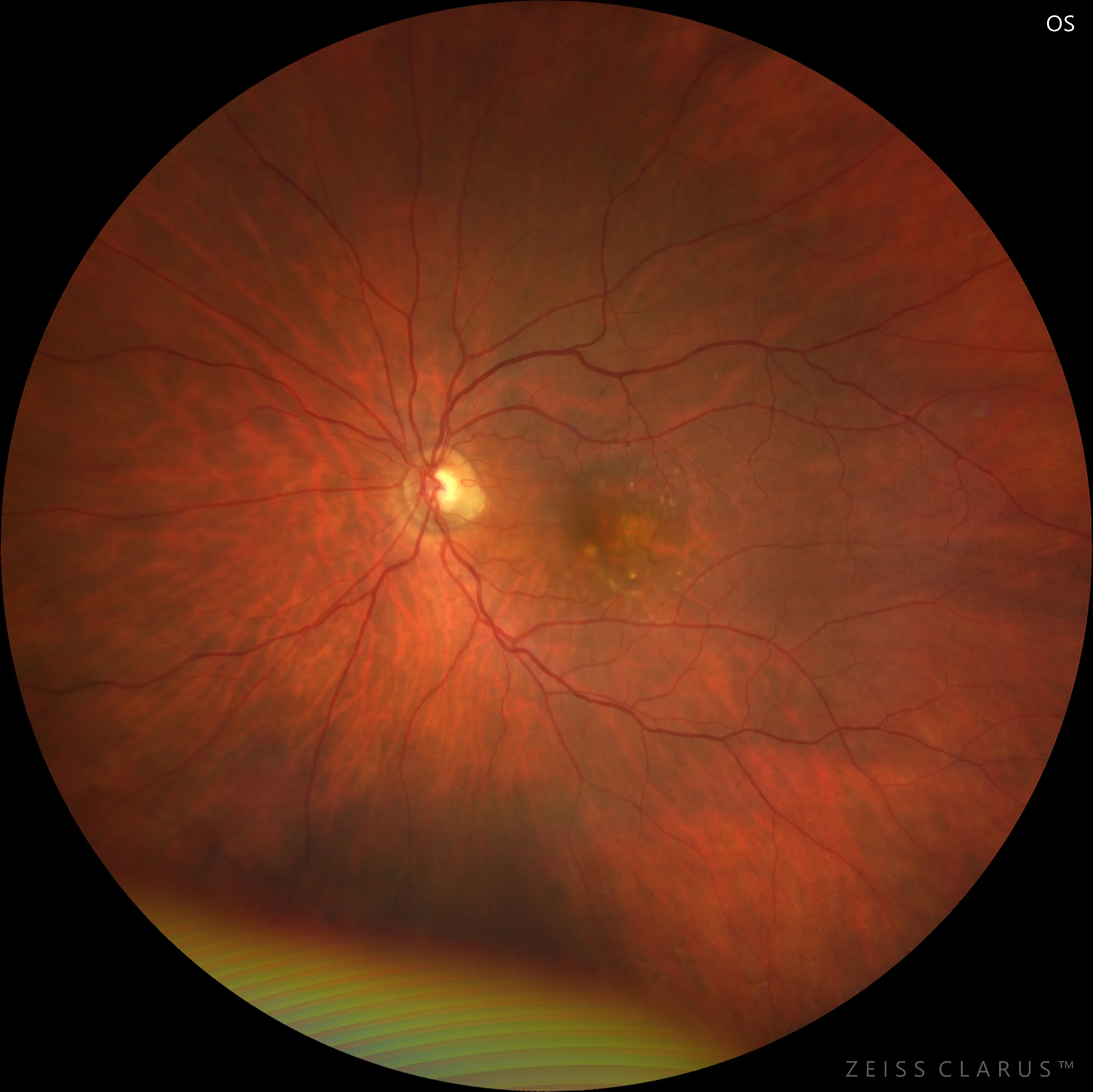
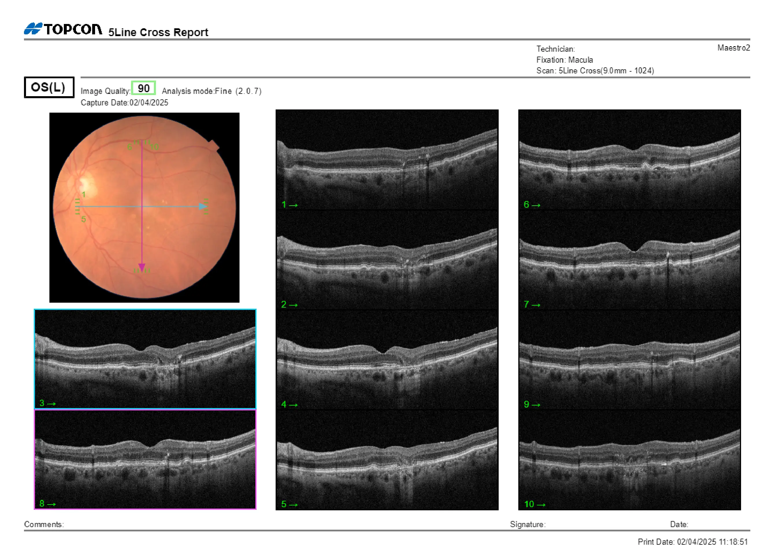
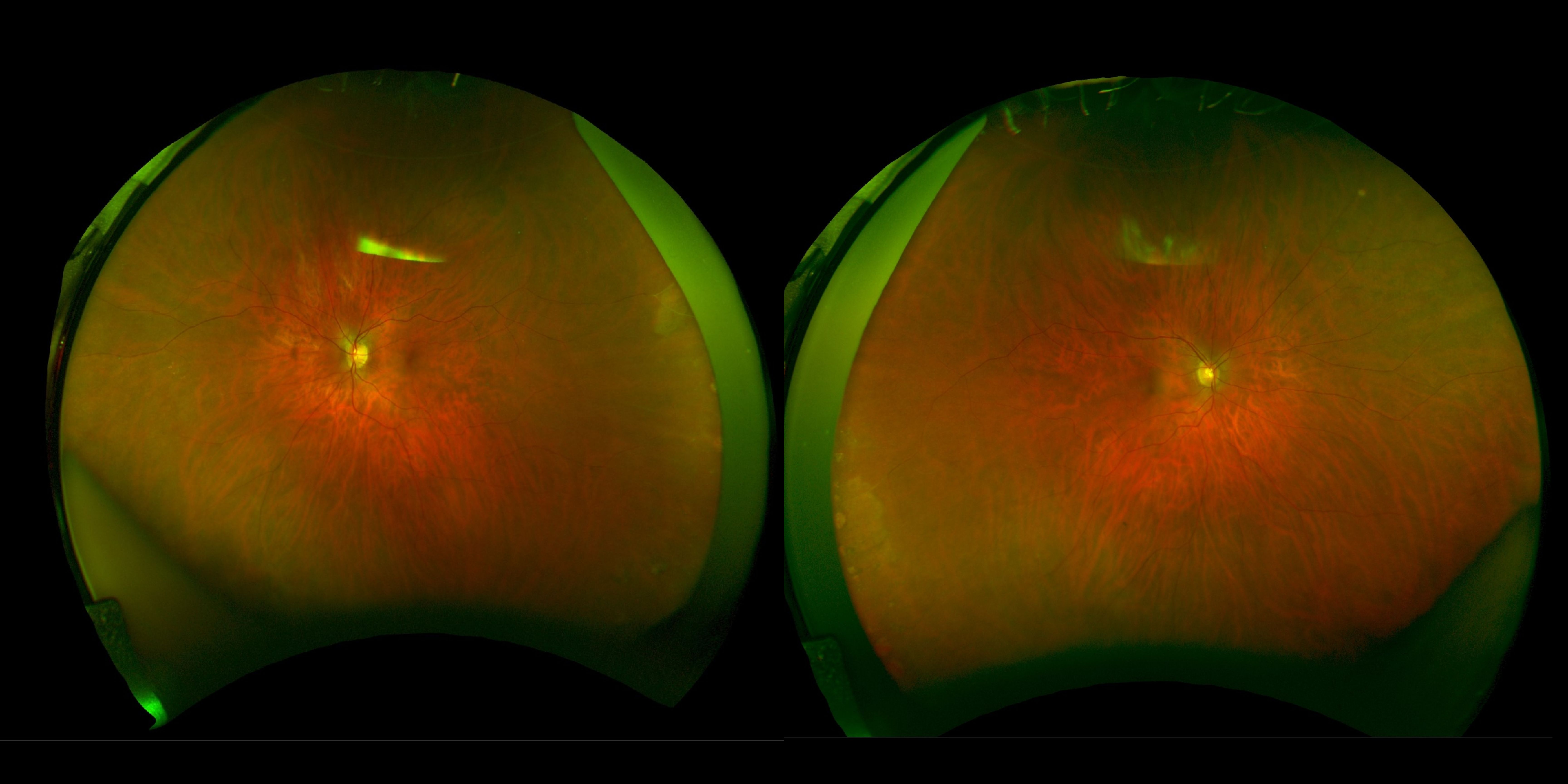
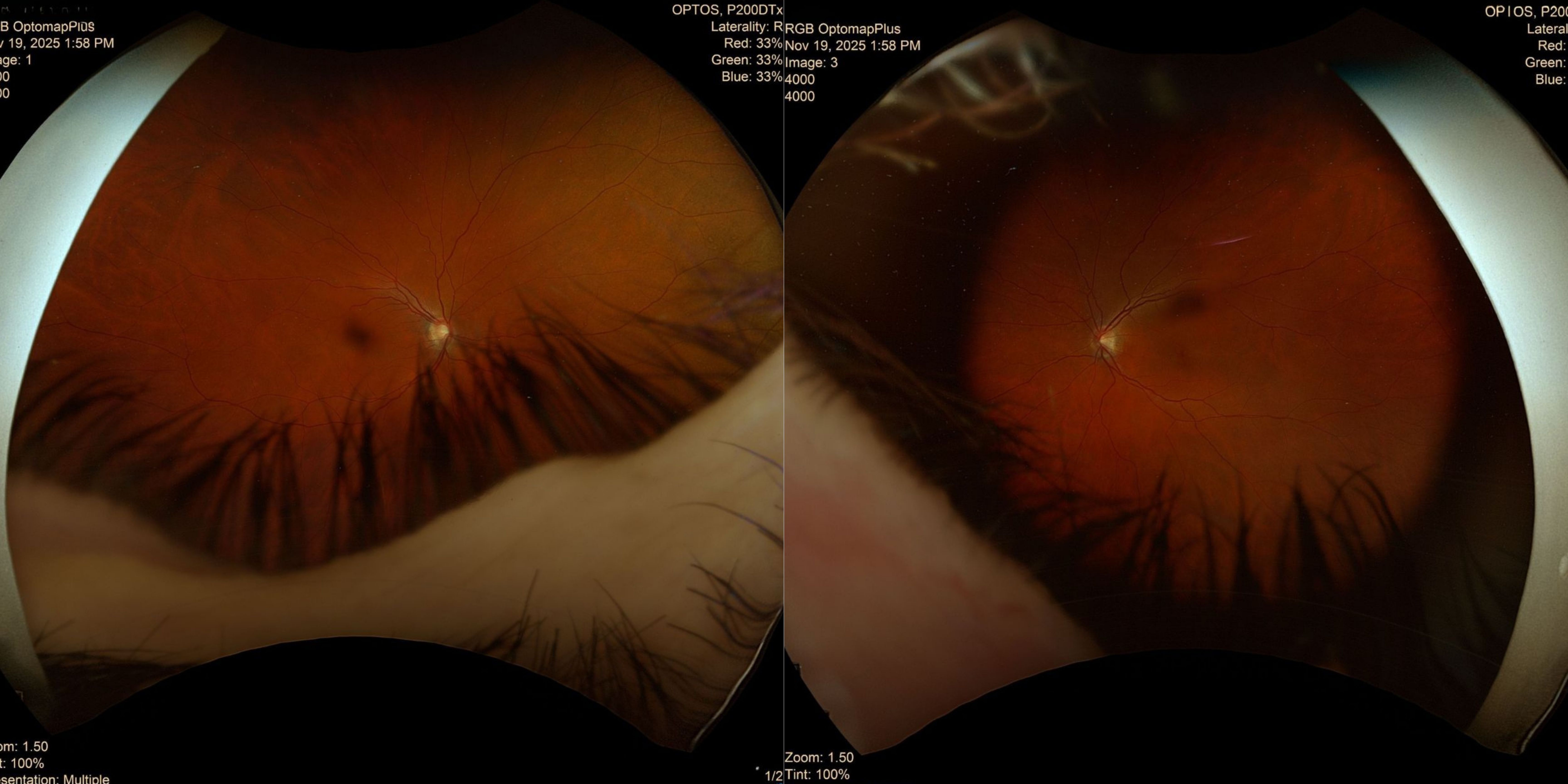
On examination, the posterior segment showed extensive scarring in the previously treated eye and mild retinal changes in the fellow eye. OCT confirmed the presence of old fibrotic changes in one eye and subtle structural alterations in the other, but no signs of active leakage or new membrane formation were noted.
Does this patient need an in-person retina consultation, or can they safely continue with periodic monitoring?
An ophthalmology subspecialist provided a virtual consult within 1-2 weeks through Care1. Scroll below to see their diagnosis.
Care1 Retina Surgeon’s Key Takeaways
Agree with your assessment. This 84-year-old patient has dry AMD with progression of geographic atrophy, more advanced in the right eye. OCT shows atrophic cysts in the right eye and justafoveal geographic atrophy in the left eye, with no signs of neovascularization or subretinal hyperreflective material. AREDS2 vitamins and continued monitoring are recommended.
If there are signs of conversion to wet AMD, an in-person retina consultation would be required.
Continue with observation and follow-up as scheduled. I will continue to assist with management via review of the patient’s file and diagnostic testing.
Care1 AI’s Clinical Insight
Geographic atrophy (GA) is a late stage of age-related macular degeneration (AMD) characterized by sharply defined areas of atrophy in the retinal pigment epithelium, photoreceptors, and choriocapillaris, often revealing larger underlying choroidal vessels. Its development is associated with drusen, reticular pseudodrusen, and both genetic and environmental risk factors, including variants in CFH and ARMS2. GA progresses slowly over time, leading to gradual visual decline, and individuals with GA remain at risk for developing choroidal neovascularization. Inflammation, oxidative stress, and immune system dysfunction are thought to play significant roles in its pathogenesis.
Did You Know?
The term “geographic atrophy” comes from the fact that the patches of retinal cell loss in this condition resemble islands or continents on a map due to their sharply defined borders and visible underlying choroidal vessels.