A 52-year-old patient presents with optic nerve changes and subtle visual field defects; evaluation insights included.
Case Study: Glaucoma Suspect with Strong Family History and Thick Corneas
An optometrist uploaded this case to Care1.
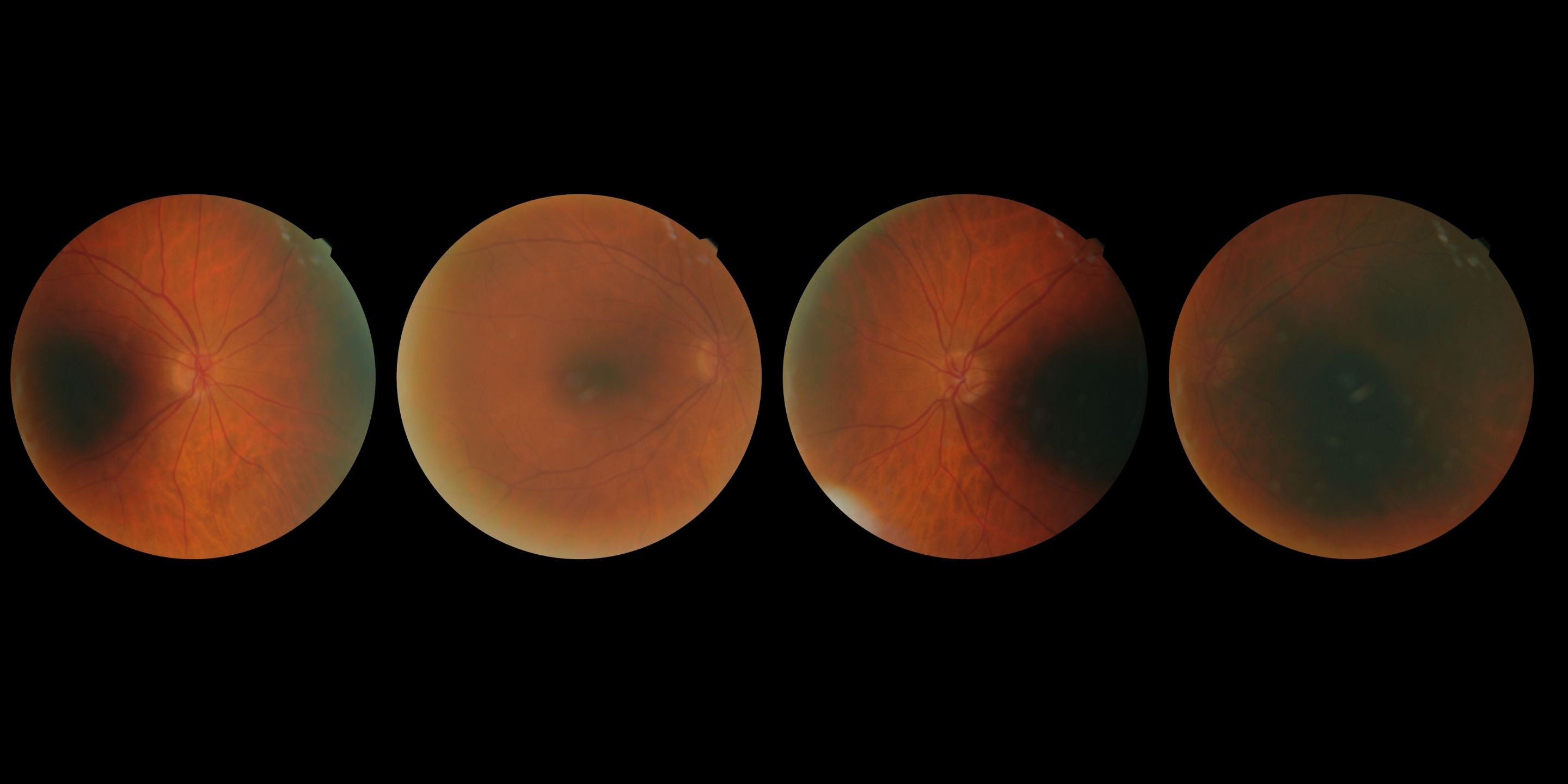
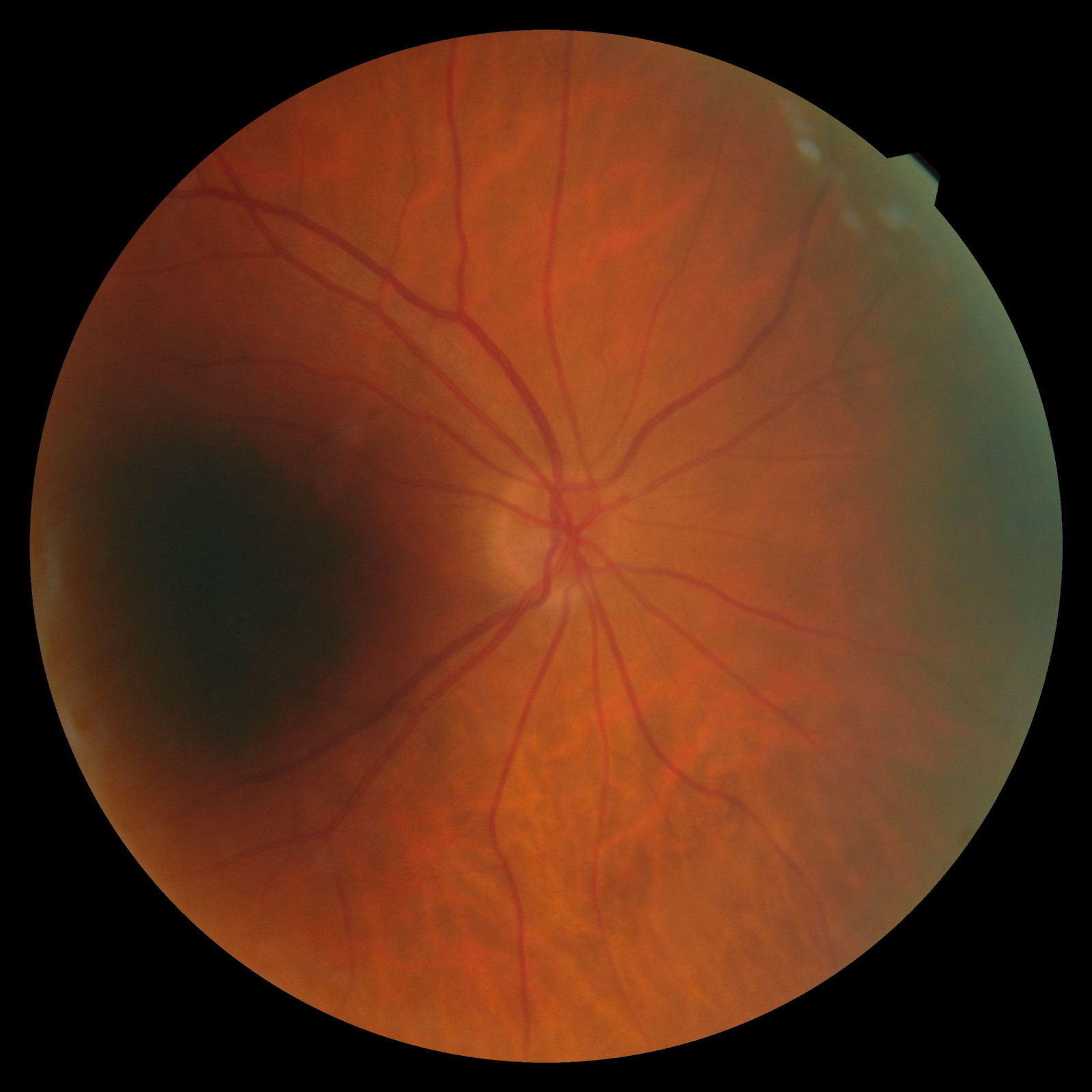
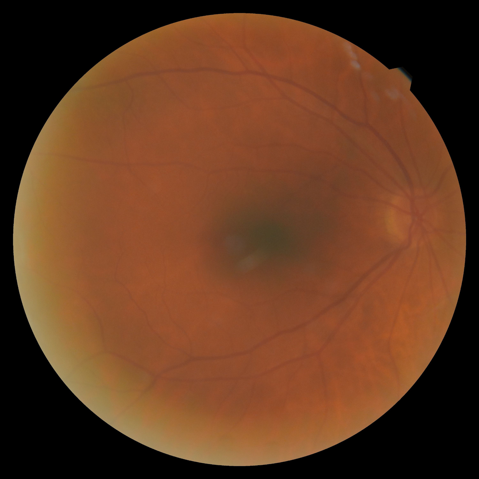
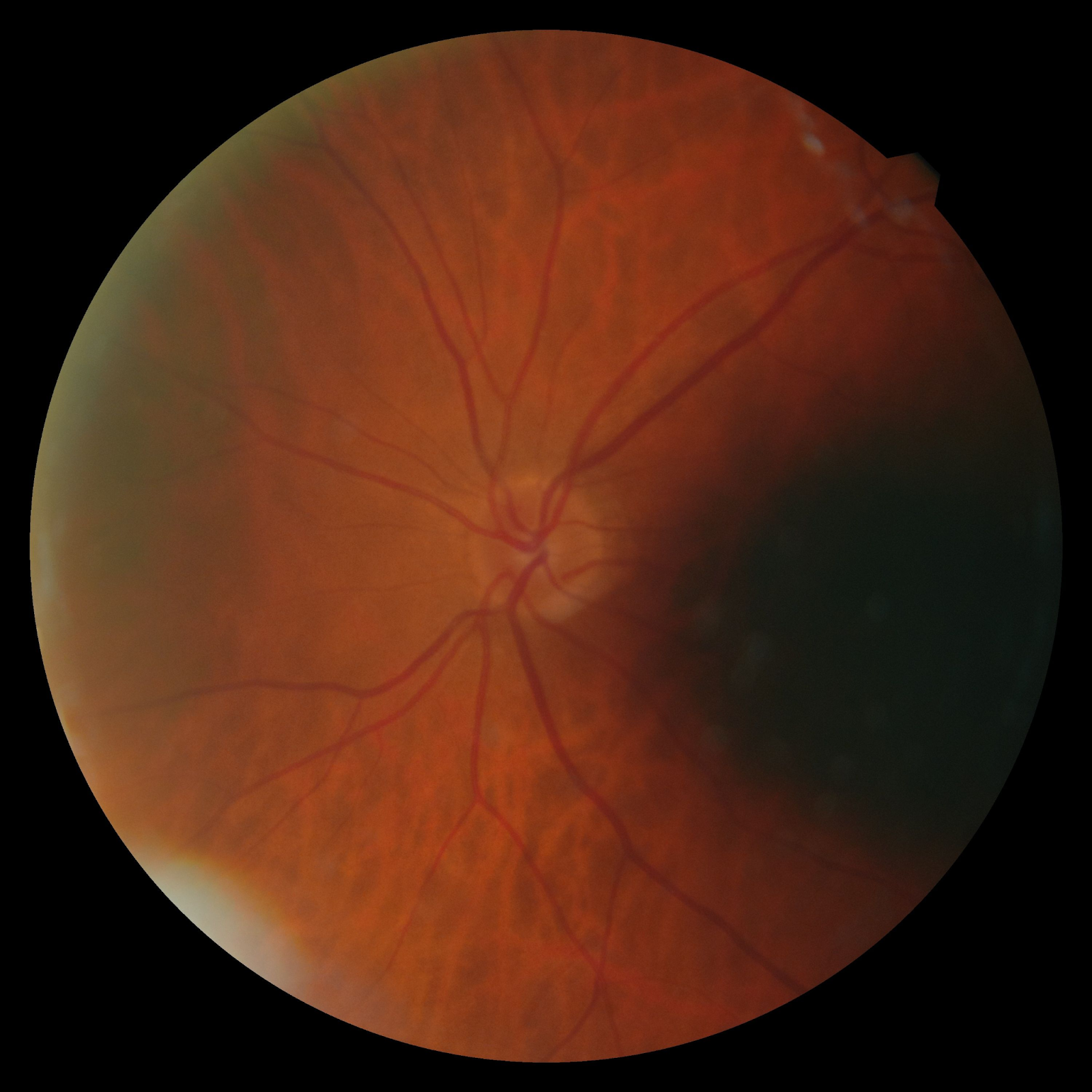
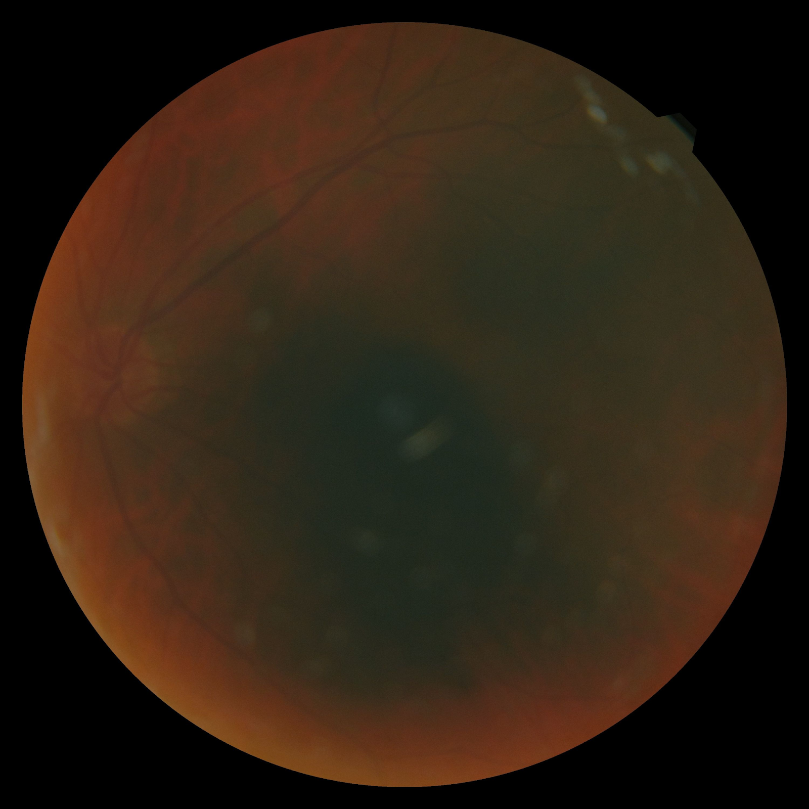
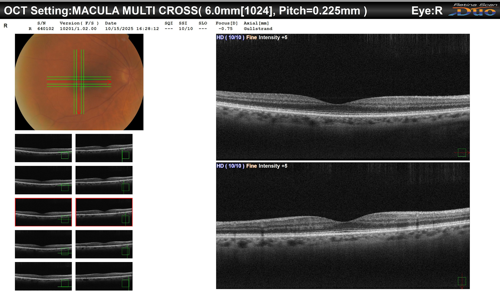
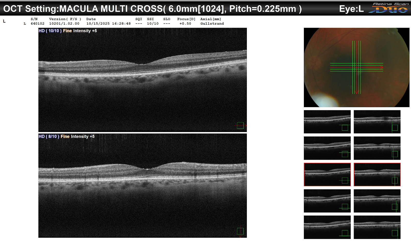
A 52-year-old patient was referred for evaluation due to optic nerve abnormalities noted during a routine exam. BCVA was 20/25 OD and 20/20 OS. IOPs were 18 mmHg OD and 16 mmHg OS. Anterior segment examination was unremarkable. Posterior segment evaluation revealed enlarged optic nerve cups and subtle nerve fiber layer thinning. Visual field testing showed early peripheral defects. OCT imaging confirmed structural changes consistent with optic nerve stress.
What would be your next step in assessing this patient’s optic nerve status?
An ophthalmology subspecialist provided a virtual consult within 1-2 weeks through Care1. Scroll below to see their diagnosis.
Care1 Subspecialist’s Key Takeaways
The reviewing glaucoma specialist emphasized careful assessment of optic nerve morphology and correlated structural-functional analysis. High-quality OCT imaging is essential to detect early changes before significant visual field loss occurs. The importance of repeated, consistent IOP measurements and attention to family history was highlighted. Clinicians should maintain vigilance for subtle peripheral defects on visual field testing. Early identification supports timely specialist referral and patient counseling.
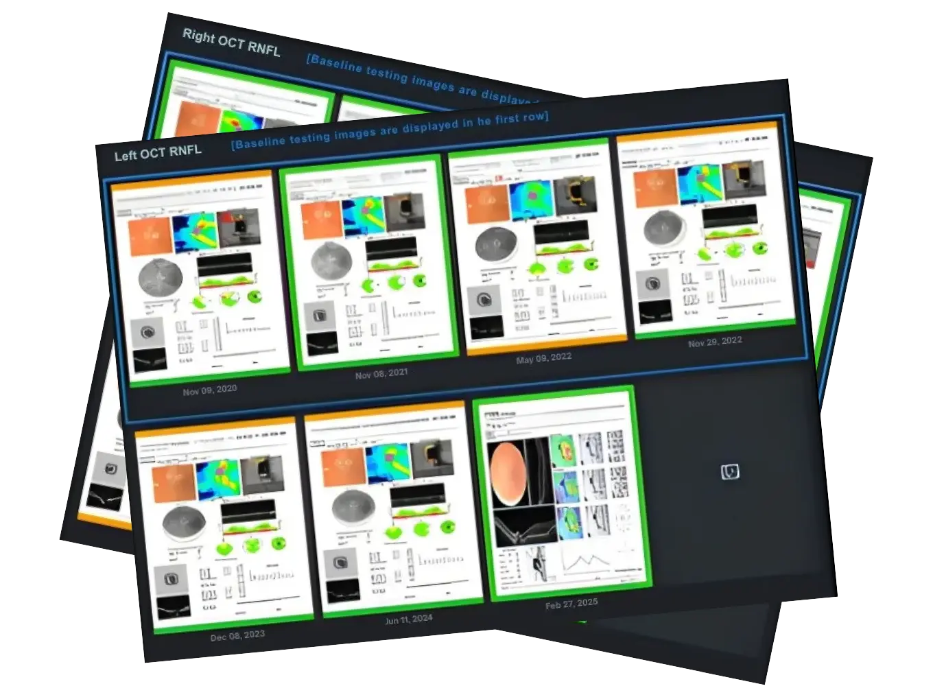
Care1 also provides AI-powered RNFL trend analysis to help clinicians detect subtle structural progression over time, supporting earlier identification of glaucomatous change. Learn more about this approach in our article on AI-powered OCT RNFL trend analysis for glaucoma monitoring.
Care1 AI’s Clinical Insight
Optic nerve cupping can result from a range of chronic or acute stressors. OCT is a sensitive tool for detecting early thinning of the retinal nerve fiber layer and ganglion cell layer. Structural changes often precede functional loss, making early imaging crucial. Monitoring trends over time is essential to differentiate physiologic variations from pathologic progression.
Unlock earlier glaucoma detection with AI-powered trend analysis.
Up to 30% of retinal ganglion cells can be lost before visual field defects are clinically detectable.
Reference: Quigley HA, Addicks EM. Quantitative studies of retinal ganglion cell and optic nerve fiber loss in experimental glaucoma. Arch Ophthalmol. 1980;98(5): 826–831. doi:10.1001/archopht.1980.01020010826008
Clinical Pearls
Subtle optic nerve cupping can indicate early structural stress.
OCT is critical for detecting retinal nerve fiber layer thinning.
Visual field testing may reveal peripheral defects before patient symptoms.
Family history and consistent IOP monitoring are important risk indicators.
Early specialist referral helps optimize patient outcomes.
Structural-functional correlation is key for longitudinal monitoring.