79-year-old female with stable optic nerve and visual field changes. Should current therapy continue, or is adjustment needed?
Case Study: Stable Optic Nerve with Mild Field Loss
An optometrist uploaded this case to Care1.
A 75-year-old female from a care home presented for a routine follow-up. She has a history of Alzheimer’s disease, arthritis, and prior spinal injury, and her ocular medications are managed by care staff. She is compliant with nightly topical drops and uses glasses for distance vision.
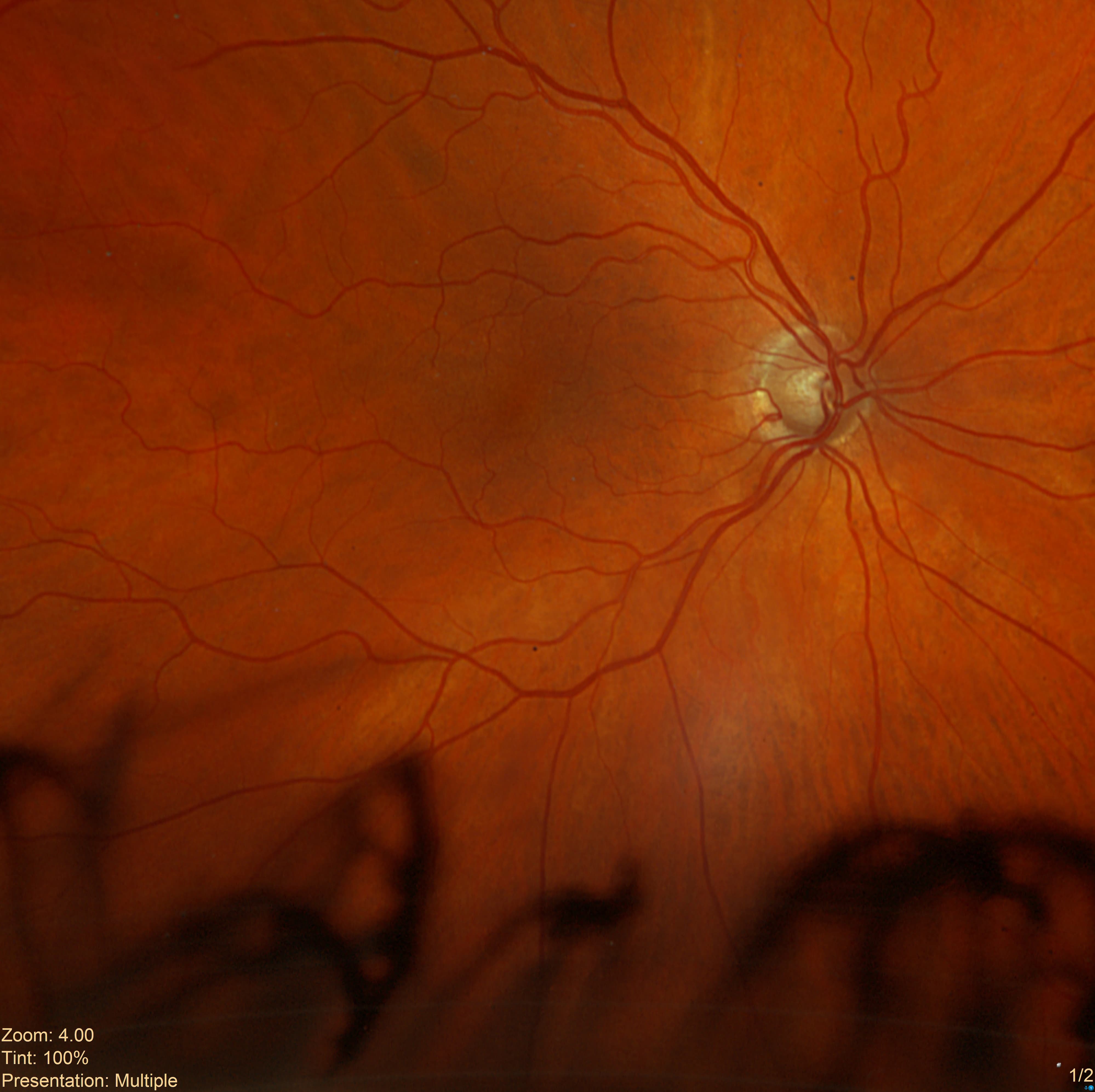
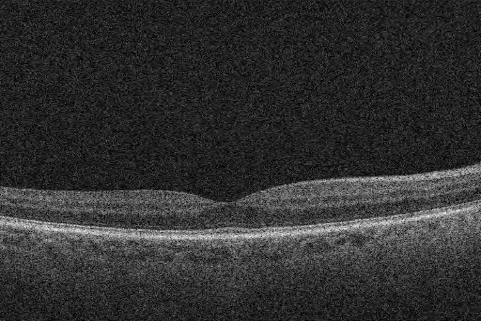
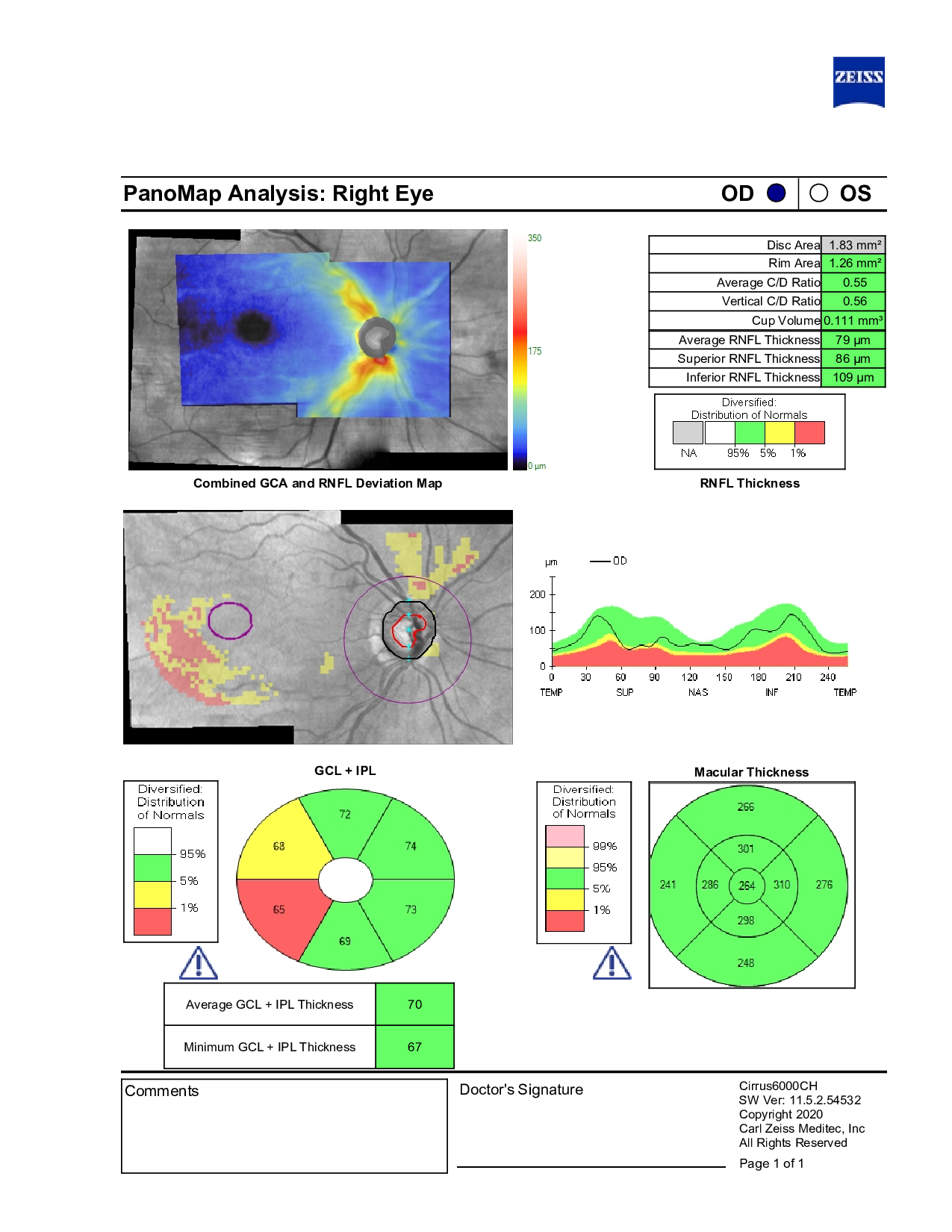
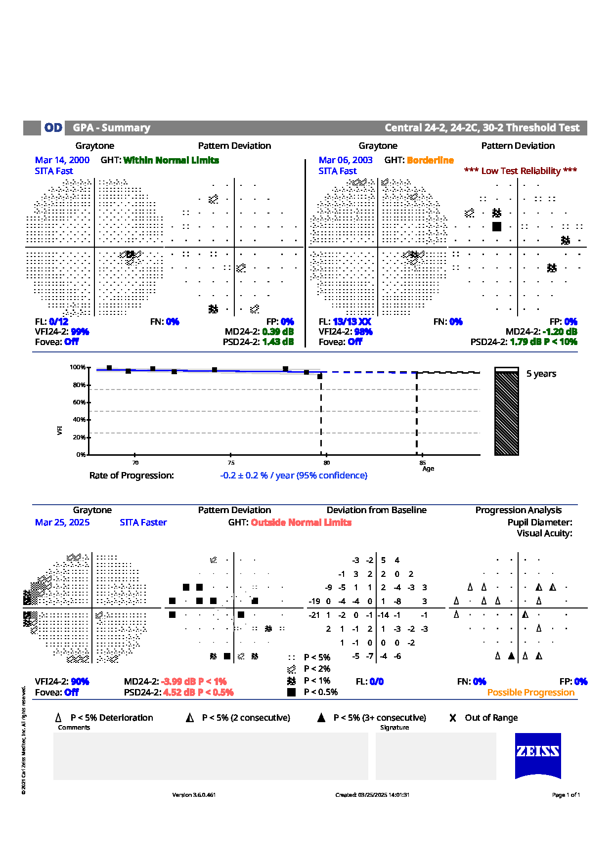
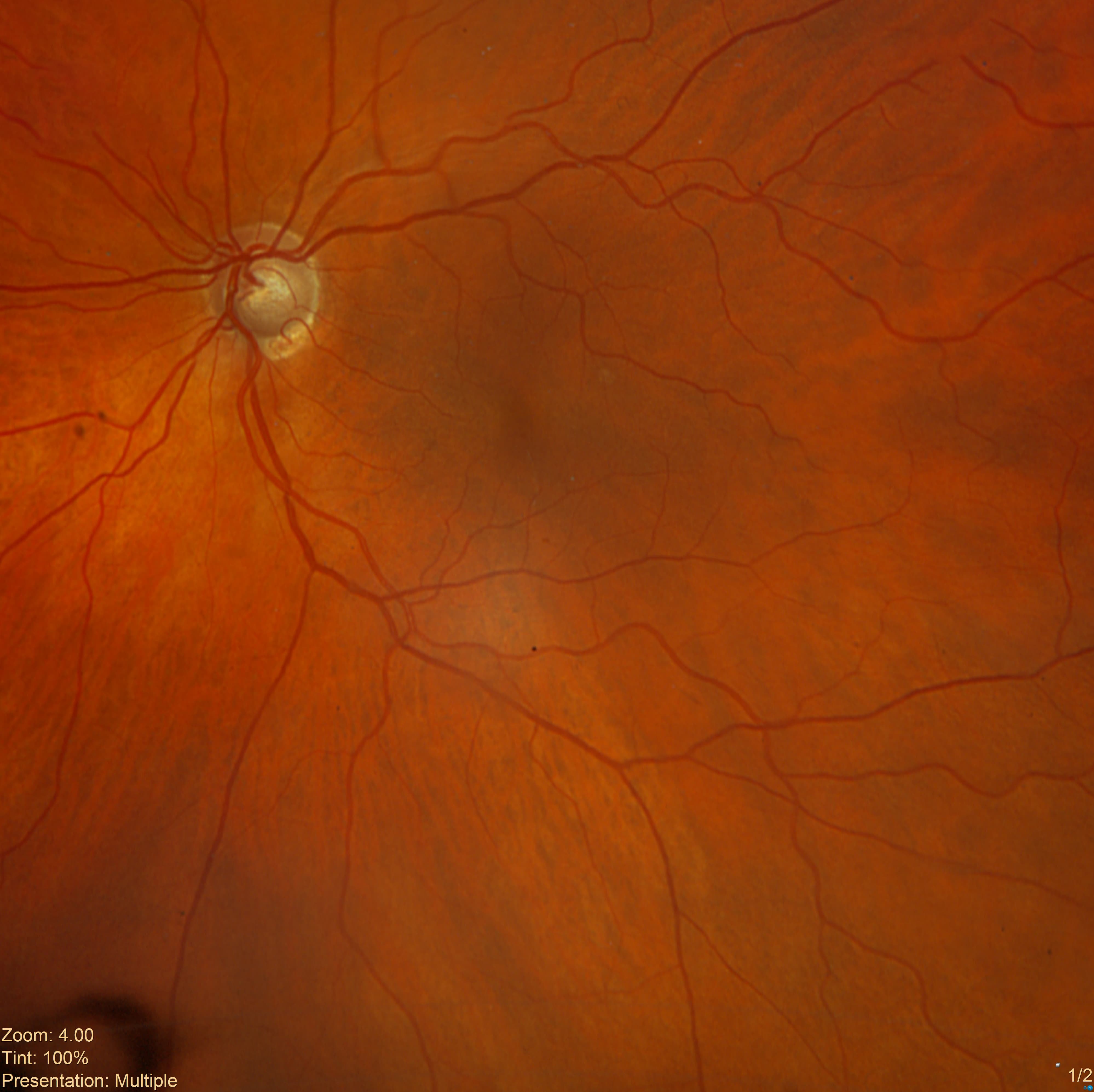
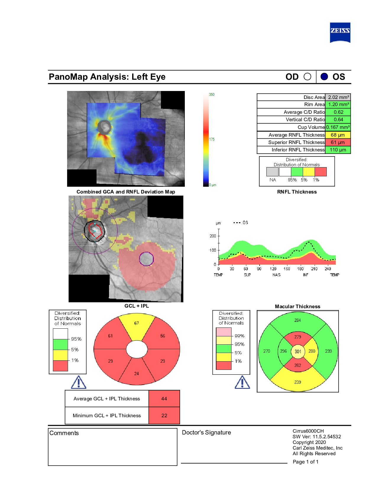
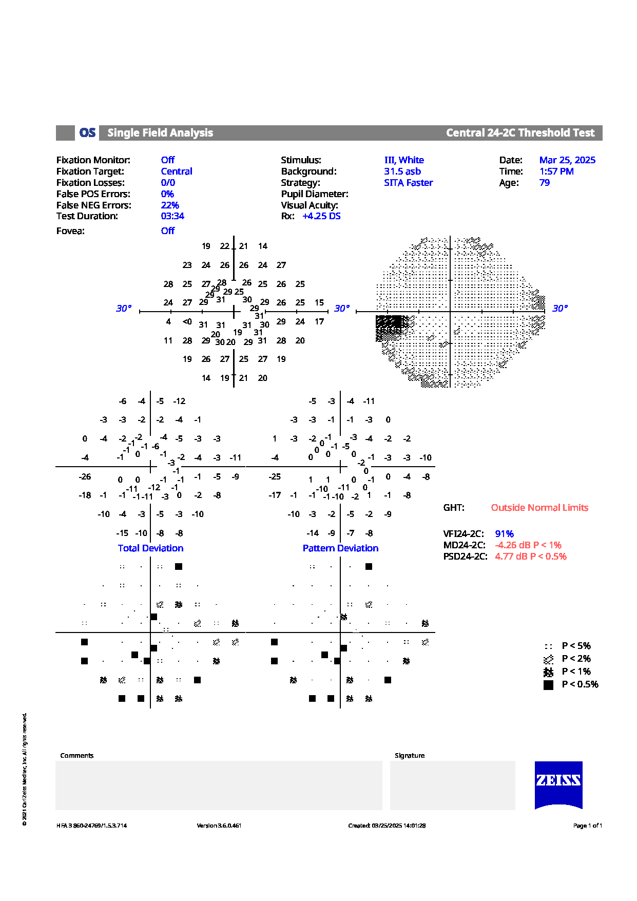
Examination showed intraocular pressures of 16 mm Hg in both eyes. Optic nerve evaluation revealed slightly asymmetric cupping with thinning of the inferior neuroretinal rim in both eyes. OCT demonstrated minor superior nerve fiber layer thinning, relatively stable since prior visits. Visual field testing revealed superior nasal defects in the right eye and some paracentral defects in the left eye.
Structural and functional assessments remain stable under current therapy.
Given these findings, should ongoing management continue as is, or is there a need for any adjustment in therapy or monitoring frequency?
An ophthalmology subspecialist provided a virtual consult within 1-2 weeks through Care1. Scroll below to see their diagnosis.
Care1 Retina Surgeon’s Key Takeaways
A patient on Travatan OU shows well-controlled IOP (OD 12–26, OS 14–26 mm Hg). C/D asymmetry is noted (0.40 OD, 0.70 OS), with possible RNFL thinning and mild progression on visual fields in the left eye. The right eye remains stable.
Follow-up every 6–12 months with OCT and visual field testing is recommended. Escalation of therapy may be considered if left-eye changes progress. Reassuring features include stable pressures, advanced age, and a normal right eye.
Care1 AI’s Clinical Insight
Asymmetric optic nerve cupping with retinal nerve fiber layer (RNFL) thinning reflects localized loss of retinal ganglion cell axons, often associated with glaucomatous optic neuropathy. Mechanical stress from intraocular pressure and vascular factors can lead to remodeling of the lamina cribrosa and progressive axonal loss. Structural changes include focal or diffuse thinning of the RNFL, enlargement of the optic cup, and notching of the neuroretinal rim. These changes often precede detectable visual field defects and are key early indicators of optic nerve damage.
Did You Know?
Early thinning of the retinal nerve fiber layer (RNFL), detectable by OCT, often occurs before visual field loss becomes apparent, making RNFL measurement a powerful early indicator of optic nerve damage.
Swaminathan SS, Jammal AA, Berchuck SI, Medeiros FA. Rapid initial OCT RNFL thinning is predictive of faster visual field loss during extended follow‑up in glaucoma. American Journal of Ophthalmology. 2021;229:100‑107.