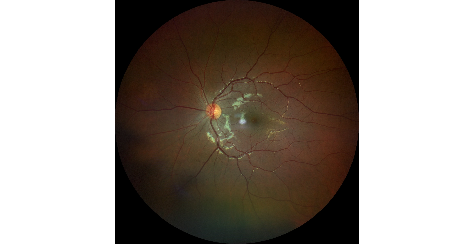
An optometrist uploaded this case to Care1.
A 12-year-old girl was uploaded for a suspected optic pit OD. Refractive error showed low hyperopia/astigmatism OU with no amblyopia. Pupils were equal OU.

A 12-year-old girl was uploaded for a suspected optic pit OD. Refractive error showed low hyperopia/astigmatism OU with no amblyopia. Pupils were equal OU.
An ophthalmology subspecialist provided a virtual consult within 1-2 weeks through Care1. Scroll below to see their diagnosis.


This appears to be an optic nerve pit in the right eye (OD). Optic nerve pits are rare conditions, found in fewer than 1 in 10,000 people. They do not typically affect visual acuity unless there is macular involvement (e.g., serous retinal detachment, retinal schisis, or CME). From a pathophysiology perspective, fluid from the vitreous or cerebrospinal fluid (CSF) may travel through the pit into the macula via a communication with the subarachnoid space. For this particular patient, as there are no current signs of macular involvement, we would recommend continued observation and follow-up with the optometrist. Care1 OMDs can remain involved remotely.
Optic nerve pits are congenital excavations of the optic disc that are typically asymptomatic but can occasionally lead to significant visual disturbances if fluid migrates into the macula. Early detection and monitoring are key. OCT plays a crucial role in identifying any macular changes, especially in pediatric cases where diagnosis may be delayed. Management is typically conservative unless macular involvement occurs, at which point surgical intervention may be necessary.
✔ Deliver the highest standard of care
✔ Increase patient satisfaction and retain patients
✔ Stimulate revenue
While often asymptomatic, about half of patients who develop symptoms experience vision changes in their 20s or 30s due to fluid accumulation under the macula.
*Kranenburg EW. Congenital pits of the optic nerve head. I. Clinical aspects. Arch Ophthalmol. 1960;64(6):912–924. doi:10.1001/archopht.1960.01840010914010