73-year-old with blurred vision and macular elevation on OCT showing central changes and subretinal features requiring further assessment.
Case Study: Blurred Vision With Macular Elevation
An optometrist uploaded this case to Care1.
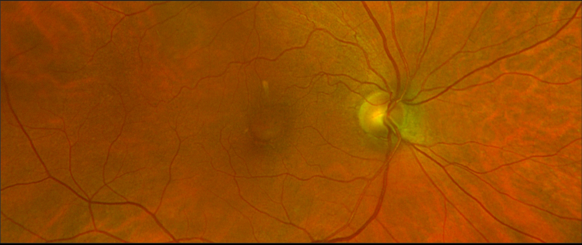
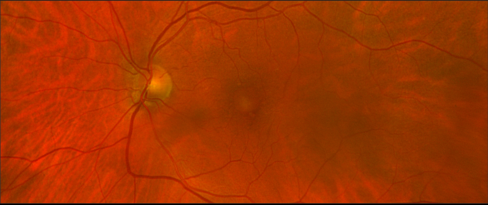
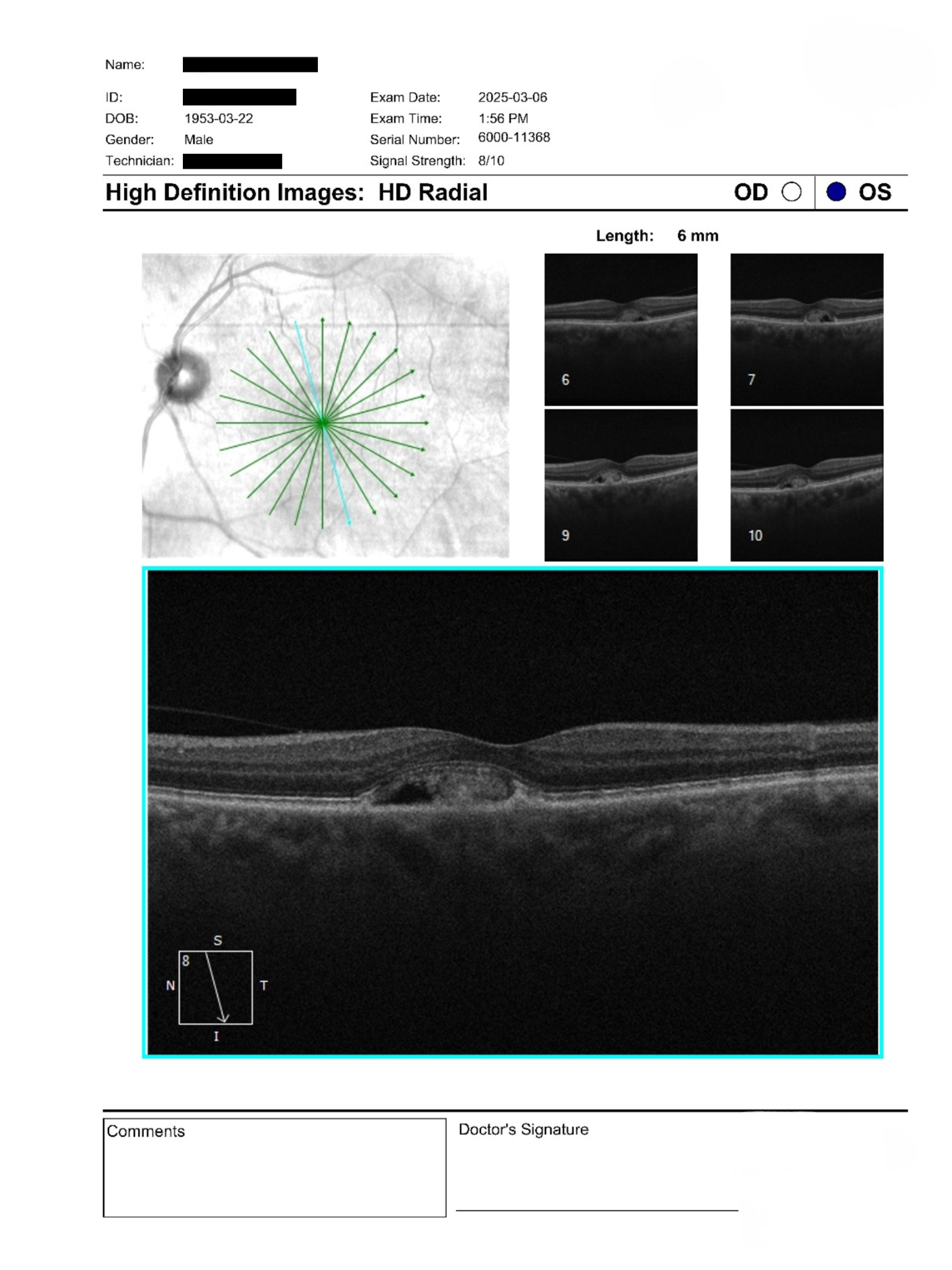
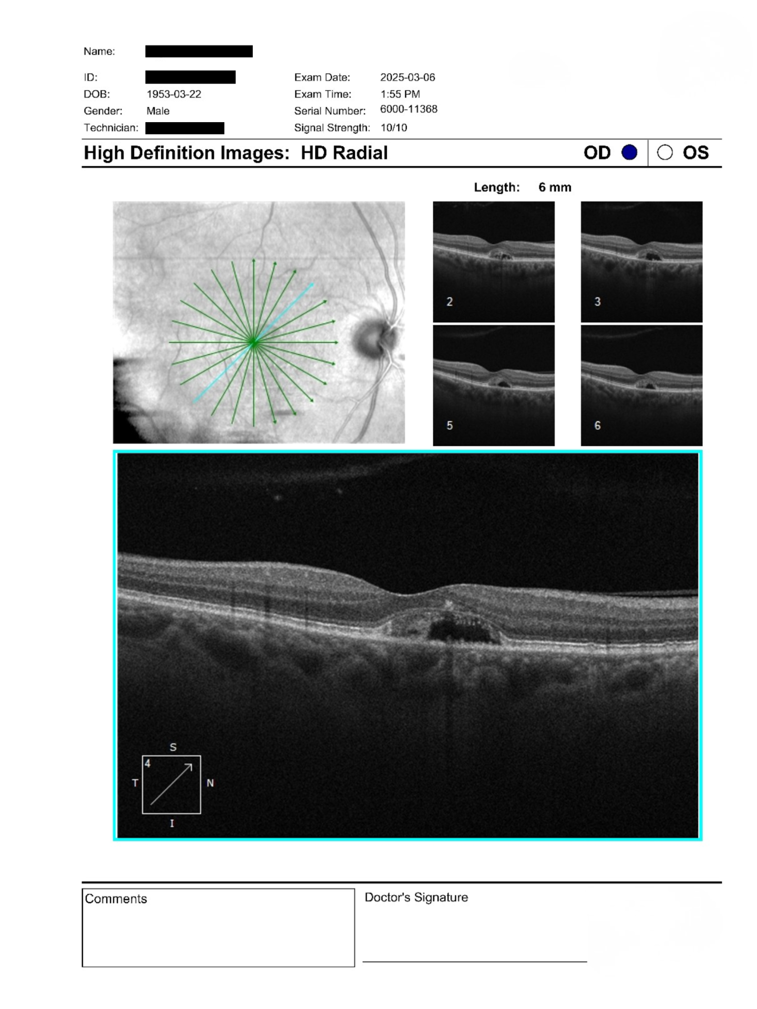
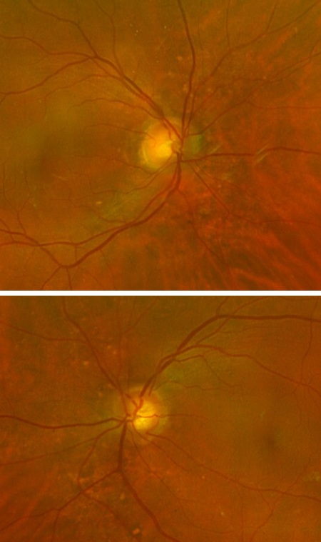
A 73-year-old male was referred for reduced near vision and suspected macular changes. BCVA was 20/50 OU and IOPs were 20 and 21 mmHg. Anterior segment exam showed mild nuclear sclerosis cataracts. Posterior segment exam revealed subtle macular pallor with OCT demonstrating central elevation and cystic spaces suggestive of subretinal pathology. The patient denied acute visual changes.
Should this patient be monitored or referred for further retinal imaging to rule out neovascular complications?
A retina specialist provided a virtual consult within 1-2 weeks through Care1. Scroll below to see their diagnosis.
Care1 Ophthalmologist's Teleconsult
The imaging demonstrates bilateral vitelliform-like deposits with signs of partial reabsorption and associated basal laminar changes. There is no clear evidence of active exudation, though subtle subretinal alterations warrant caution. The likelihood of neovascular activity appears low, but cannot be definitively excluded based on current imaging alone.
Referral for advanced imaging such as IVFA and OCTA is recommended to further evaluate for occult neovascularization. In the interim, conservative management with nutritional supplementation, UV protection, and home monitoring using an Amsler grid is appropriate. Ongoing follow-up is essential to detect progression.
Care1 AI’s Clinical Insight
Vitelliform lesions are characterized by subretinal deposits of lipofuscin-like material, often associated with retinal pigment epithelium dysfunction. These findings can overlap with other macular conditions and may evolve over time, including partial resorption stages. Multimodal imaging is key for accurate characterization and differentiation.
Unlock your full revenue potential
✔ Specialist consults within 1-2 weeks ✔ AI-powered clinical support ✔ Greater confidence in complex decision making
Vitelliform lesions can demonstrate spontaneous partial or complete resorption over time, sometimes leading to atrophic changes in the retinal pigment epithelium.
Reference: Ikuno Y, Tano Y. Ophthalmology. 2009;116(12):2315–2321. doi:10.1016/j.ophtha.2009.05.035
Clinical Pearls
Subretinal deposits may mimic multiple macular diseases
OCT essential for detecting subtle macular elevation
Consider OCTA when neovascularization is uncertain