78-year-old diabetic with unilateral retinal hemorrhages raises concern for vascular occlusion versus retinopathy.
Case Study: Unilateral Retinal Hemorrhages
An optometrist uploaded this case to Care1.
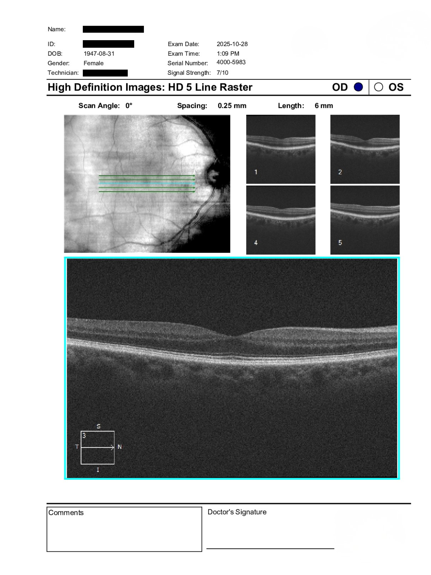
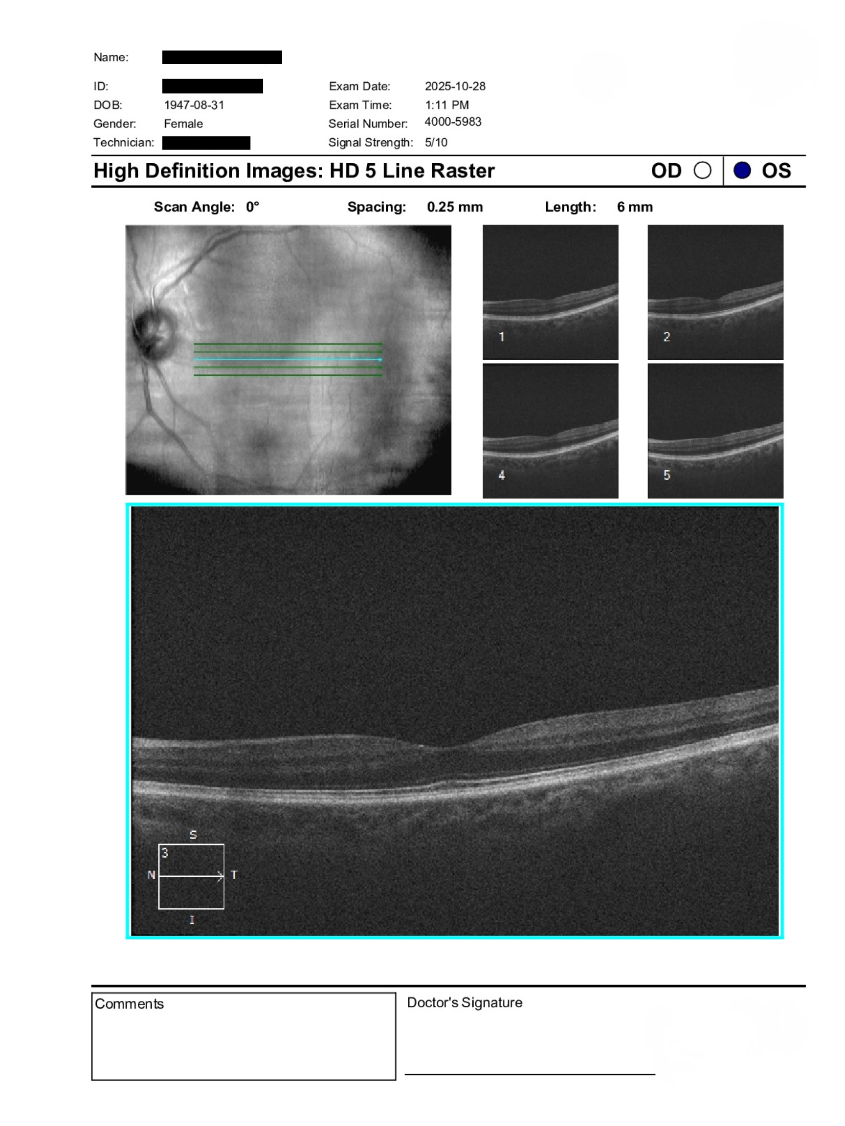
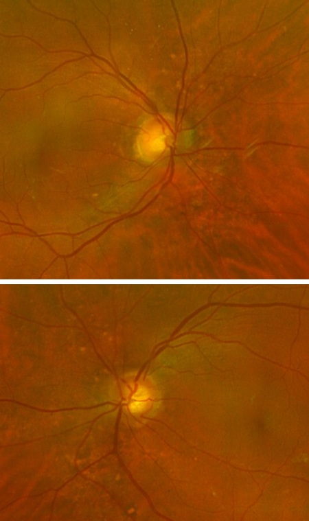
A 78-year-old patient with diabetes presented for assessment. BCVA was not documented. IOPs were not recorded. Anterior segment exam showed mild nuclear sclerosis in both eyes. Posterior segment exam revealed retinal hemorrhages in the right eye and none in the left. The differential included diabetic retinopathy versus retinal vein occlusion.
What is the most likely cause of these unilateral retinal hemorrhages?
A retina specialist provided a virtual consult within 1-2 weeks through Care1. Scroll below to see their diagnosis.
Care1 Ophthalmologist's Teleconsult
The findings in the right eye are concerning for a retinal vein occlusion, which remains high on the differential. However, the absence of optociliary shunt vessels and limited venous tortuosity raises concern for ocular ischemic syndrome. Further diagnostic evaluation is required to distinguish between these possibilities.
Urgent in-person assessment is recommended, including intravenous fluorescein angiography and carotid ultrasound. Timely referral is important to guide diagnosis and management.
Care1 AI’s Clinical Insight
Retinal vein occlusion is commonly associated with systemic vascular conditions such as hypertension and diabetes. It typically presents with retinal hemorrhages, venous dilation, and variable vision loss depending on macular involvement. Ocular ischemic syndrome, in contrast, is often linked to carotid artery stenosis and may show less venous tortuosity with broader ischemic signs.
Unlock your full revenue potential
✔ Specialist consults within 1-2 weeks ✔ AI-powered clinical support ✔ Greater confidence in complex decision making

.png)