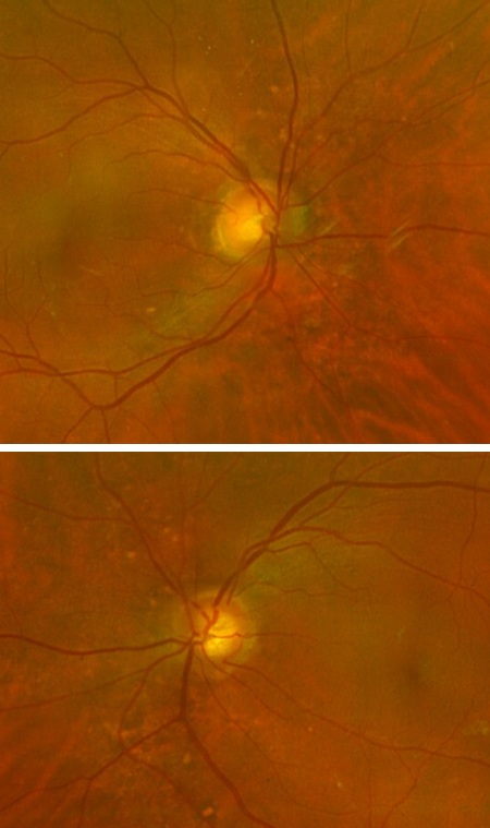
A 46-year-old male with isolated temporal blot hemorrhage OD and possible surrounding microaneurysms; systemic versus myopic etiology considered.
Case Study: Temporal Blot Hemorrhage with Possible Microaneurysms
An optometrist uploaded this case to Care1.
A 46-year-old male presented for a routine eye exam with no visual complaints. Past ocular history included nasolacrimal duct surgery OD (2020) and a stable choroidal nevus OS. Vision was 20/20 OU, intraocular pressures normal, and the anterior segment showed mild lid telangiectasia consistent with ocular rosacea.
Fundus exam revealed healthy optic nerves and maculae, but a temporal blot hemorrhage OD with possible surrounding microaneurysms was noted. The left eye’s choroidal nevus remained stable. Superotemporal microcystic degeneration OD was also observed.
What is your impression, and what next steps would you consider?
A retina specialist provided a virtual consult within 1-2 weeks through Care1. Scroll below to see their diagnosis.
Care1 Subspecialist’s Key Takeaways
This appears most consistent with a small peripheral retinal hemorrhage with possible adjacent microaneurysms. In a younger patient without a known history of diabetes or hypertension, systemic causes must be considered, even if medical history is reportedly unremarkable.
Peripheral capillary remodeling, particularly in myopic patients, may present with small hemorrhages. However, vascular etiologies such as early diabetic or hypertensive retinopathy should be ruled out.
A fluorescein angiogram may help determine whether there is active leakage or peripheral vascular abnormality, but initial management should focus on confirming systemic status. Blood pressure measurement and laboratory screening for diabetes and dyslipidemia are recommended.
Close follow-up with repeat dilated examination is advised to ensure stability or resolution. Referral for in-person retinal consultation may be considered if findings progress or if vascular leakage is confirmed.
Care1 AI’s Clinical Insight
Peripheral retinal blot hemorrhages may result from microvascular leakage, capillary remodeling, or localized venous congestion. Microaneurysms represent focal capillary outpouchings and are commonly associated with diabetic retinopathy but may occasionally be seen in other vascular conditions. Identifying whether such findings are isolated or part of a broader systemic process is essential in determining appropriate monitoring and referral.
Did You Know?
Many retinal microaneurysms form at sites of weakened capillary walls and can be visualized not only on clinical exam but with imaging before patients notice symptoms. Their presence correlates with early microvascular damage and can help clinicians identify systemic vascular stress, even before overt diabetic changes or vision loss occur.
Source: Wong TY, et al. “Microaneurysms in diabetic retinopathy: a marker of vascular injury.” Prog Retin Eye Res. 2008;27(5):628–656.
Key Clinical Takeaway
A 46-year-old male with 20/20 vision was found to have an isolated peripheral blot retinal hemorrhage with possible microaneurysms OD during a routine exam, with additional findings of a stable choroidal nevus OS and peripheral microcystic degeneration OD.
Even in asymptomatic patients with excellent acuity, peripheral hemorrhage may represent early microvascular disease or localized vascular remodeling.
Initial management should prioritize systemic evaluation, including blood pressure measurement and metabolic screening for diabetes and dyslipidemia.
Close follow-up with repeat dilated examination is appropriate; fluorescein angiography may be considered if progression or leakage is suspected.