63-year-old with asymptomatic nasal retinal changes and suspected progression on imaging, raising questions about monitoring vs referral.
Case Study: Nasal Retinal Change Progression
An optometrist uploaded this case to Care1.
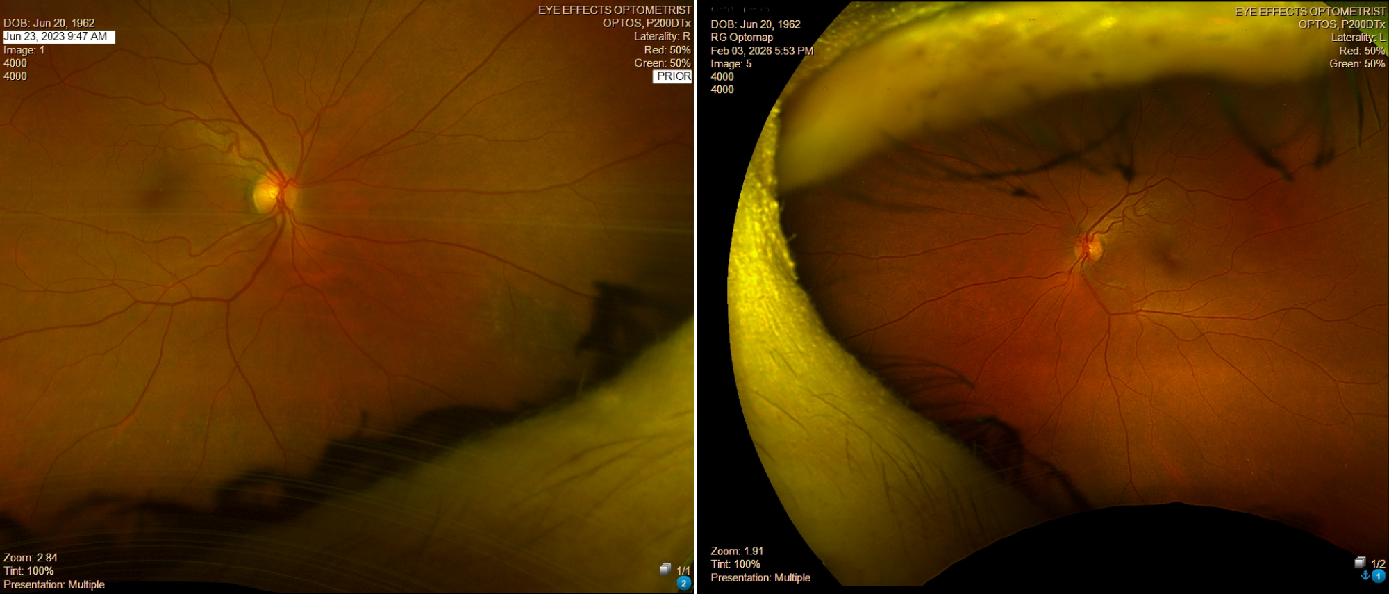
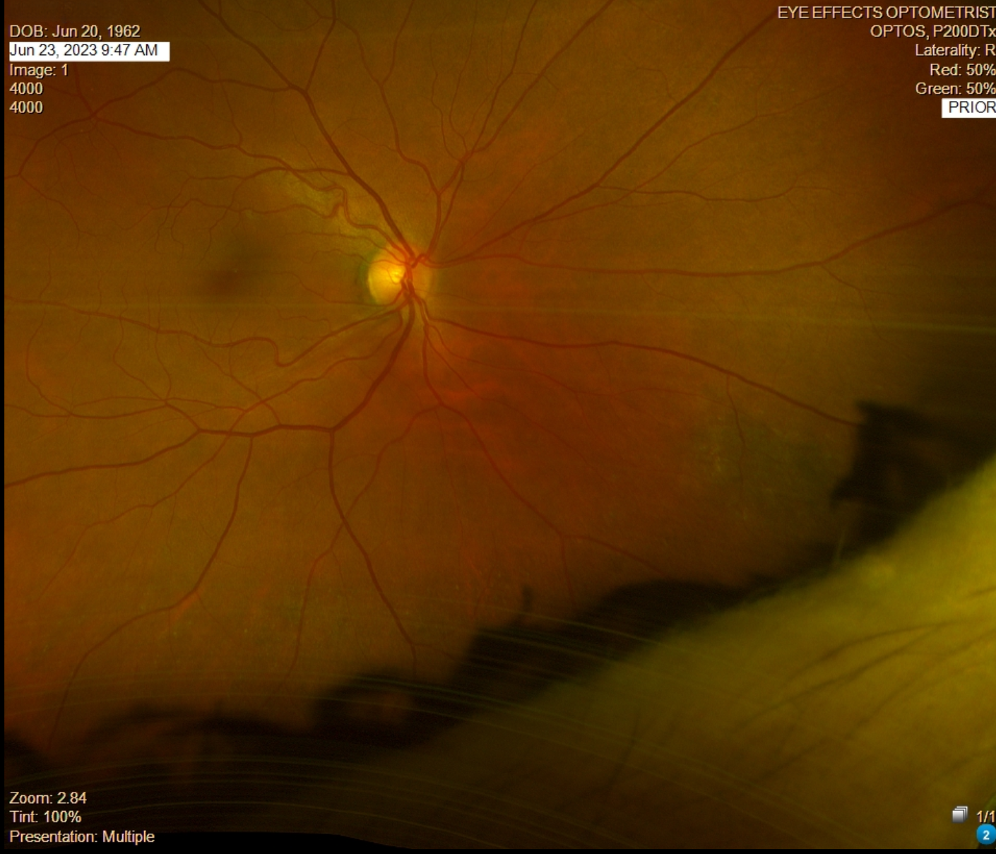
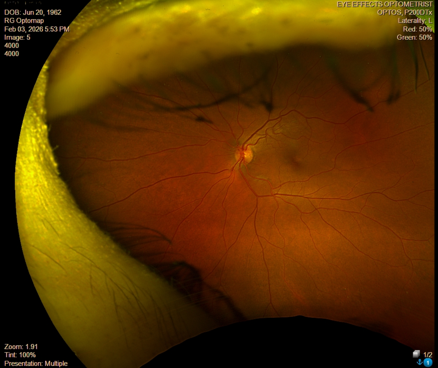
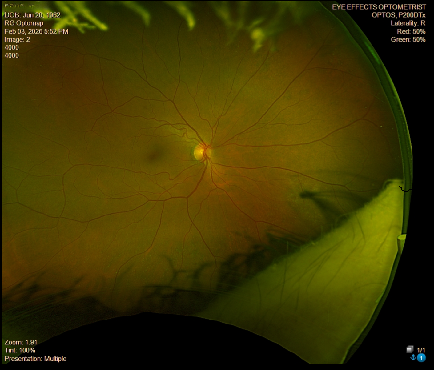
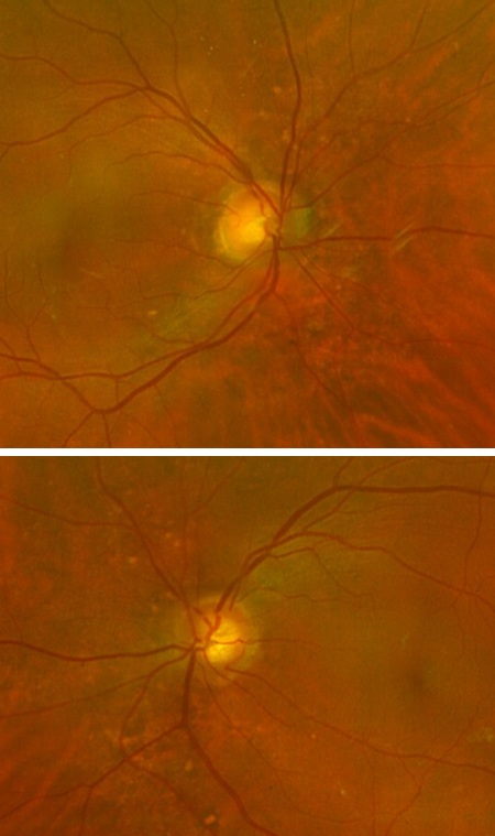
A 63-year-old patient was referred for evaluation of a nasal retinal change in the right eye that appeared to have progressed compared to prior imaging. The patient is asymptomatic with no reported visual complaints. BCVA and IOPs were not documented. Anterior segment findings were not noted. Posterior segment imaging revealed localized retinal atrophy along the nasal border of the right eye.
What is the most appropriate next step for evaluating a potentially evolving nasal retinal lesion in an asymptomatic patient?
A retina specialist provided a virtual consult within 1-2 weeks through Care1. Scroll below to see their diagnosis.
Care1 Ophthalmologist's Teleconsult
The reviewed lesion does not demonstrate features suggestive of a concerning or aggressive process. It may represent a subtle pigmented lesion with associated drusen rather than true progressive atrophy. While there is no urgency, further in-person evaluation and multimodal imaging can help better characterize the finding and confirm stability.
Referral for a non-urgent ophthalmology assessment is reasonable to establish a baseline and guide longitudinal monitoring.
Care1 AI’s Clinical Insight
Choroidal nevi are common benign pigmented lesions that may demonstrate overlying drusen, indicating chronicity. Multimodal imaging including OCT and fundus autofluorescence can help differentiate benign lesions from those with malignant potential. Risk assessment is typically based on features such as thickness, subretinal fluid, and associated symptoms.
Deepen your doctor-patient relationships
✔️ Shorter wait times for specialist input
✔️ More confident, transparent patient conversations
Choroidal nevi with overlying drusen are generally considered longstanding lesions, as drusen formation reflects chronic retinal pigment epithelium changes over time.
Reference: Shields CL, Furuta M, Thangappan A, et al. Metastasis of uveal melanoma millimeter-by-millimeter in 8033 consecutive eyes. Ophthalmology. 2009;116(5):917–923. doi:10.1016/j.ophtha.2008.12.031
Clinical Pearls
Subtle nasal retinal changes may indicate slow, chronic progression
Drusen over lesions usually suggest longstanding, benign pathology
Non-urgent referral helps establish baseline and monitor changes