79F follow-up: Macular changes stable, vision excellent (20/20 OD, 20/25 OS). Is it time for closer monitoring or keep up with Amsler testing?
Case Study: Retinal Changes and Vision Monitoring
An optometrist uploaded this case to Care1.
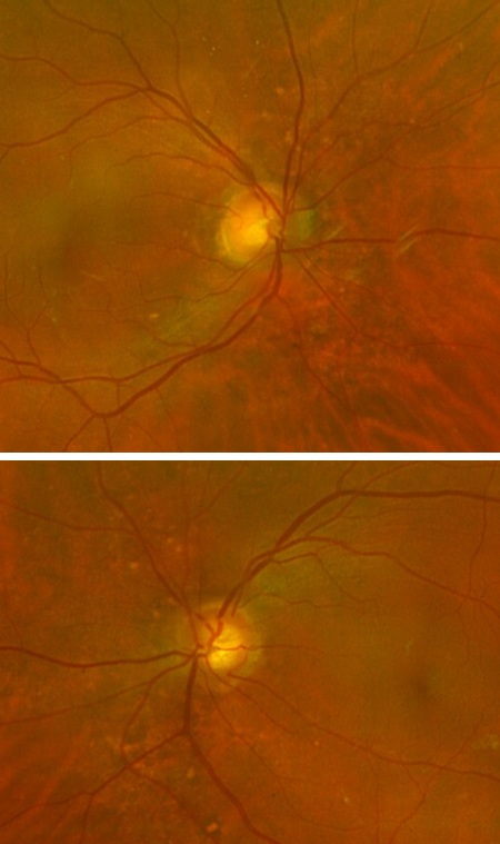
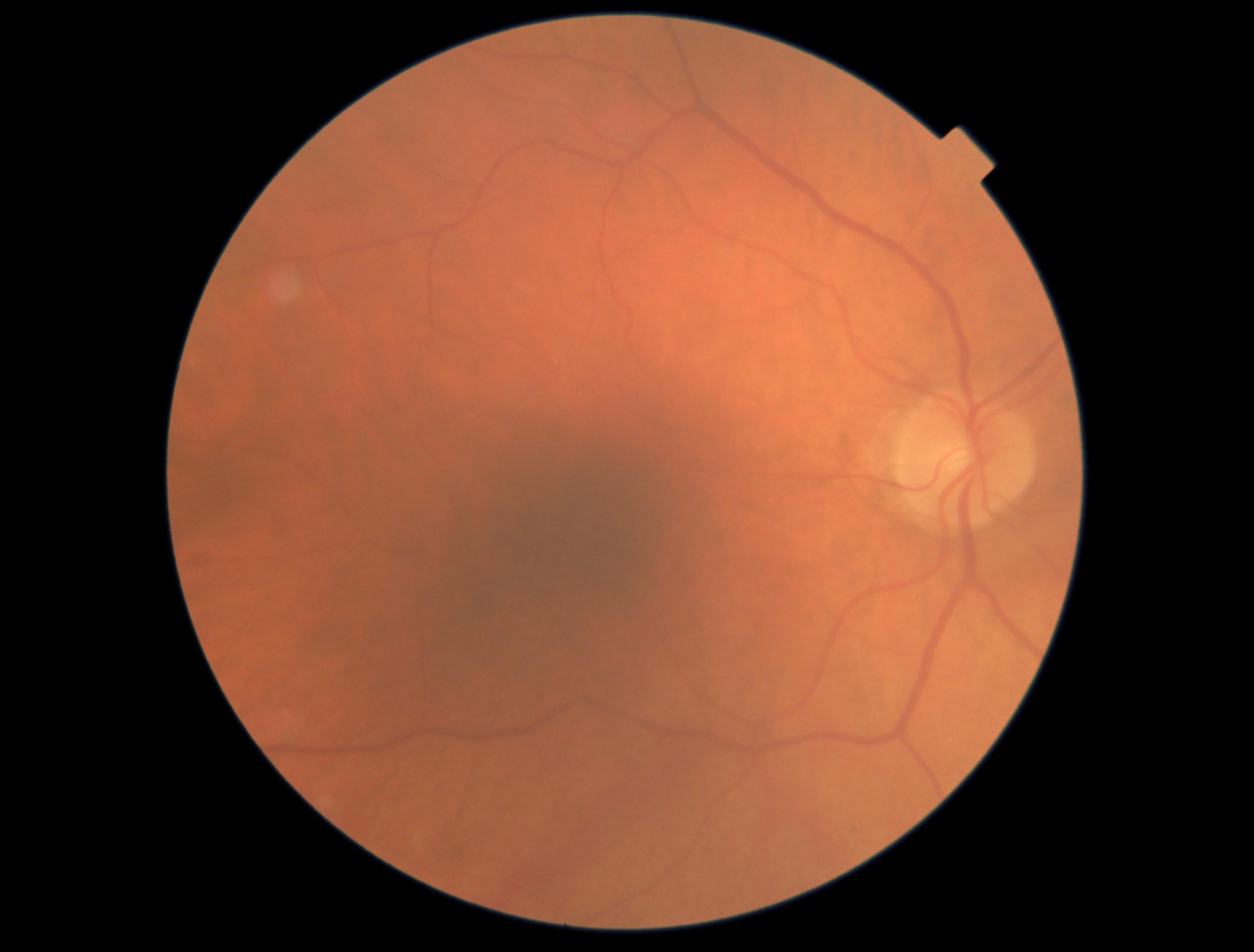
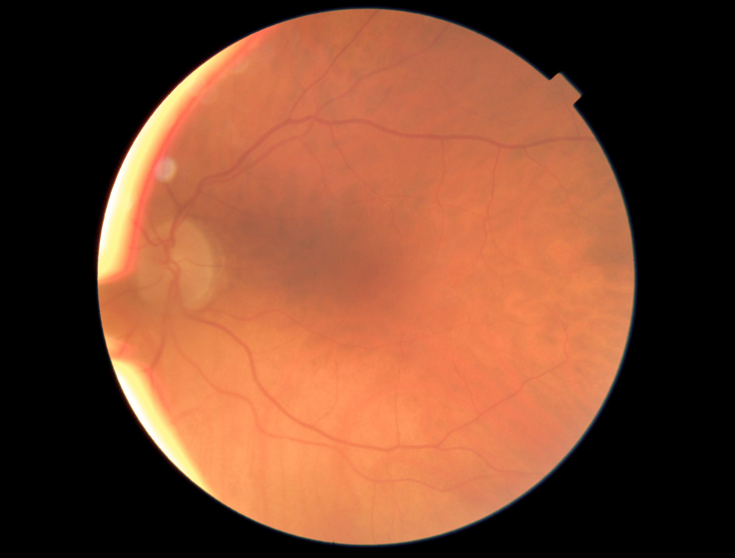
A 79-year-old female came in for a routine follow-up, with no new visual concerns. OCT imaging revealed moderate macular changes in the right eye and mild changes in the left, both causing distortion. Despite this, her vision remains excellent (20/20 OD, 20/25 OS).
She’s continuing at-home Amsler monitoring.
Given these findings, would you recommend staying on track with follow-up in six months, or should we consider closer monitoring?
An ophthalmology subspecialist provided a virtual consult within 1-2 weeks through Care1. Scroll below to see their diagnosis.
Care1 Subspecialist’s Key Takeaways
This patient has an epiretinal membrane with a lamellar hole in the right eye and mild ERM in the left. OCT imaging shows traction and schisis in the right eye, with a cyst in the left. Visual acuity remains excellent.
Given the minimal impact on vision, continued observation is recommended, as surgery may not improve vision despite membrane removal.
Continue with observation and follow-up as scheduled. I will continue to assist with management via review of the patient’s file and diagnostic testing.
Care1 AI’s Clinical Insight
A lamellar macular hole is a partial-thickness retinal defect affecting the macula, often associated with traction from an epiretinal membrane (ERM). It typically causes central visual distortion but preserves the outer retinal layers. The condition may lead to subtle visual symptoms, with varying degrees of impact on vision. Management options are generally conservative, focusing on observation unless the hole progresses or vision is significantly impaired.
Did You Know?
Lamellar macular holes can occur without full-thickness retinal disruption, which differentiates them from complete macular holes. This partial defect in the retina may cause significant visual distortion, despite not affecting the entire macula.
Cheng L, Chan YK, Lam DS. Lamellar macular hole: A review of pathogenesis, clinical features, and management. Ophthalmology. 2010;117(10):1822-1830. doi:10.1016/j.ophtha.2010.03.010.

.jpg)
.jpg)