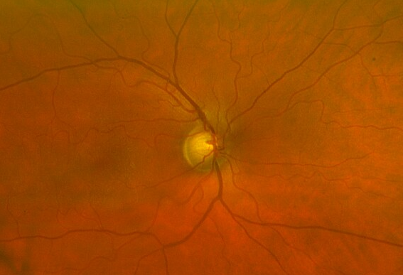
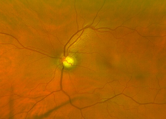
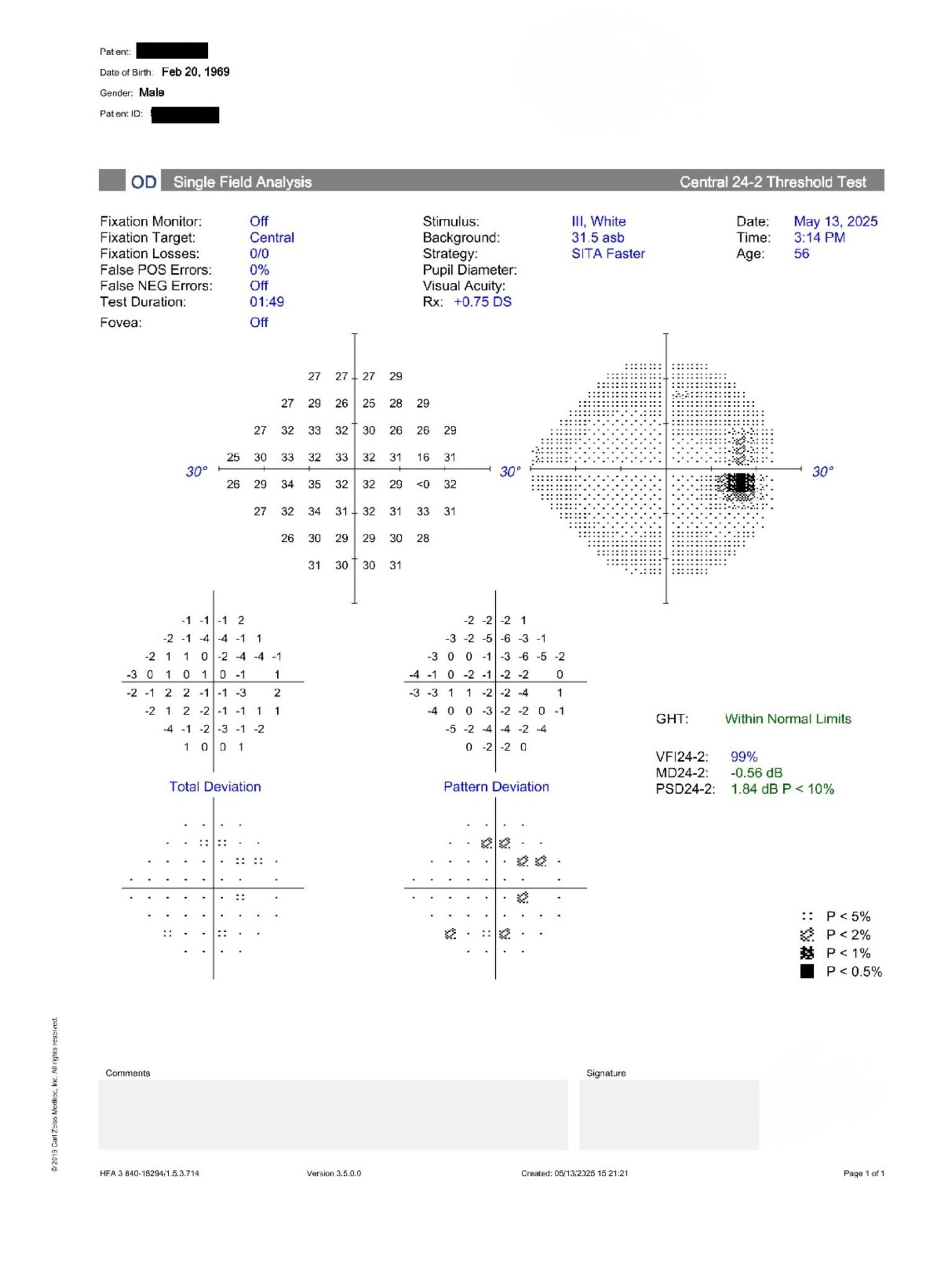
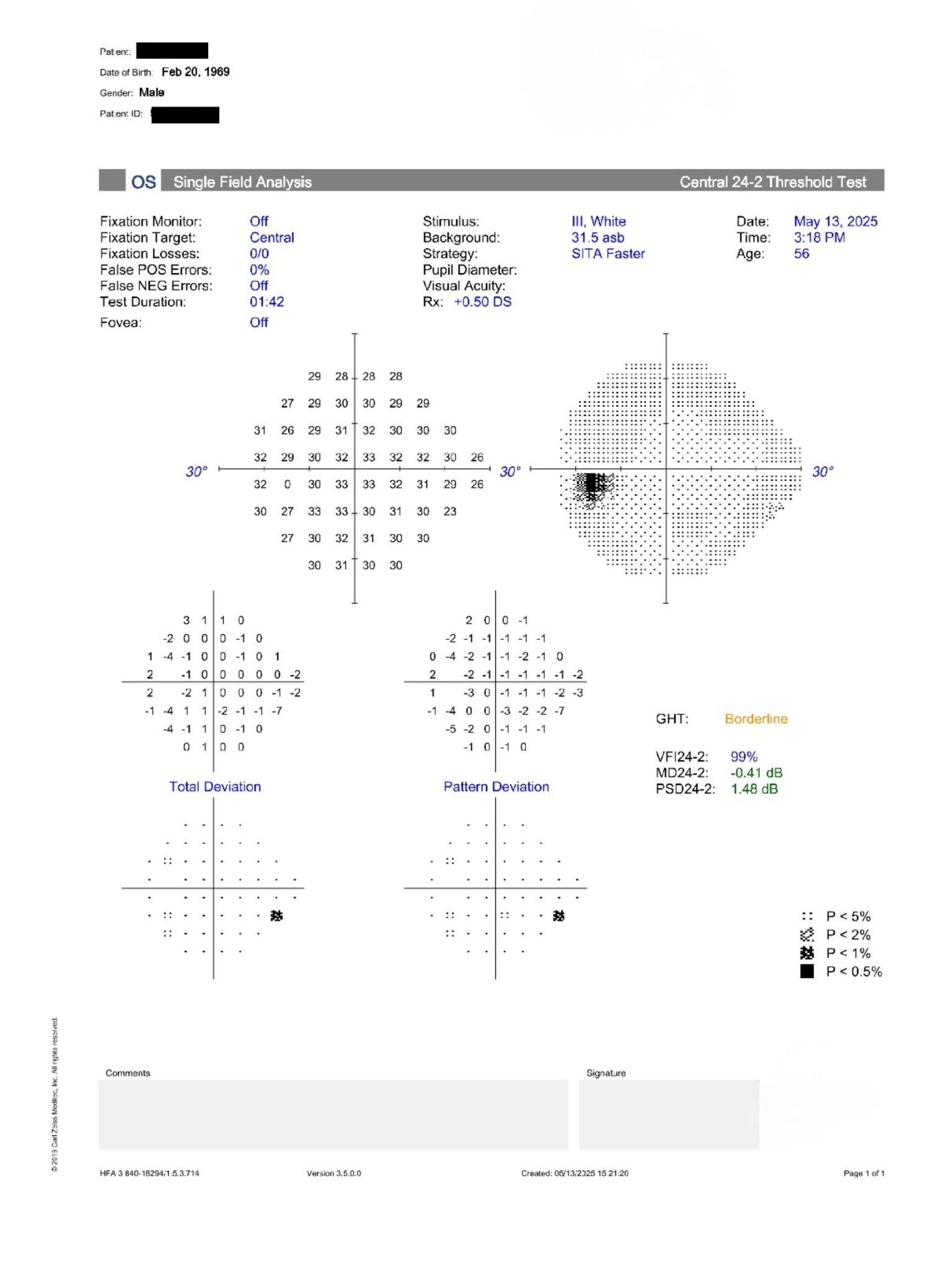
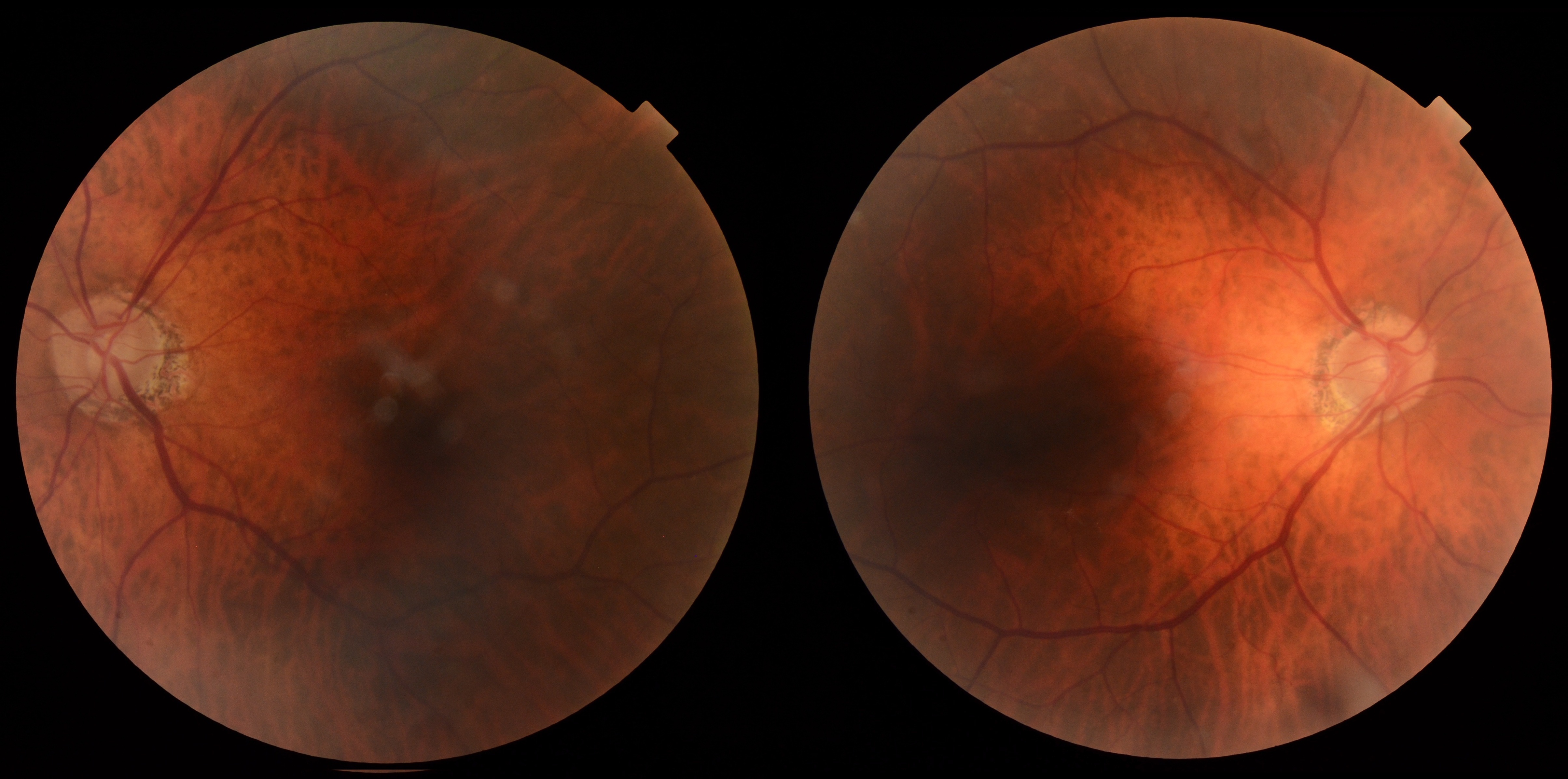
57-year-old male with large cup-to-disc ratios, iris transillumination defects, normal visual fields, and stable IOPs.
Case Study: Large Cupping With Normal VF
An optometrist uploaded this case to Care1.
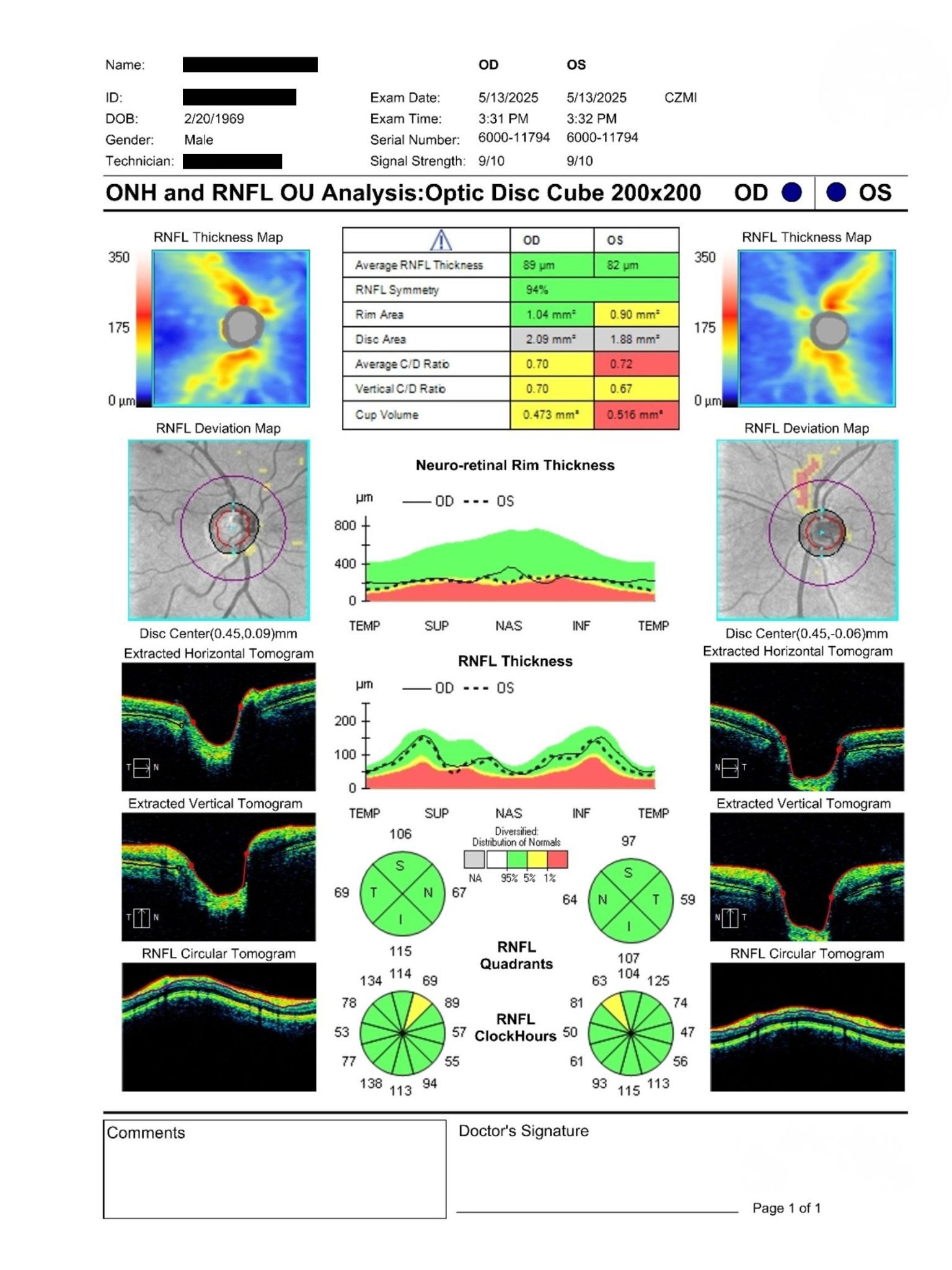
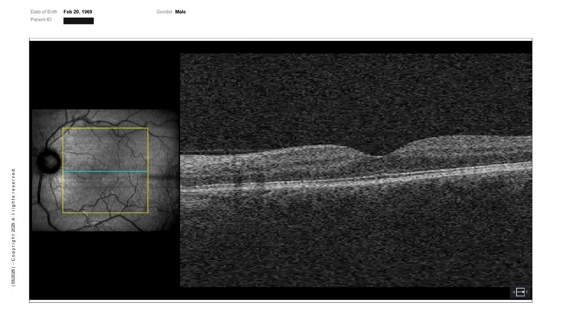
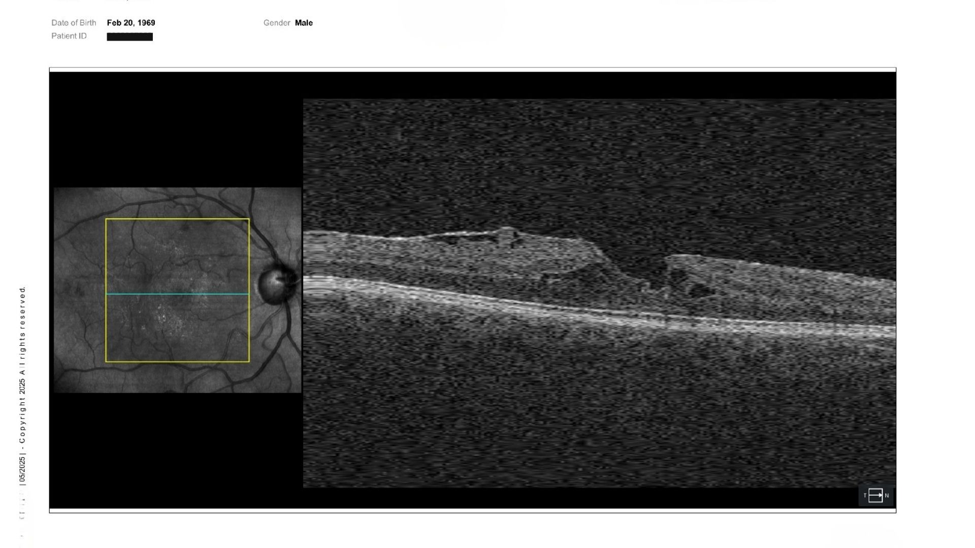
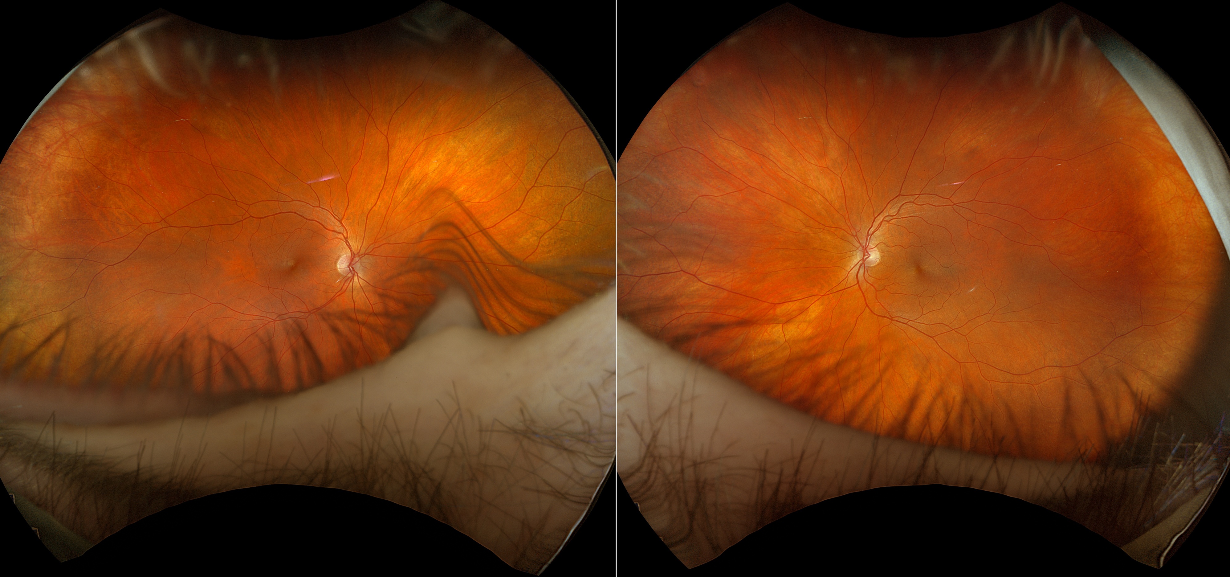
A 57-year-old male was referred for glaucoma assessment due to large cup-to-disc ratios, pigment dispersion findings, and glaucoma risk factors. BCVA was 20/20 OU. IOP measured 13 mmHg OD and 16 mmHg OS. Anterior segment examination revealed iris transillumination defects and Krukenberg spindles OU. Posterior segment examination showed large optic nerve cupping OU, with an epiretinal membrane and lamellar macular hole OD. OCT RNFL and visual fields were within normal limits.
Given the combination of large optic nerve cupping, thin corneas, and pigment dispersion findings, is observation with serial OCT RNFL and visual field testing the most appropriate management strategy?
An ophthalmology subspecialist provided a virtual consult within 1-2 weeks through Care1. Scroll below to see their diagnosis.
Care1 Ophthalmologist's Teleconsult
The ophthalmologist noted stable intraocular pressures and normal visual field testing. Baseline OCT RNFL imaging demonstrated robust retinal nerve fiber layer thickness in both eyes without structural evidence of glaucomatous damage. Although several risk factors were present, including pigment dispersion syndrome, thin corneas, younger age at initial presentation, and large cup-to-disc ratios, the normal OCT RNFL and visual field results were considered reassuring. Repeat glaucoma testing within 6 to 12 months was recommended to monitor for future change.
Care1 AI’s Clinical Insight
Unique Feature:
Pigment dispersion syndrome is characterized by the release of iris pigment throughout the anterior segment, producing findings such as iris transillumination defects, Krukenberg spindles, and trabecular meshwork pigmentation. The condition occurs most commonly in myopic individuals and may remain asymptomatic for years. Clinical evaluation focuses on identifying characteristic examination findings and monitoring for structural or functional changes in the optic nerve over time.
Practice at the highest level of medicine
✔ Specialist consults within 1-2 weeks ✔ AI-powered clinical support ✔ Greater confidence in complex decision making
A population-based study cited by the AAO EyeWiki reported annual incidences of only 4.8 per 100,000 for pigment dispersion syndrome and 1.4 per 100,000 for pigmentary glaucoma, highlighting how relatively uncommon these conditions are despite their prominence in glaucoma education. This finding emphasizes the importance of recognizing subtle clinical signs during routine examinations.
Reference: Ritch R, Steinberger D, Liebmann JM. Prevalence of pigment dispersion syndrome in a population undergoing glaucoma screening. Am J Ophthalmol. 1993;115(6):707-710.
Clinical Pearls
• Large C/D alone does not confirm disease • OCT RNFL and VF guide risk assessment • Thin corneas increase glaucoma suspicion