70-year-old with vision loss in one eye shows lens and macular changes; is retina referral needed or safe to monitor?
Case Study: Progressive Vision Loss and Macular Changes in One Eye
An optometrist uploaded this case to Care1.
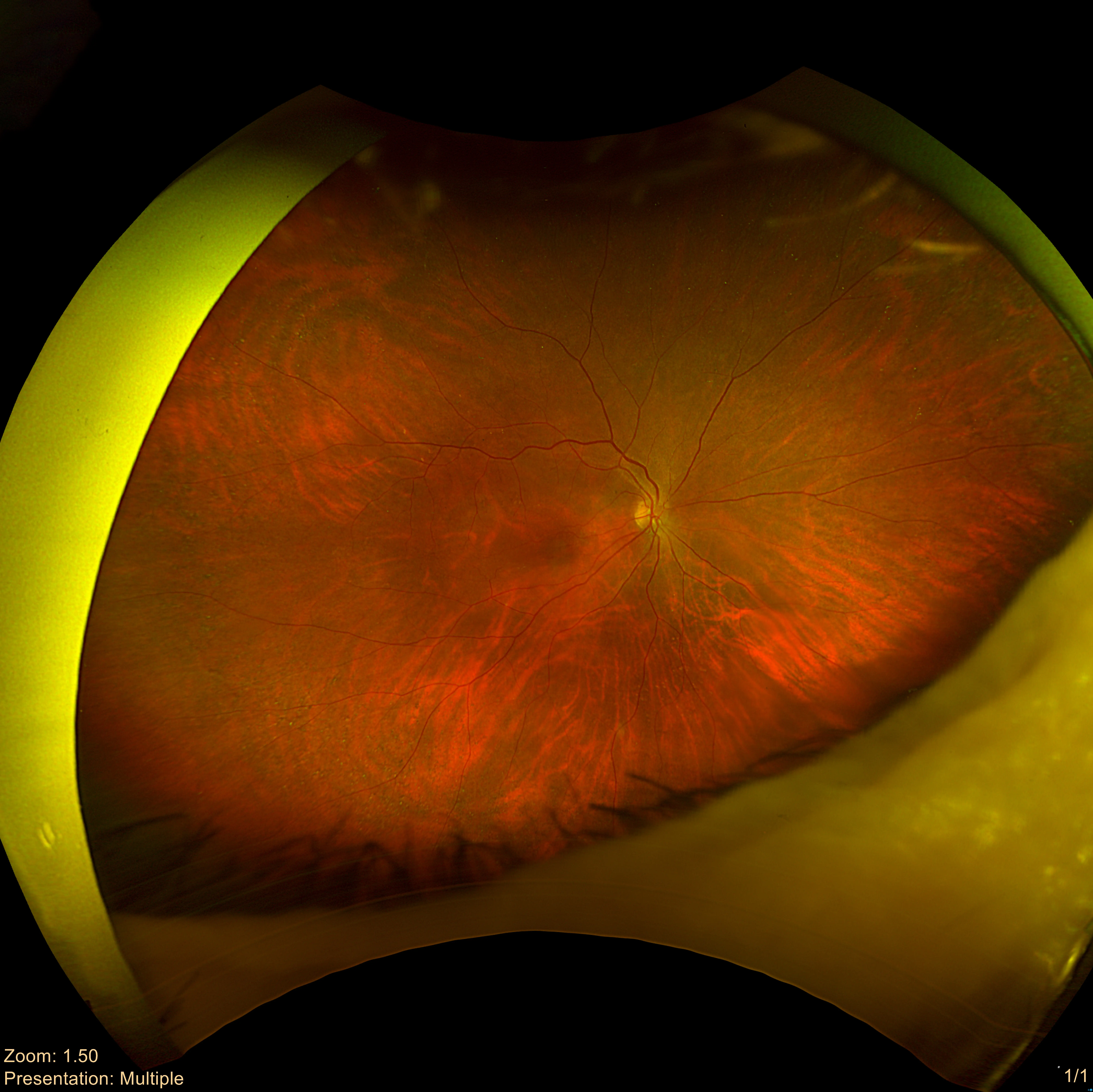
A 70-year-old patient was seen for a routine eye examination after noticing reduced vision in one eye over several months, while the other eye maintained good vision. The patient’s ocular history included prior cataract surgery in both eyes, and the ocular surfaces were healthy and quiet.
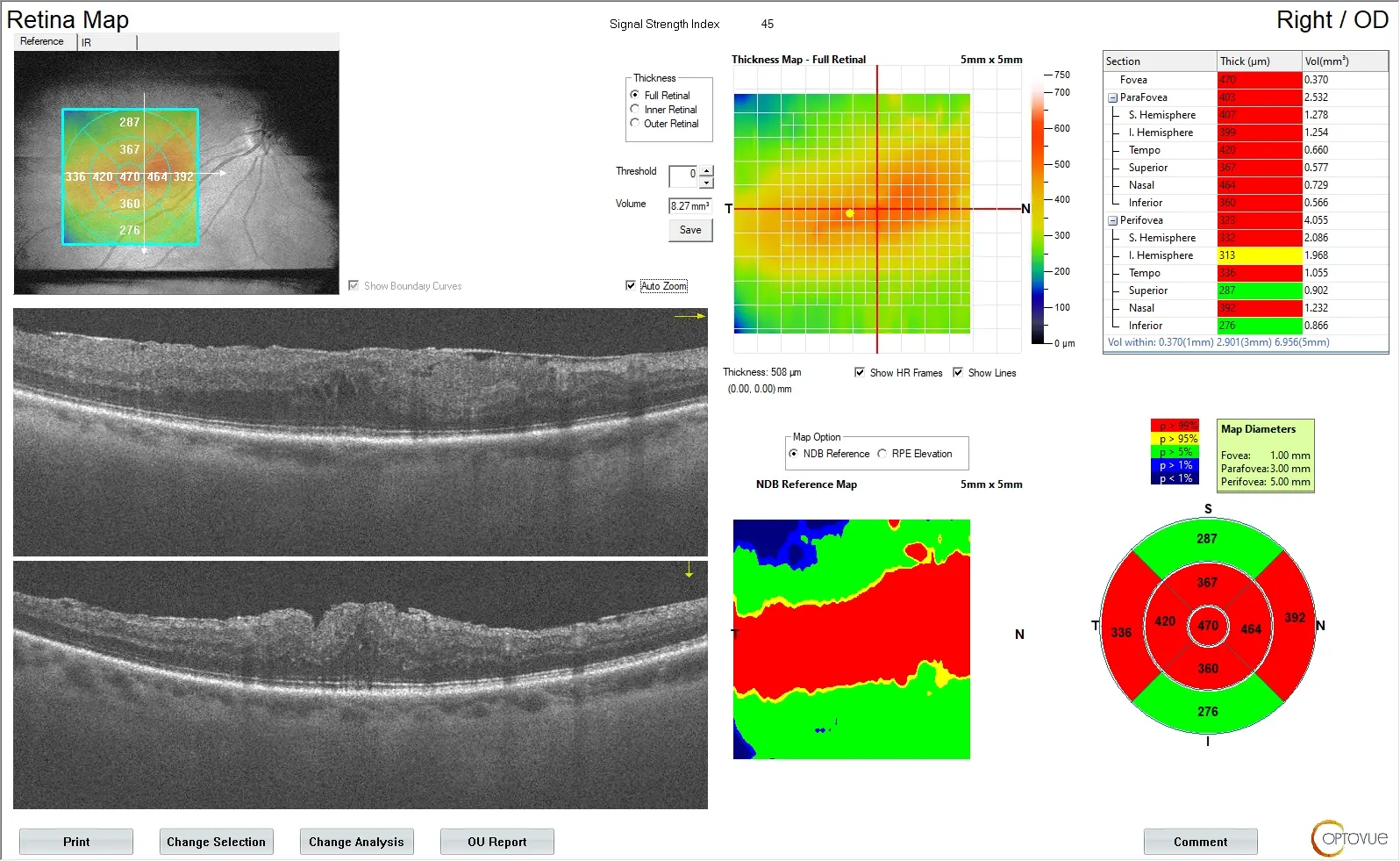
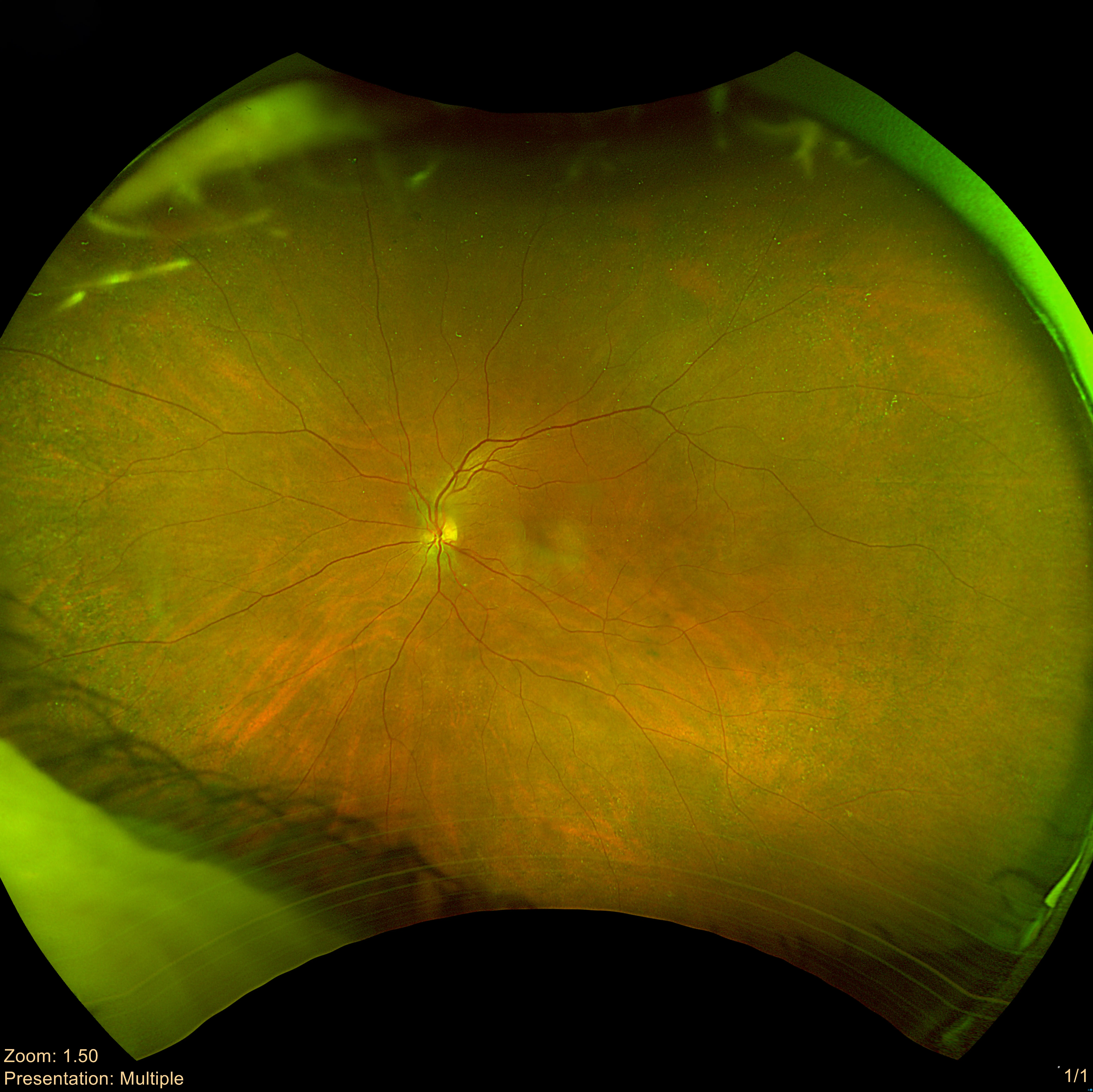
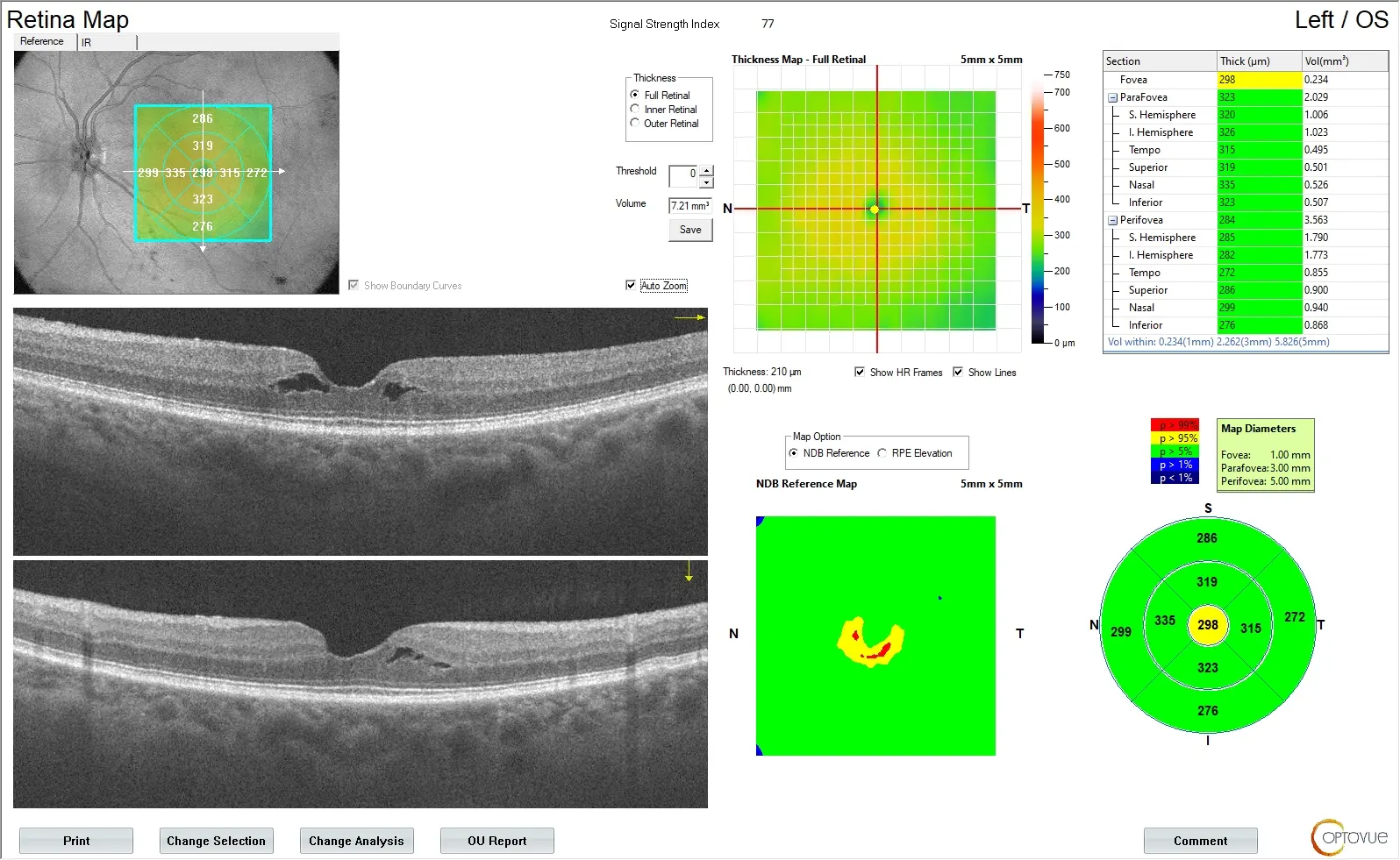
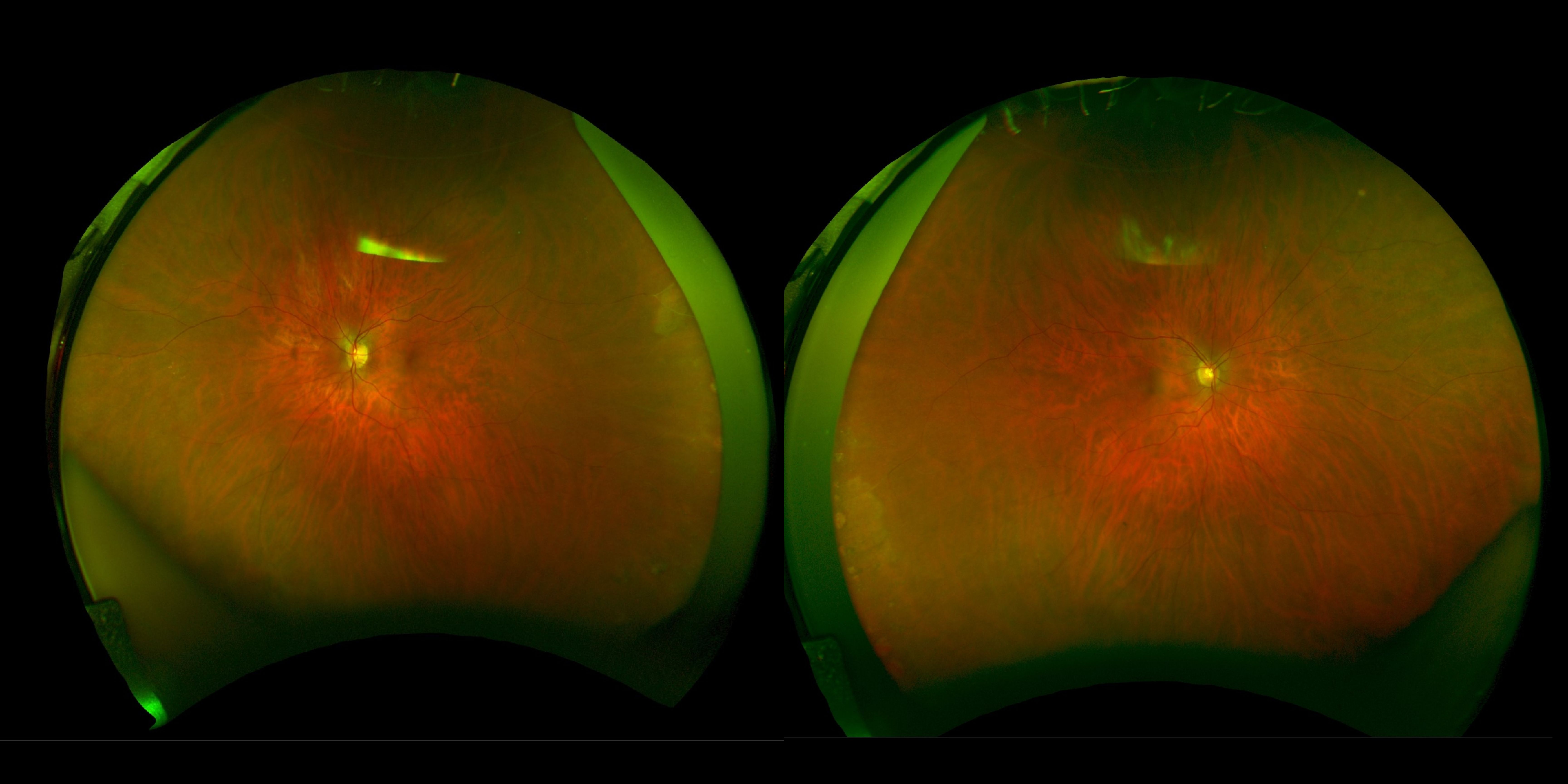
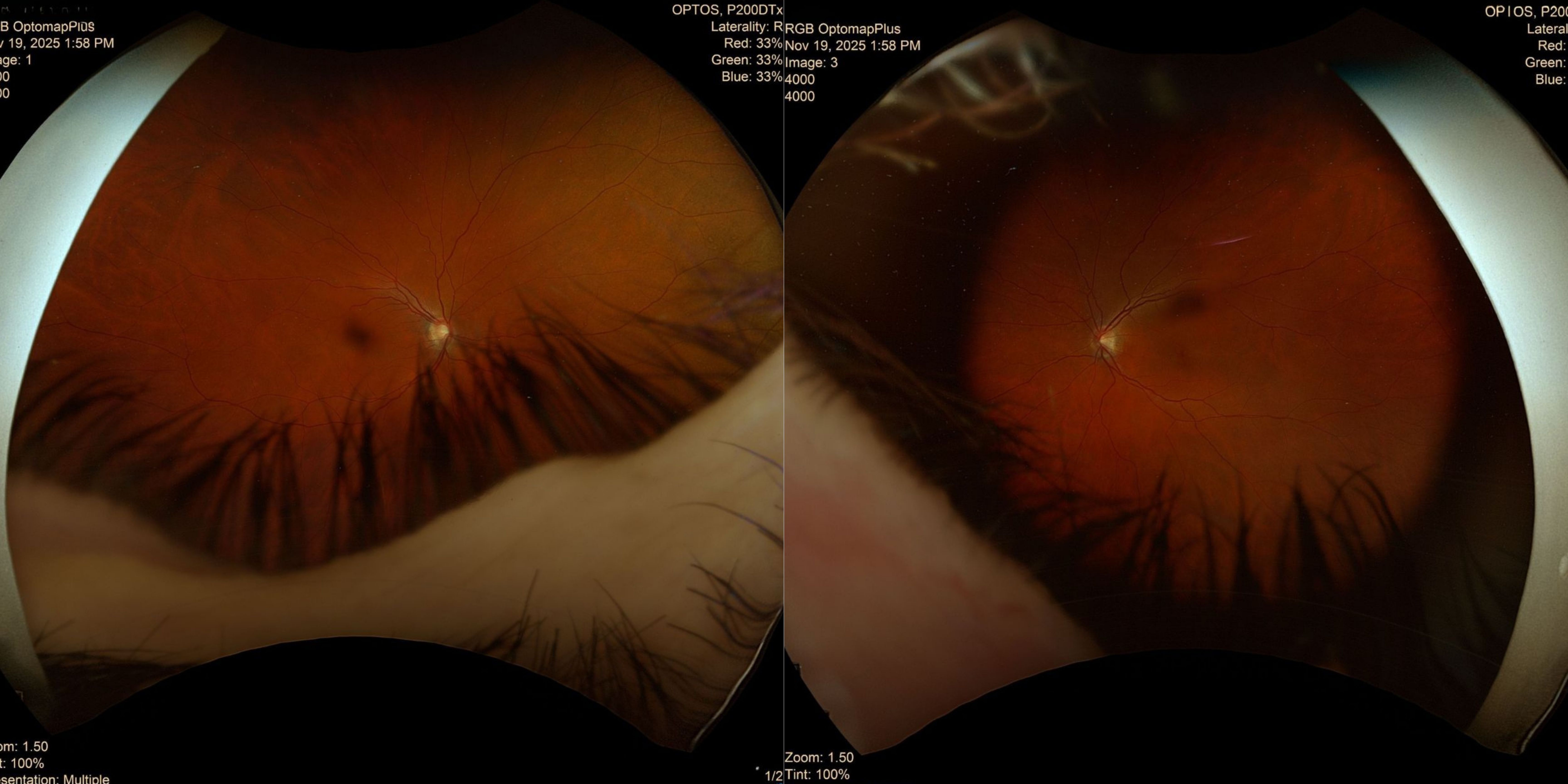
On examination, the affected eye showed notable changes in the lens and subtle structural alterations at the macula, while the fellow eye remained largely stable. OCT imaging highlighted significant changes in the affected eye and milder alterations in the other eye.
Would this patient benefit from an in-person consultation with a retina specialist, or can the case safely continue with periodic monitoring?
An ophthalmology subspecialist provided a virtual consult within 1-2 weeks through Care1. Scroll below to see their diagnosis.
Care1 Retina Surgeon’s Key Takeaways
This 70-year-old patient has reduced vision in the right eye with macular changes and a posterior capsule opacity. OCT shows significant structural alterations in the affected eye and milder changes in the fellow eye.
A YAG capsulotomy could be attempted to see if visual acuity improves. If vision does not improve, surgical intervention for the macular changes may be considered.
Has the patient been evaluated by a retina specialist for further consultation?
Continue with observation and follow-up as scheduled. I will continue to assist with management via review of the patient’s file and diagnostic testing.
Care1 AI’s Clinical Insight
Posterior capsule opacification (PCO) is a common long-term complication of cataract surgery that occurs when residual lens epithelial cells on the posterior capsule proliferate, migrate, and differentiate abnormally, leading to opacification. The process is influenced by multiple cytokines and growth factors, including TGF-β, FGF-2, and hepatocyte growth factor, as well as extracellular matrix remodeling. PCO can present in two forms: fibrous, characterized by wrinkles and folds on the capsule, and pearl (proliferative), with clusters of swollen, opacified lens epithelial cells known as Wedl cells. Both forms can impair vision by reducing clarity through the posterior capsule.
Did You Know?
Posterior capsular opacification is caused mainly by remnant lens epithelial cell proliferation and migration, epithelial‑mesenchymal transition, collagen deposition, and lens fiber generation.
Awasthi N, Guo S, Wagner BJ. Posterior capsular opacification: a problem reduced but not yet eradicated. Archives of Ophthalmology. 2009;127(4):555‑562.