Stable IOP and visual fields with a longstanding defect. Should this 70-YO continue routine monitoring or adjust management?
Case Study: Longstanding Field Defect With Stable Low Pressures
An optometrist uploaded this case to Care1.
A 70-YO male presented for a follow-up visit, reporting no new visual concerns. They use a nightly topical pressure-lowering medication in both eyes and have been stable on this regimen.
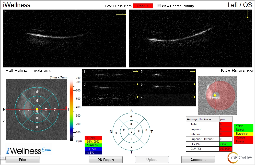
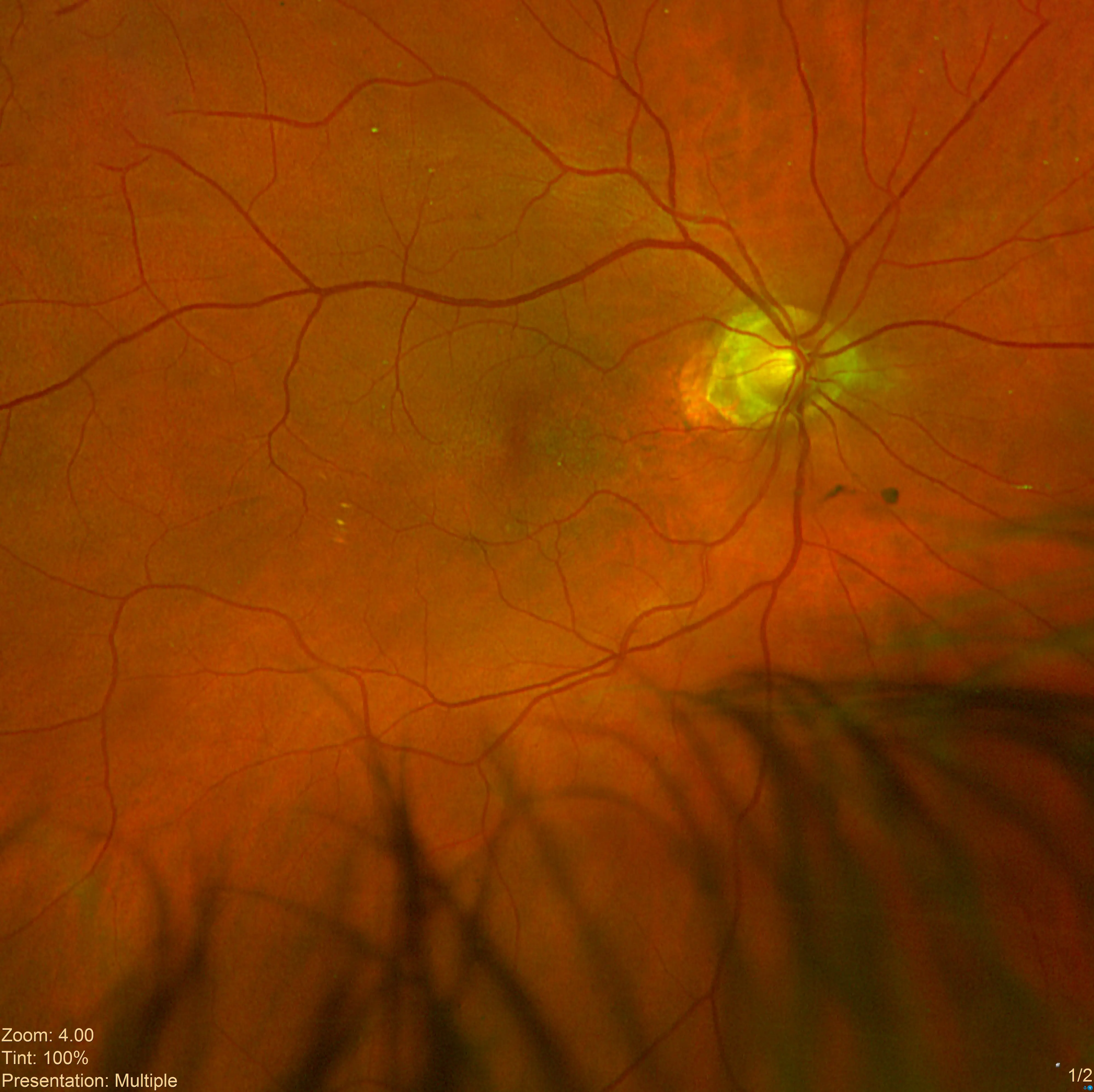
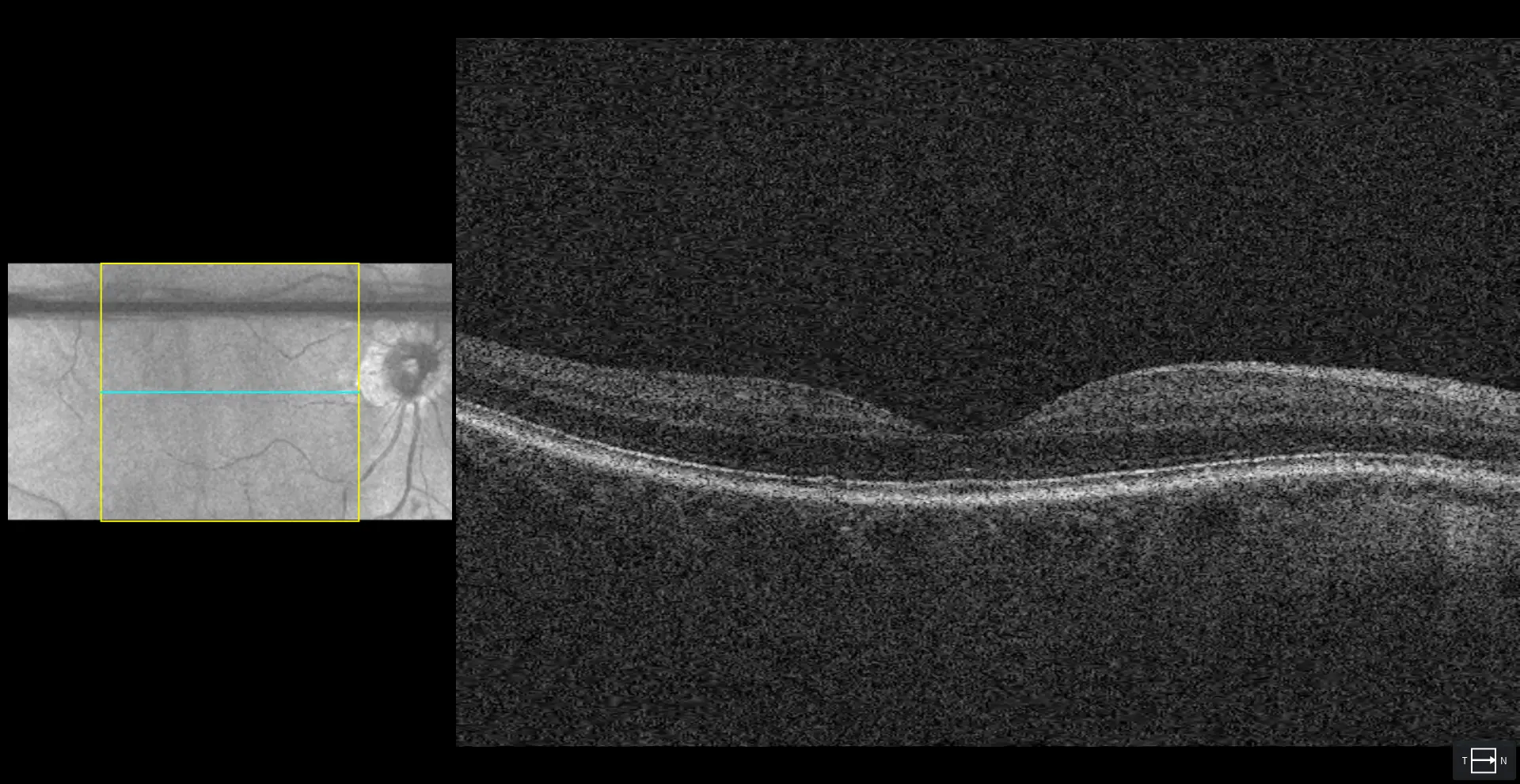
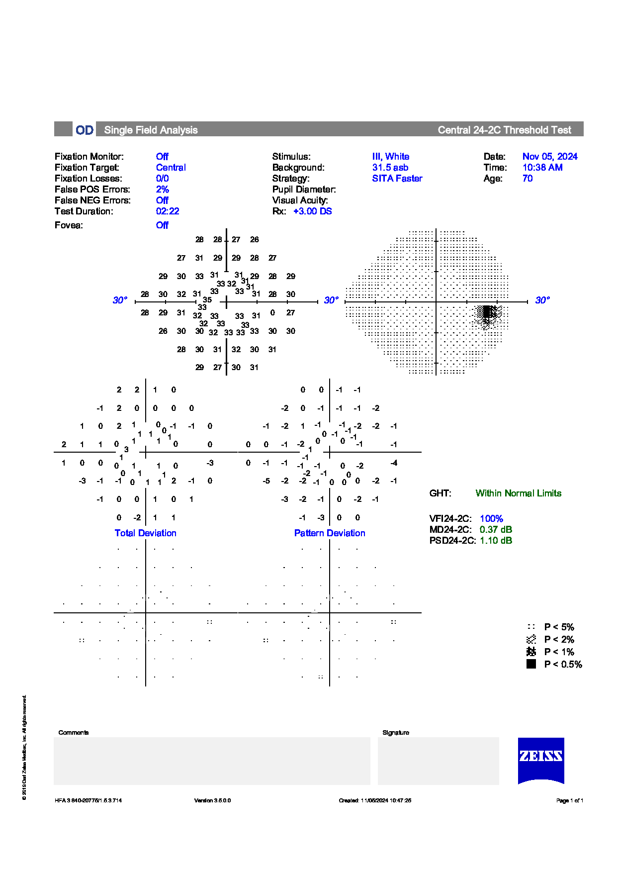
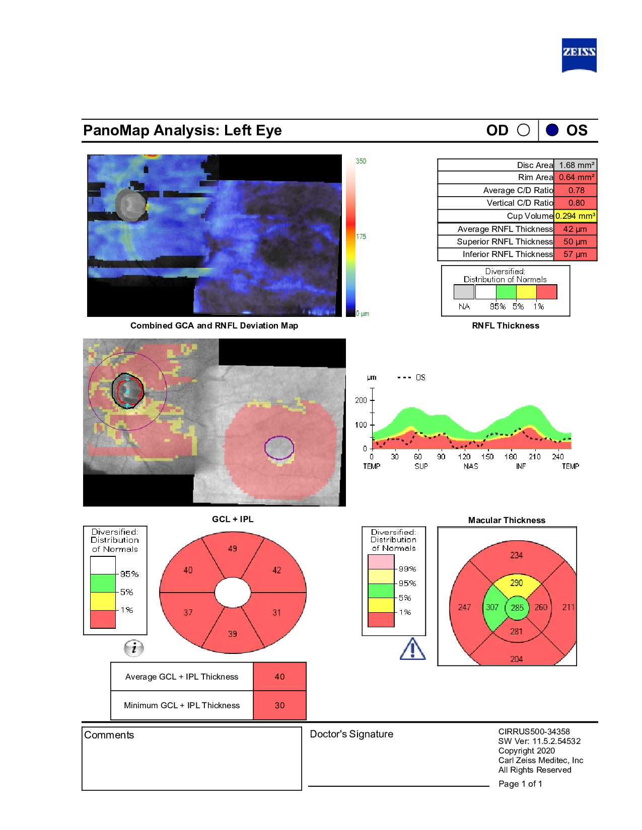
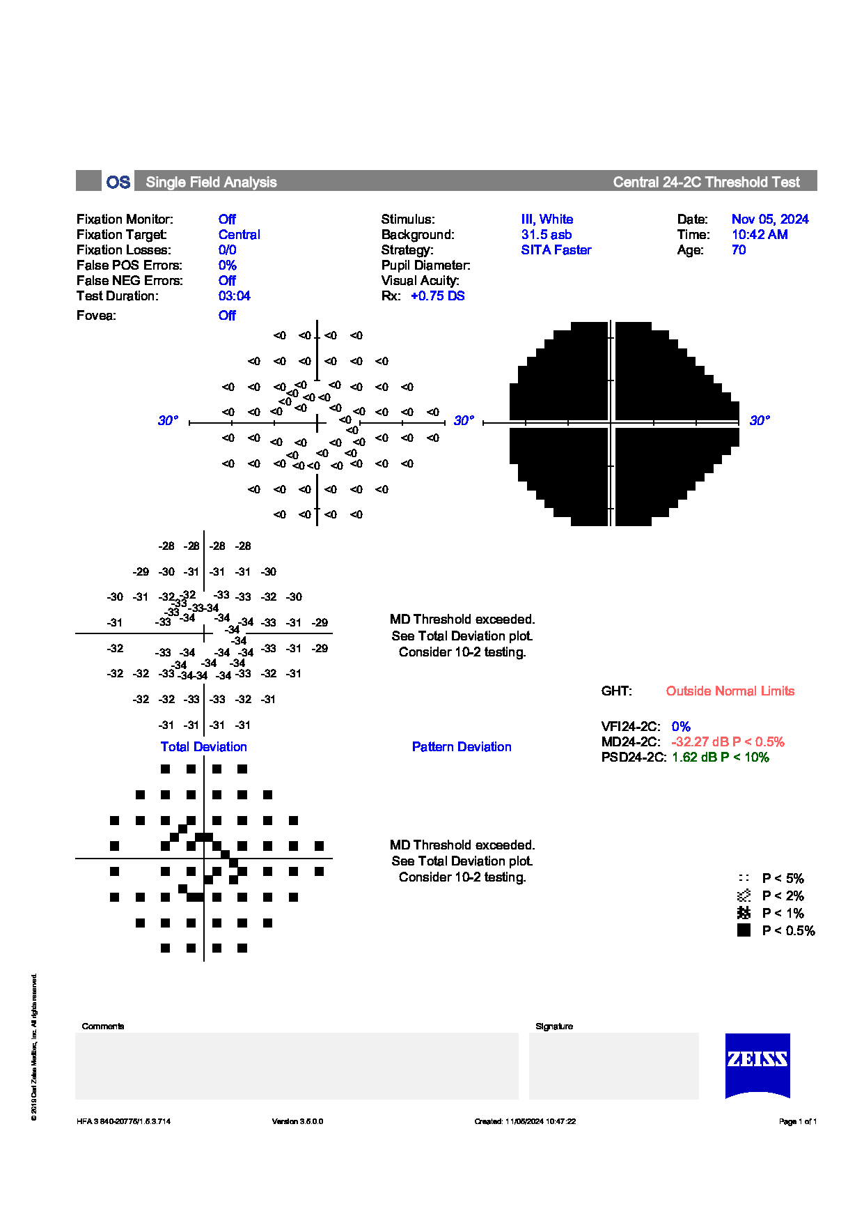
In-office testing showed low intraocular pressures in both eyes. Visual field testing of the right eye remained stable. The left eye showed a longstanding, dense superior defect consistent with previous macular and optic nerve changes from prior surgery, with no indication of progression. Ocular health was otherwise stable, and the patient was scheduled for routine monitoring with semiannual visual fields, OCT imaging, and pressure checks.
Would this patient benefit from any changes to their current management plan, or is continued routine surveillance appropriate at this stage?
An ophthalmology subspecialist provided a virtual consult within 1-2 weeks through Care1. Scroll below to see their diagnosis.
Care1 Subspecialist’s Key Takeaways
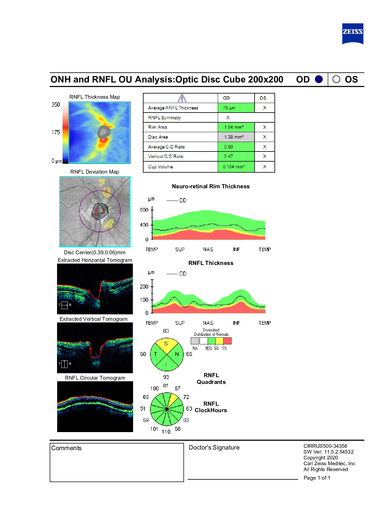
This patient has a complex ocular history, including prior macular hole repair in the left eye, LASIK in both eyes, and retinal surgery in the right eye. Current eye pressures are well controlled on topical therapy, with C/D ratios showing asymmetry (0.30 OD, 0.80 OS). OCT imaging shows some superior nerve thinning in the right eye, while visual fields remain normal.
Ongoing follow-up is recommended every 6–12 months, including OCT and visual field testing at each visit. Areas of note include asymmetry in optic nerve appearance and borderline OCT findings, while reassuring features include stable pressures and advanced age. The patient’s care continues collaboratively, with communication to their family physician and regular monitoring at the primary office.
Care1 AI’s Clinical Insight
Primary open-angle glaucoma (POAG) is a chronic optic neuropathy characterized by an open anterior chamber angle and progressive retinal ganglion cell loss that typically leads to optic nerve cupping and visual field defects. It is usually asymptomatic until advanced, and risk increases with age, elevated intraocular pressure, family history, and certain racial backgrounds; all current treatments focus on lowering intraocular pressure to slow progression.
Did You Know?
Primary open-angle glaucoma often progresses silently, with patients remaining asymptomatic until significant peripheral visual field loss has occurred, which is why it is frequently diagnosed at a later stage during routine eye examinations.