77-year-old man with stable OCT, thin RNFL, and subtle visual field changes. An optometrist uploaded this case to Care1.
Case Study: Stable RNFL With Cupping
An optometrist uploaded this case to Care1.
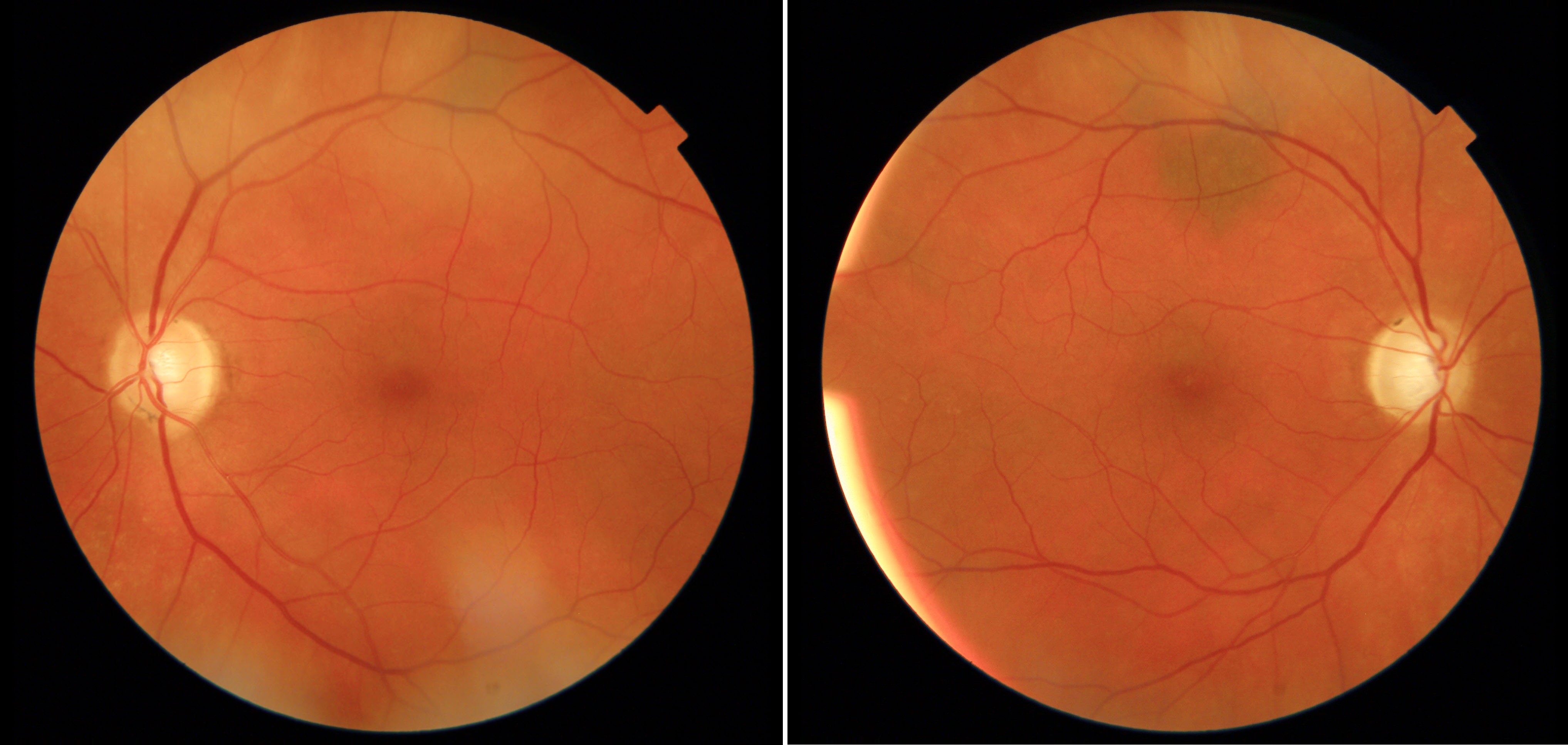
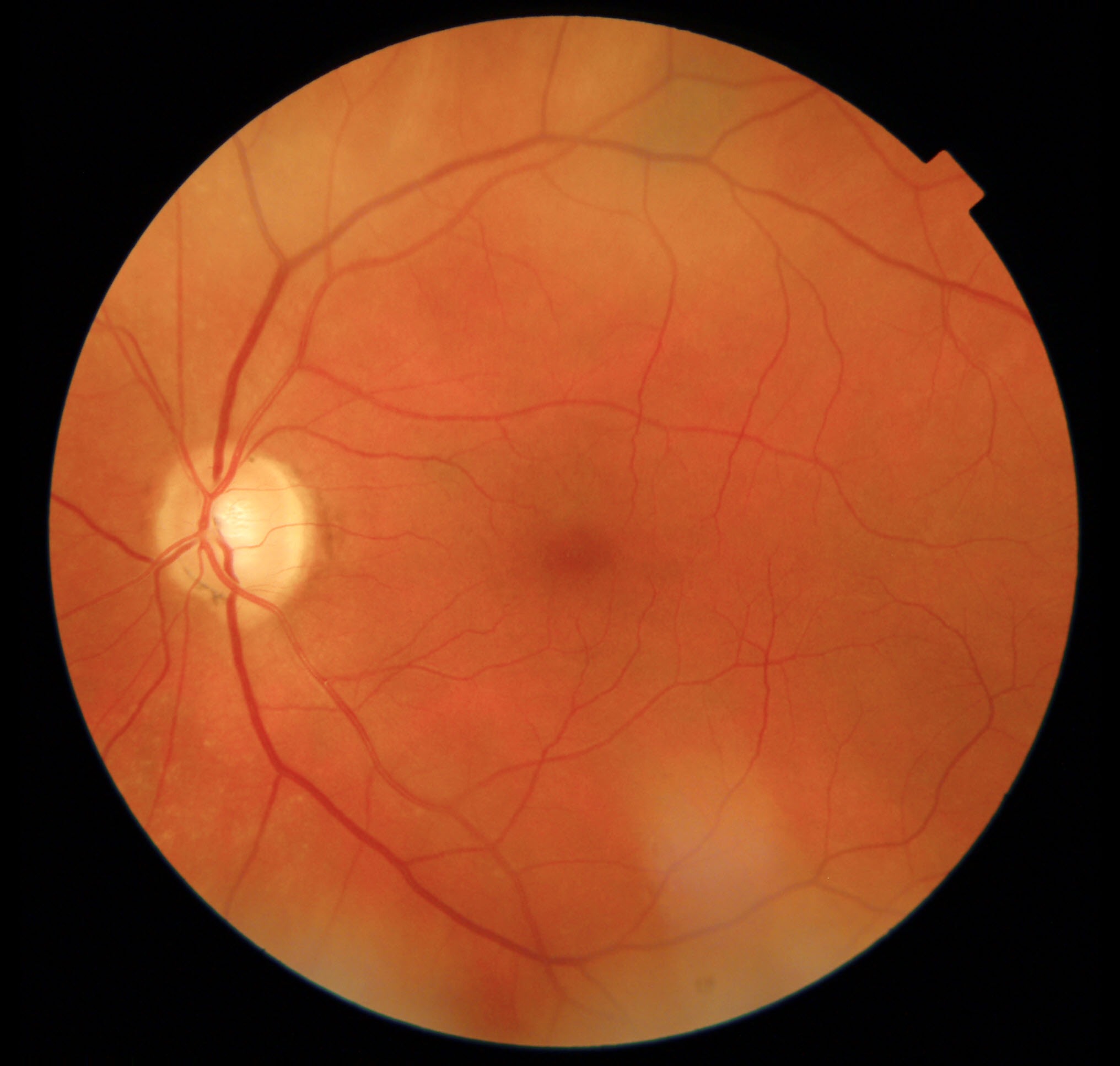
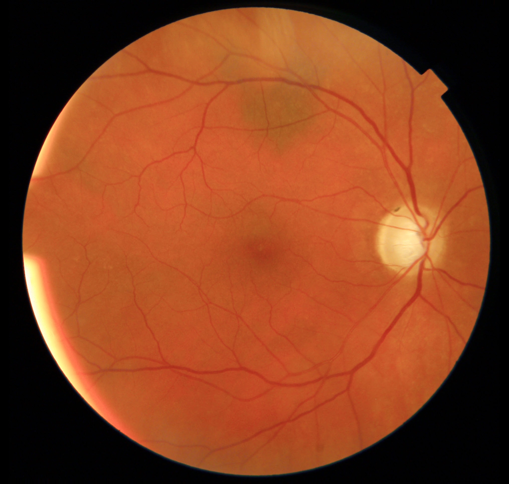
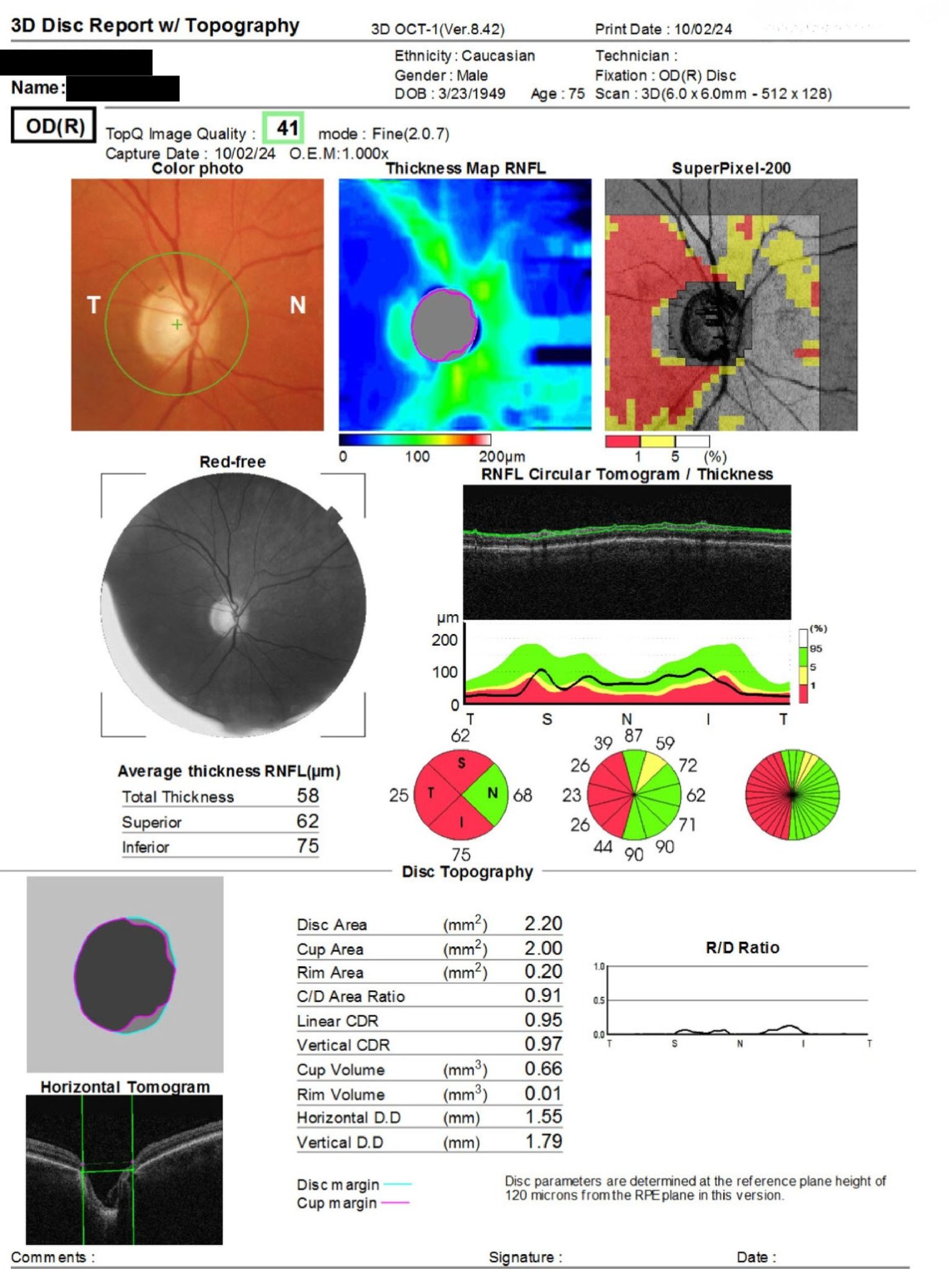
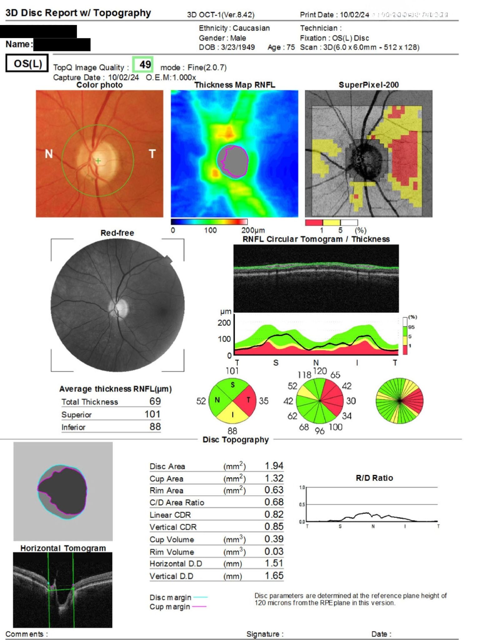
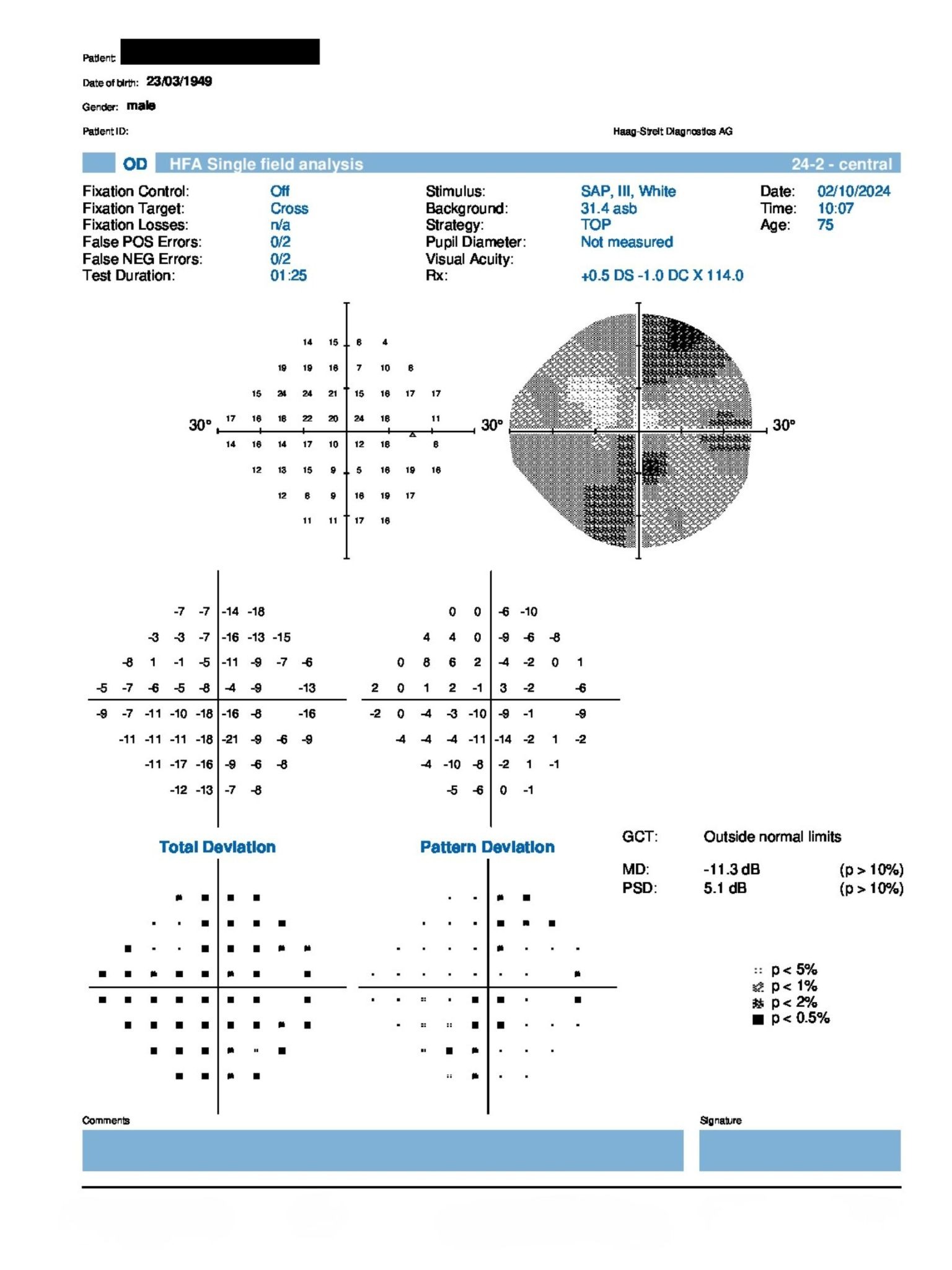
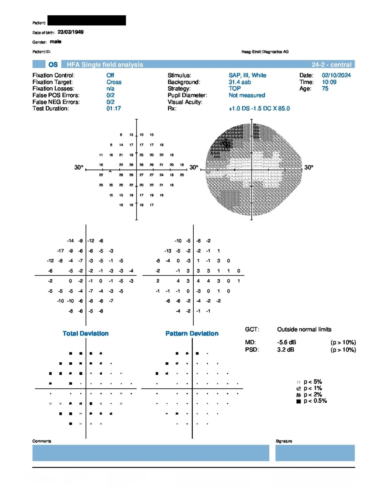
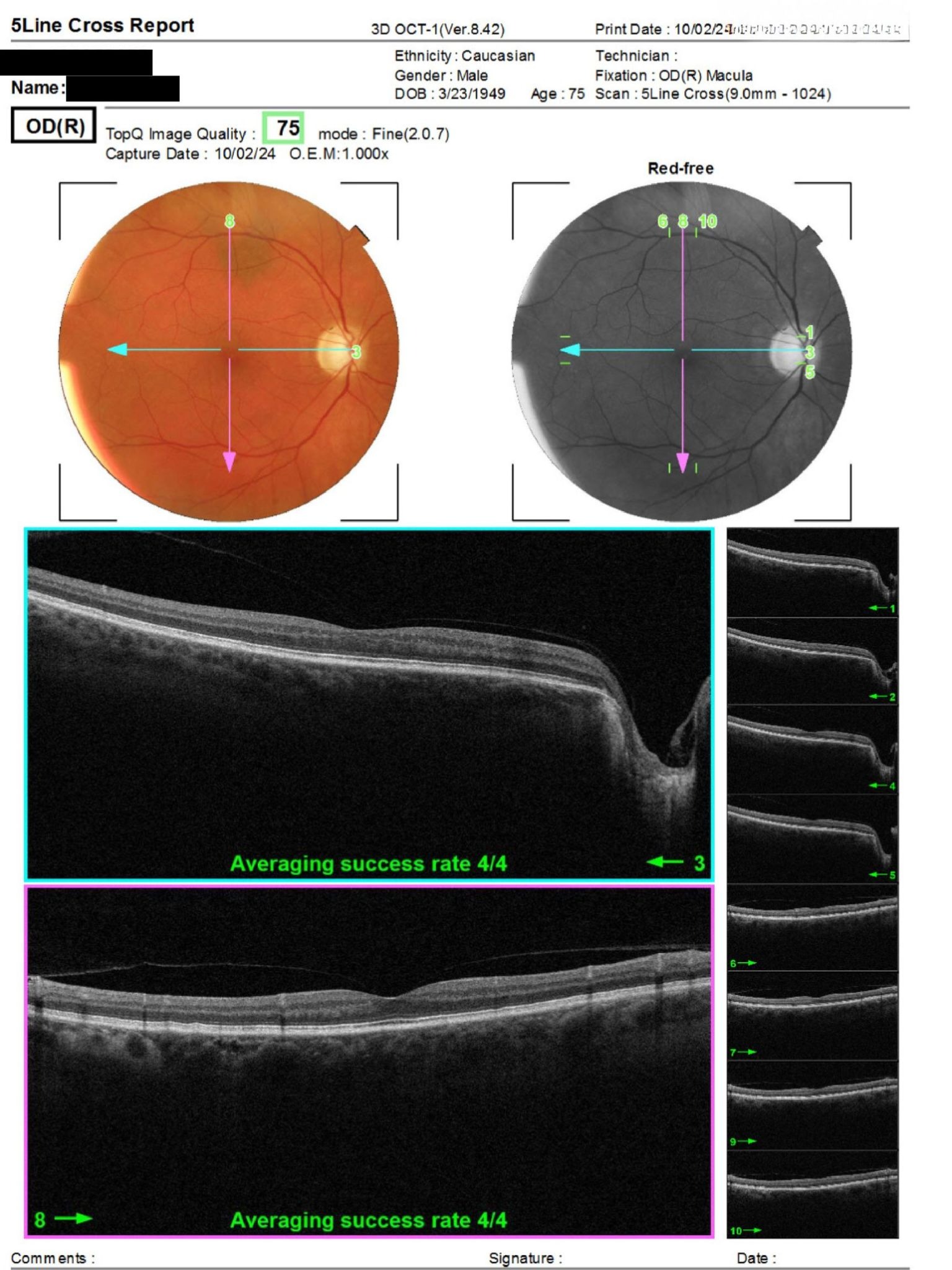
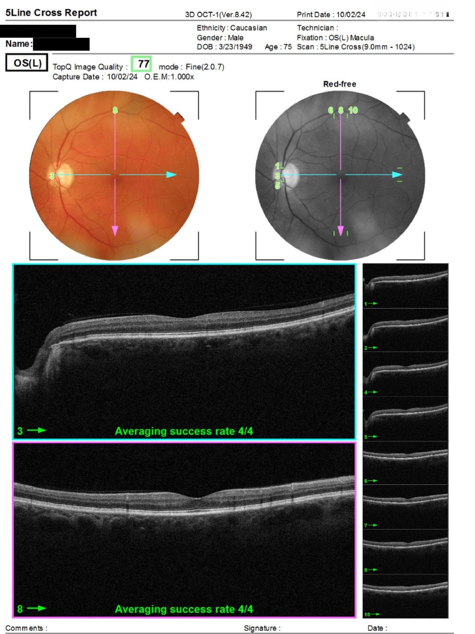
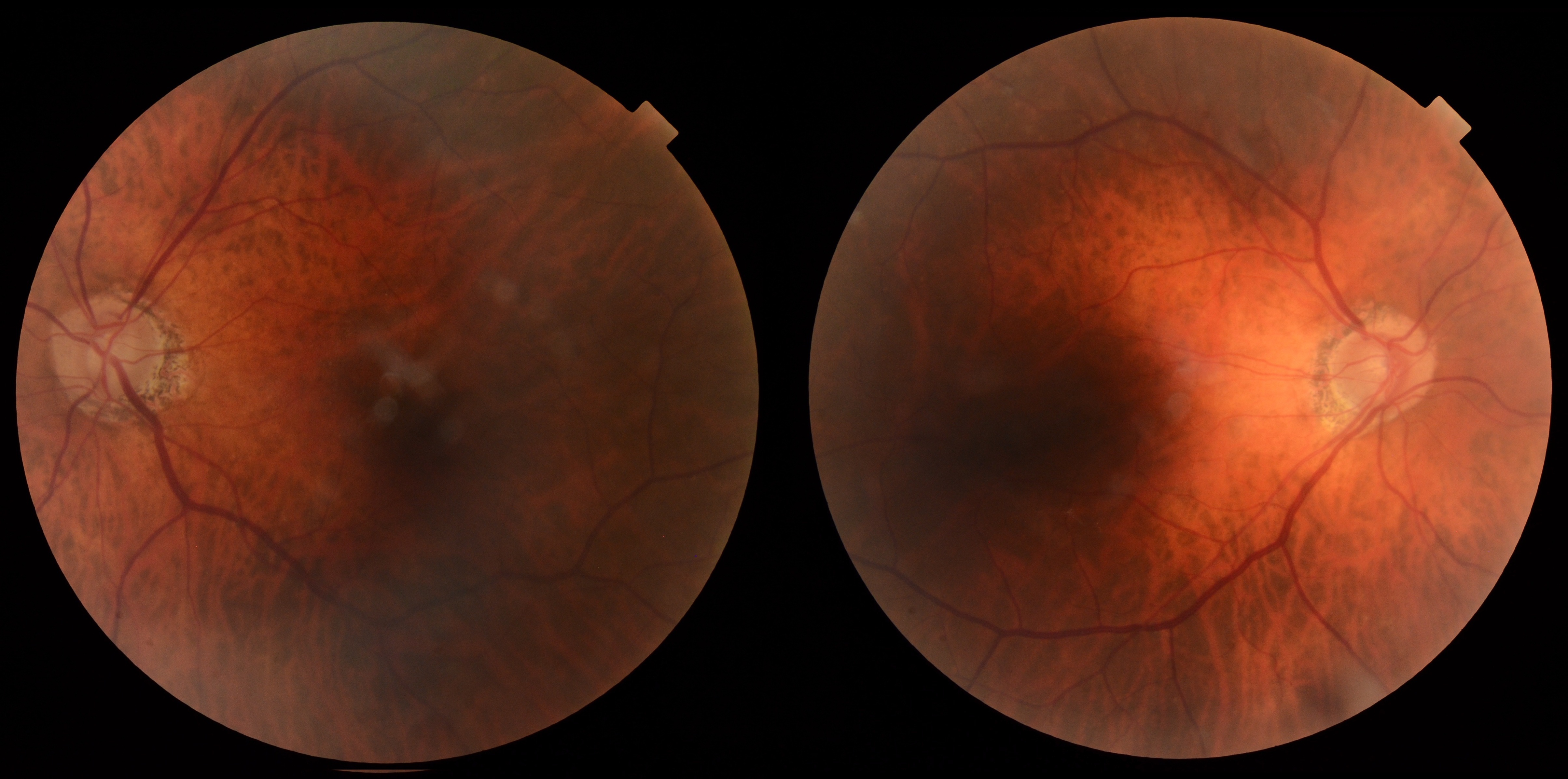
A 77-year-old man was referred for glaucoma follow-up after intraocular pressures trended slightly higher despite excellent medication compliance. BCVA was 20/20 OU with IOPs of 16 mmHg OU. The anterior segment was unremarkable following previous cataract surgery. Posterior examination showed asymmetric optic disc cupping, RNFL thinning greater in the right eye, and stable visual field defects without progression.
With stable structural and functional testing despite slightly increasing IOP, should the current management plan be maintained?
An ophthalmology subspecialist provided a virtual consult within 1-2 weeks through Care1. Scroll below to see their diagnosis.
Care1 Ophthalmologist's Teleconsult
The OCT demonstrated stable superior and inferior RNFL thinning in the right eye and inferior thinning in the left eye. Visual fields remained stable with paracentral defects in the right eye and superior defects in the left eye. Intraocular pressures were within the individualized target range while using Duotrav, and the optic nerve appearance remained unchanged with greater cupping in the right eye. Continued monitoring with repeat IOP assessment and routine OCT and visual field testing was considered appropriate.
Care1 AI's Clinical Insight
Structure-function stability
Structural assessment with OCT RNFL and functional assessment using standard automated perimetry provide complementary information when monitoring glaucoma progression. Progressive loss of the retinal nerve fiber layer may precede visual field changes, while visual field defects can fluctuate over time, making serial testing essential for accurate longitudinal assessment.
Practice at the highest level of medicine
✔ Specialist consults within 1-2 weeks ✔ AI-powered clinical support ✔ Greater confidence in complex decision making
Longitudinal data from the Diagnostic Innovations in Glaucoma Study found that eyes with faster RNFL thinning on OCT were significantly more likely to develop measurable visual field progression over time, underscoring the value of trend-based structural analysis even when functional testing appears stable.
Reference: Yu M, Lin C, Weinreb RN, et al. Risk of visual field progression in glaucoma patients with progressive retinal nerve fiber layer thinning: a 5-year prospective study. Ophthalmology. 2016;123(6):1201-1210. doi:10.1016/j.ophtha.2016.01.045