81-year-old with progressive visual field loss, stable OCT RNFL, and reduced vision OS requiring glaucoma reassessment.
Case Study: Progressive Visual Field Loss
An optometrist uploaded this case to Care1.
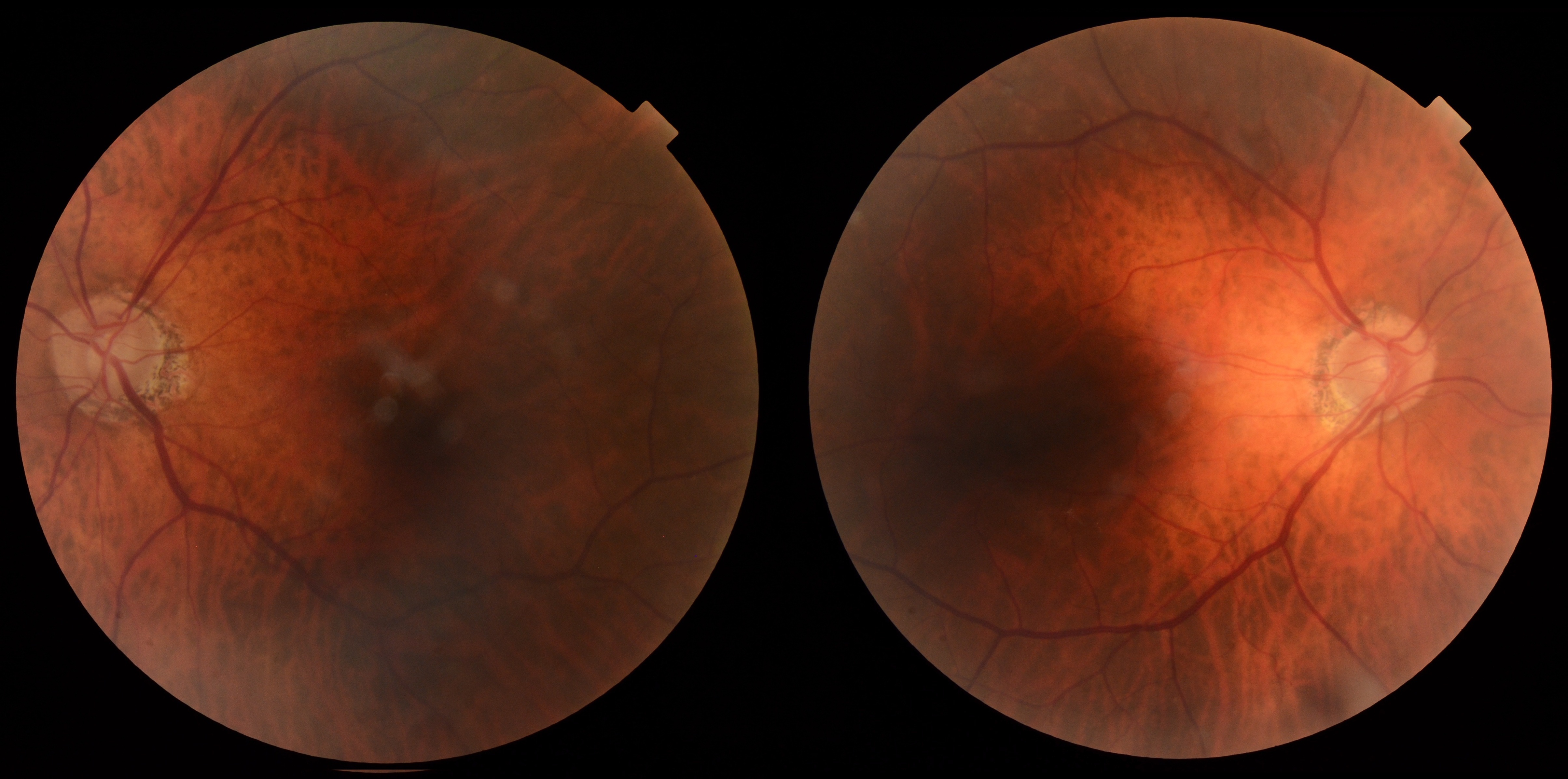
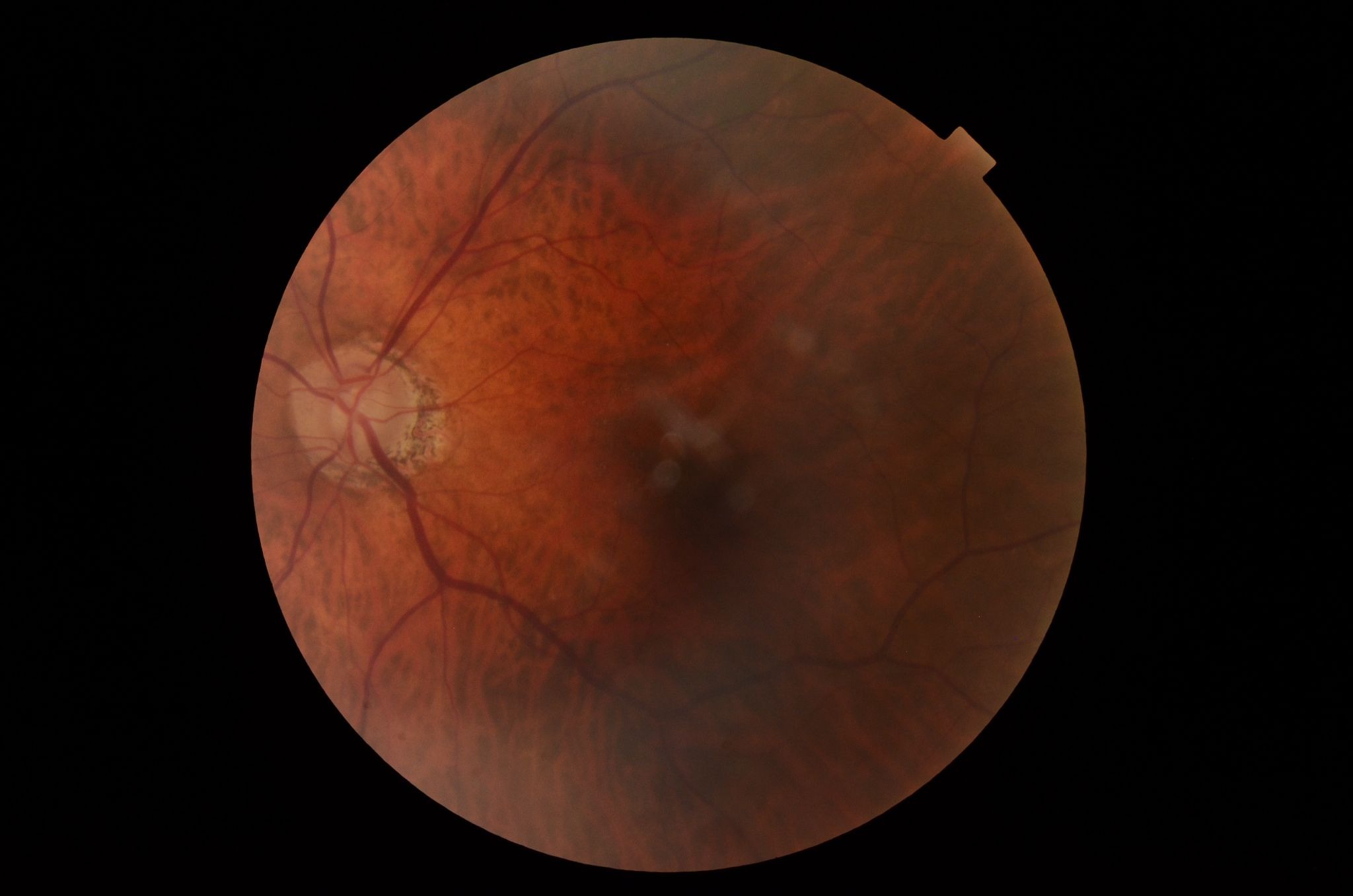
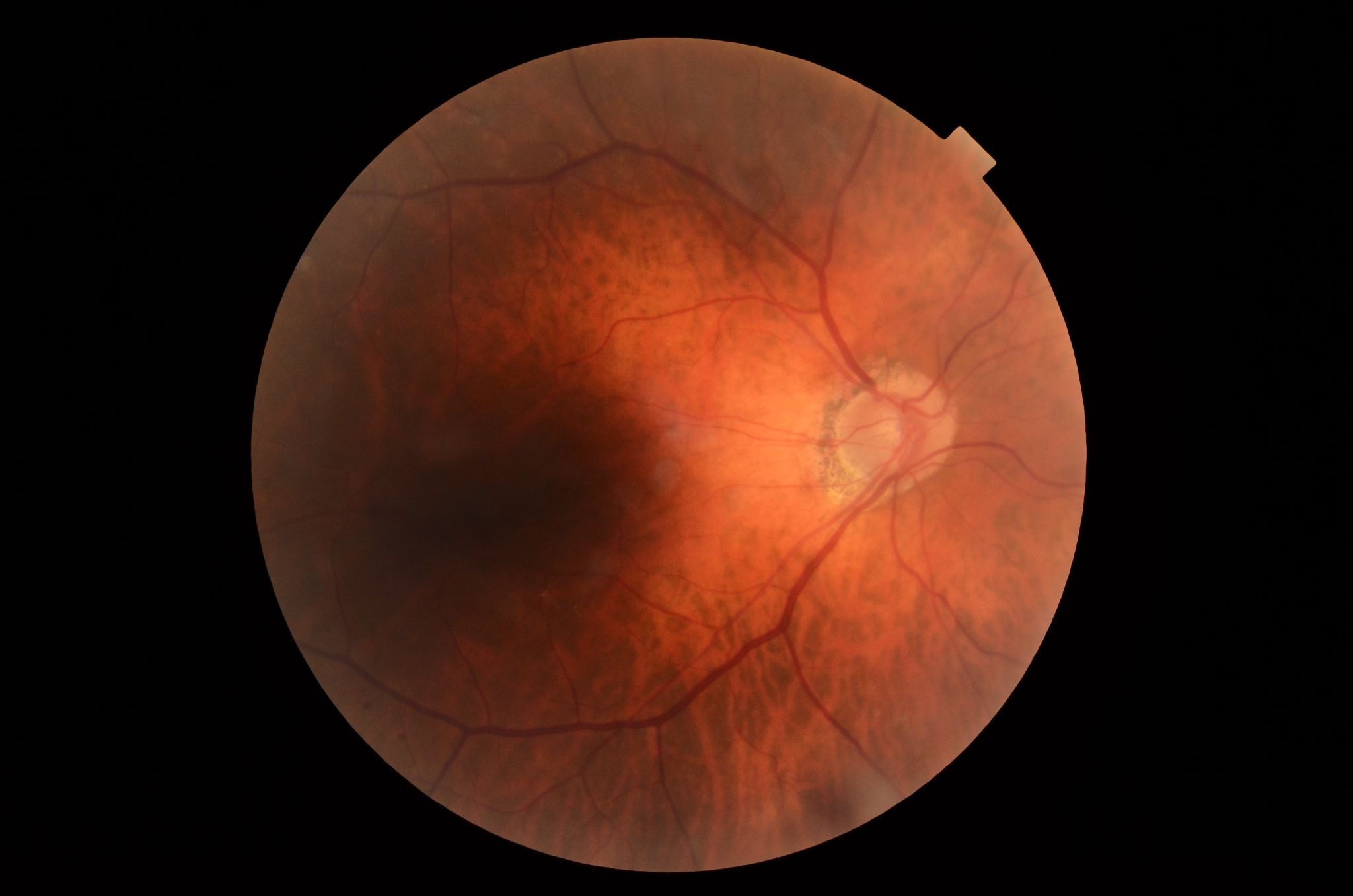
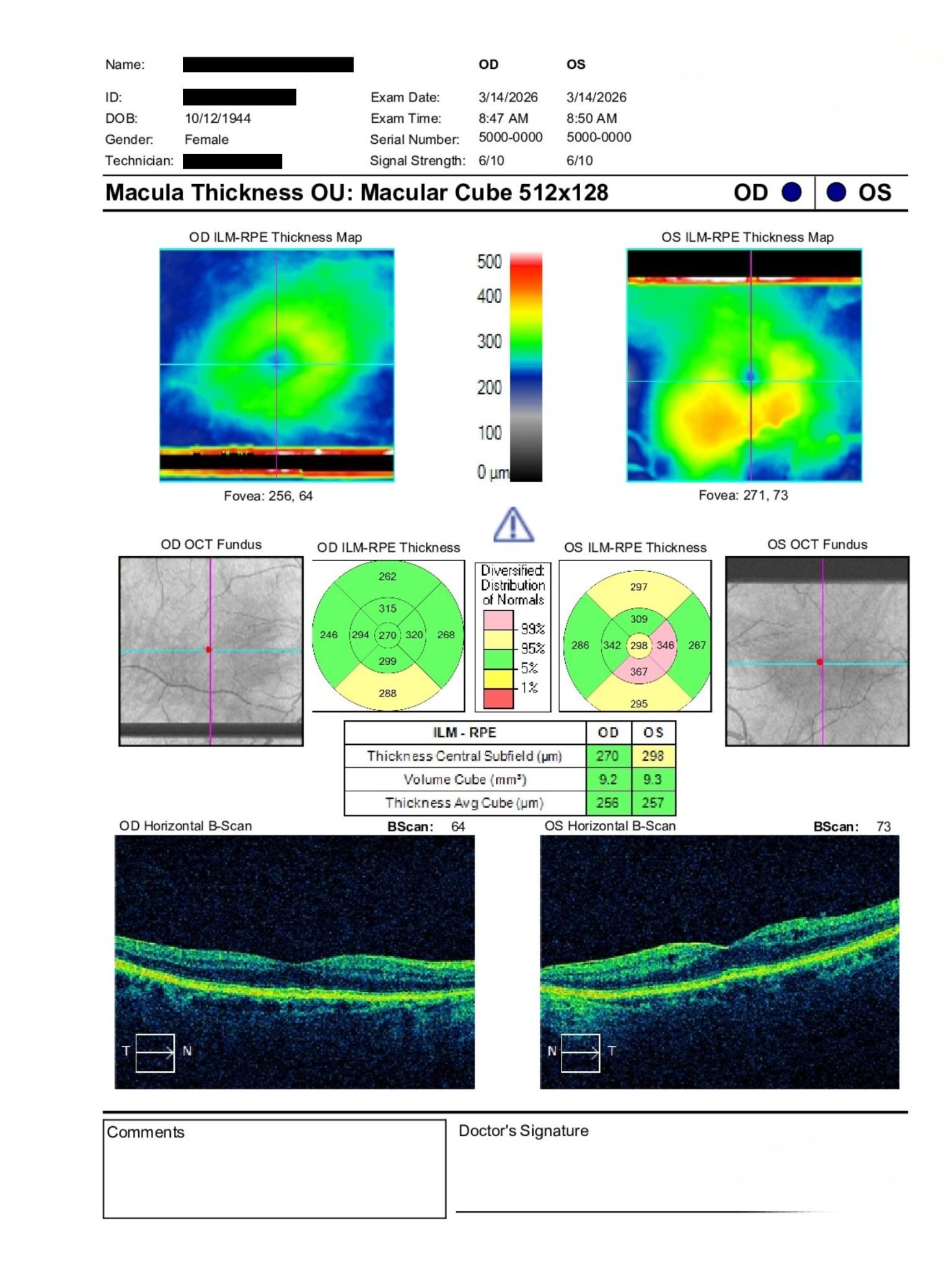
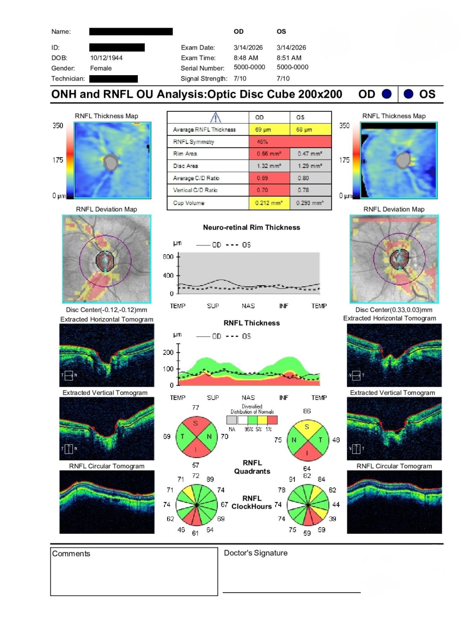
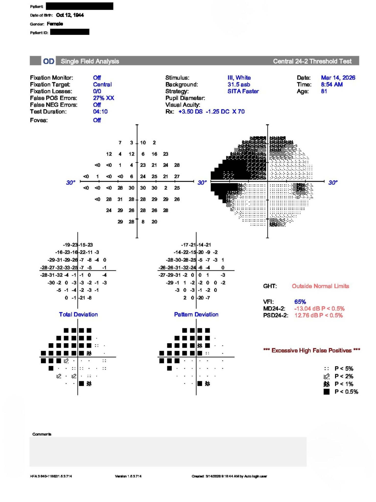
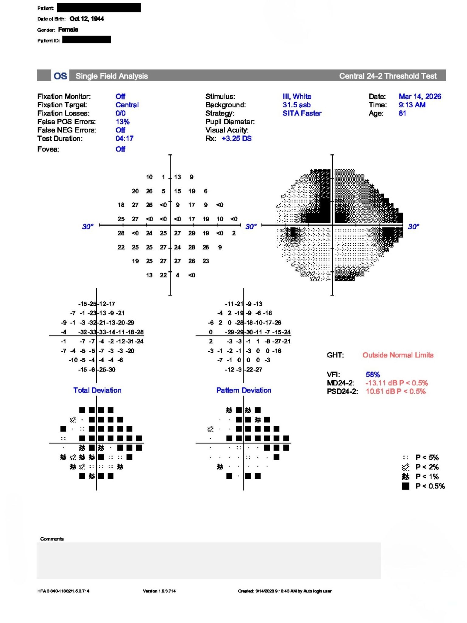
An 81-year-old woman with known glaucoma was referred for teleconsult due to concern for progression. BCVA was 20/25 OD and 20/200 OS with IOPs of 13 and 15 mmHg. The anterior segment was notable for advanced cataracts OU. Posterior examination showed large cup-to-disc ratios with OCT RNFL relatively stable compared with prior imaging, while visual fields demonstrated progression and cystic macular changes were noted OS.
How should these findings influence further glaucoma assessment and management?
An ophthalmology subspecialist provided a virtual consult within 1-2 weeks through Care1. Scroll below to see their diagnosis.
Care1 Ophthalmologist's Teleconsult
The OCT RNFL remained relatively stable compared with previous imaging, but visual fields demonstrated progression in both eyes. Reduced visual acuity in the left eye, together with cystic macular changes, suggested additional pathology contributing to the clinical picture. Because medication history was incomplete, treatment recommendations could not be finalized. An in-person ophthalmology assessment was recommended to clarify current therapy, establish a new glaucoma baseline, and reassess structural and functional progression.
Care1 AI's Clinical Insight
Stable OCT, worsening visual fields
Glaucoma progression may occur despite relatively stable retinal nerve fiber layer measurements because structural and functional changes do not always progress simultaneously. Visual field testing remains essential for detecting functional decline, particularly in advanced disease where OCT measurements may reach a measurement floor. Careful correlation between optic nerve appearance, OCT RNFL, and perimetry provides a more complete assessment of disease status.
Unlock your full revenue potential
✔ Specialist consults within 1-2 weeks ✔ AI-powered clinical support ✔ Greater confidence in complex decision making
In the Advanced Glaucoma Intervention Study (AGIS), eyes maintaining an average intraocular pressure below 18 mmHg with minimal fluctuation experienced significantly less visual field deterioration over long-term follow-up, highlighting the importance of consistent pressure control rather than isolated clinic measurements.
Reference: The AGIS Investigators. The Advanced Glaucoma Intervention Study (AGIS): 7. The relationship between control of intraocular pressure and visual field deterioration. Am J Ophthalmol. 2000;130(4):429-440. doi:10.1016/S0002-9394(00)00538-9