Retinal biomarkers like fluid, SHRM, hyperreflective foci, and PEDs can provide important clues about disease activity and treatment response. Dr. Marko Popovic explores how advanced imaging, AI, and telemedicine can support clinical decision-making, monitoring, and timely referral.
From Retinal Biomarkers to AI: How Technology Is Changing the Way I Care for Patients
“I don't think the future of ophthalmology is about replacing physicians with AI. I think it is about giving physicians better tools.”
As a retina specialist, I spend much of my day looking closely at OCTs and other retinal imaging to understand what is happening beneath the surface. Small changes in fluid, pigment epithelial detachments, hyperreflective material, or other biomarkers can tell us a great deal about disease activity and how aggressively we need to treat.
At the same time, I’m increasingly interested in what comes next. How can technology, telemedicine, and artificial intelligence help us interpret these images, make better decisions, and ultimately deliver more efficient care?
I believe these technologies will play a much bigger role in ophthalmology in the years ahead.
Looking Beyond Fluid on OCT
When we think about retinal disease, particularly age-related macular degeneration (AMD) and diabetic macular edema (DME), fluid is one of the most familiar biomarkers on OCT. Intraretinal fluid (IRF) and subretinal fluid (SRF) are among the most commonly assessed findings.
The distinction matters.
Intraretinal fluid sits within the retina and tends to be more visually disruptive. Because of the way the retinal architecture is organized, fluid within the retinal layers can lead to significant distortion and symptoms such as metamorphopsia.
Subretinal fluid, on the other hand, sits underneath the retina and above the retinal pigment epithelium (RPE). While SRF can certainly affect vision, the significance of small amounts of residual SRF can be more nuanced. In some patients, a small amount may be tolerated, while many retina specialists still aim for a dry retina. The optimal approach to small amounts of residual SRF remains an active area of clinical debate.
This is an important consideration when treating patients with AMD. After initiating anti-VEGF therapy, the question is not always simply, “Is there fluid?” We also need to ask what type of fluid is present, how much there is, how the patient is seeing, and how the OCT is changing over time.
For intraretinal fluid, I generally want to see the retina dry. With small amounts of residual subretinal fluid, however, treatment decisions need to take into account the individual patient, the amount and persistence of fluid, visual acuity, and the overall clinical picture. There is ongoing discussion about how aggressively to treat these small residual pockets of SRF.
With Care1, an ophthalmology specialist can provide a virtual consult within 1-2 weeks.
Subretinal Hyperreflective Material: A Biomarker I Pay Close Attention To
Another important OCT finding is subretinal hyperreflective material, or SHRM.
SHRM can have different appearances. It may be relatively consolidated with well-defined borders, or it may have a more amorphous, fluffy appearance.
That distinction is clinically important.
SHRM can represent different things, including fluid, fibrin, or blood, and it is generally considered a poor prognostic biomarker in AMD. Importantly, I consider SHRM an independent marker of disease activity. In other words, even if I don't see new intraretinal or subretinal fluid, the presence of active SHRM can still influence my decision to treat.
One of my biggest concerns is progression to subretinal fibrosis.
Once fibrosis develops, our ability to recover vision becomes extremely limited. That is why recognizing active SHRM and treating it appropriately is so important.
Hyperreflective Foci: A Supporting Clue in DME
When I move from AMD into diabetic macular edema, another biomarker I look for is hyperreflective foci.
These are the small, bright dots that can appear within the retina on OCT. They are commonly associated with active intraretinal fluid in patients with DME.
I don't use the number of hyperreflective foci alone to determine whether a patient needs treatment. The fluid itself remains much more important to me.
Instead, I think of hyperreflective foci as a supporting biomarker. Depending on their appearance and clinical context, they may represent exudates, RPE migration, or inflammatory activity.
That context is critical. OCT findings should never be interpreted in isolation.
Pigment Epithelial Detachments Can Tell Us When Disease Is Becoming More Active
Pigment epithelial detachments, or PEDs, are another common finding in retinal practice.
There are several different types, including serous, fibrovascular, and drusenoid PEDs. Their appearance and clinical context can tell us different things about the underlying disease.
One thing I pay close attention to is change over time.
If I am following a patient with AMD who is receiving anti-VEGF therapy and I see the PED increasing in height, even before new fluid appears, that can be an important sign that the disease is becoming more active.
This is one reason why I use a treat-and-extend approach in many patients with AMD. The goal isn't simply to treat when fluid comes back. We want to maintain control of the disease while avoiding unnecessary treatment.
The retina can be thought of as a bit of a ticking time bomb. A recurrence can lead to hemorrhage and significant vision loss. Treat-and-extend gives us a way to balance those risks by gradually extending treatment intervals while continuing to monitor for signs of recurrence.
Knowing When a Patient Needs to Be Referred
One of the most practical questions I hear from optometrists is: When should I refer?
There isn't always a single number that answers that question. Clinical context and change over time are extremely important.
For example, a small to moderate PED without fluid that is stable over time can often be monitored. As a PED becomes larger, particularly when it reaches several hundred microns, the risk of an RPE tear becomes much more concerning.
The presence of fluid is another important consideration. Even a small amount of associated fluid can change how I approach the patient.
Similarly, a large submacular hemorrhage requires prompt attention. Anti-VEGF therapy may be appropriate in some cases, while larger hemorrhages may require surgical approaches such as pneumatic displacement.
The key message is that OCT interpretation isn't just about identifying an abnormality. It is about understanding what that abnormality means, how it is changing, and what action it should prompt.
Where Does AI Fit Into All of This?
Artificial intelligence is everywhere in medicine right now, but the important question for me isn't simply, Can we use AI?
It's: Will it actually help us care for patients better?
I believe the answer is yes.
We're already seeing AI being used for diabetic retinopathy screening, particularly in settings where access to eye care can be challenging. These systems can assess retinal images and determine whether diabetic retinopathy is present above a certain threshold.
That can be extremely useful for triage, particularly in rural and remote communities.
But there are limitations. A system designed specifically to detect diabetic retinopathy may not identify every other retinal condition. A patient could have a macular hole, retinal pathology, or another abnormality that falls outside the system's intended purpose.
That is why I think we need to be thoughtful about how we use AI. It should extend our capabilities, not give us a false sense of security.
Practice at the highest level of medicine
✔ Specialist consults within 1-2 weeks ✔ AI-powered clinical support ✔ Greater confidence in complex decision making
AI Is Also Coming Directly Into Our Imaging Devices
Another area I find particularly interesting is AI embedded directly into OCT and imaging platforms.
Modern OCT devices can generate an enormous amount of information. Instead of manually scrolling through every B-scan, AI can help identify images with poor quality or highlight scans where something clinically interesting may be happening.
For a busy clinic, that can be extremely useful.
Other technologies are beginning to quantify things such as geographic atrophy, ellipsoid zone loss, intraretinal fluid, and subretinal fluid. Rather than simply looking at an OCT and saying, “There is fluid,” we can start to quantify how much fluid is present and track how that measurement changes over time.
That opens the door to a much more objective way of monitoring disease progression and treatment response.
From Image Recognition to Clinical Decision Support
I'm particularly interested in where AI goes next.
Research platforms are already being developed to identify and quantify many of the biomarkers we routinely assess on OCT, fluid, PEDs, SHRM, and others.
This has obvious applications in research and clinical trials, where large numbers of scans need to be interpreted consistently.
But I think the bigger opportunity is clinical decision support.
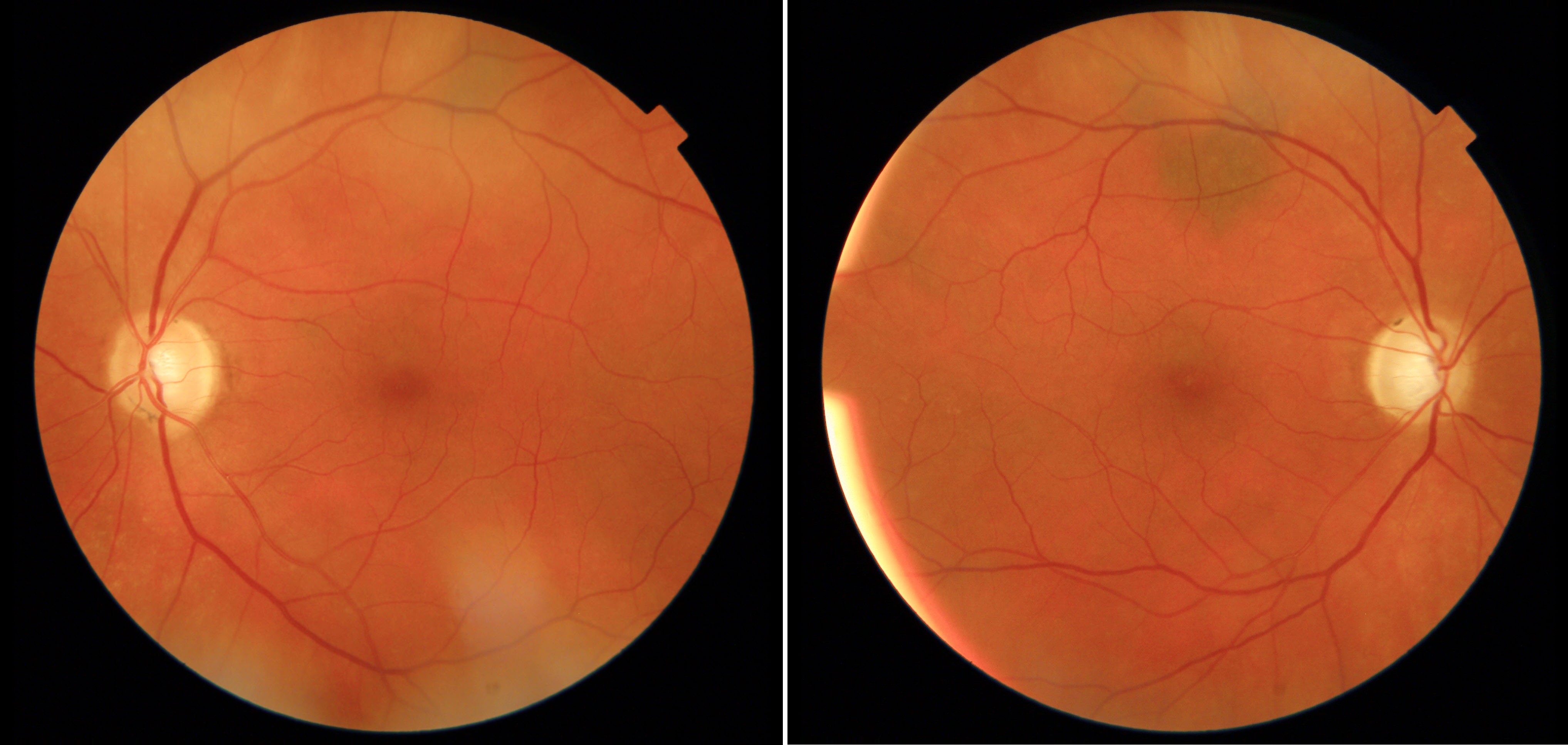
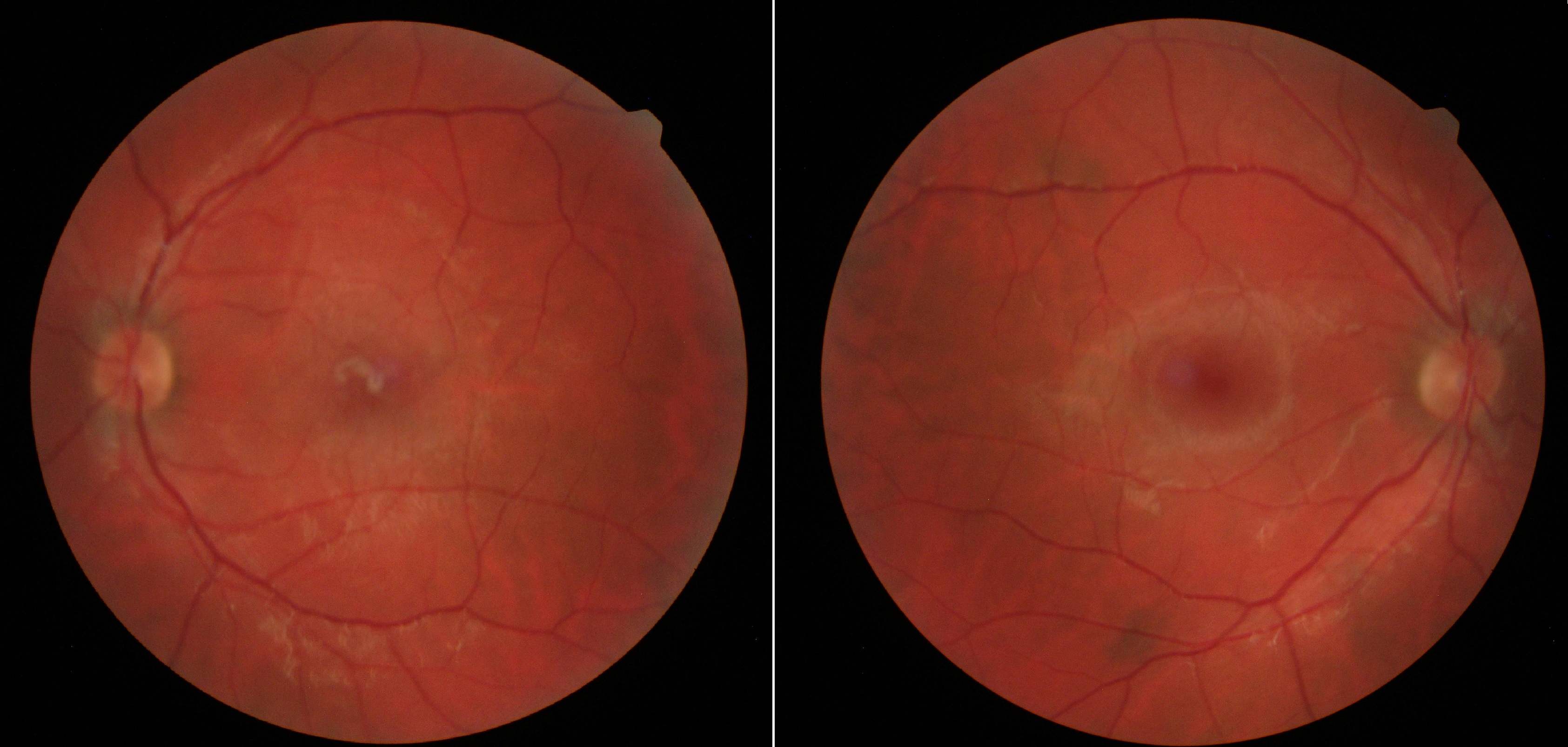
Imagine being able to provide an OCT, a fundus photograph, and relevant clinical information to a properly trained and validated system and receive additional information about the likelihood of different diagnoses or the urgency of a referral.
That doesn't replace clinical judgment.
Instead, it could give clinicians another tool to help identify something unusual, prioritize referrals, or provide another layer of confidence when managing a complex patient.
Telemedicine Can Help Connect That Technology to Patient Care
This is also where I see a lot of potential in telemedicine.
I've been using the Care1 platform for the past couple of months, and I see real potential in how it can change the way we manage patients.
Telemedicine is still in its infancy, but I think it will become increasingly important, particularly for patients who have difficulty accessing specialist care.
Consider an elderly or frail patient who has difficulty attending multiple appointments, or someone living in a rural or remote community. If we can use technology to answer a focused clinical question without requiring that patient to travel unnecessarily, why wouldn't we?
With a platform such as Care1, ophthalmic images, including OCTs, fundus photographs, and other imaging, can be securely uploaded along with a clinical question.
Is this lesion concerning?
Is this patient an appropriate surgical candidate?
Can this finding be monitored?
Does this patient need to see a retina specialist?
Having that information reviewed remotely can help streamline triage and care delivery while making specialist expertise more accessible.
The Future Is About Combining Expertise With Technology
I don't think the future of ophthalmology is about replacing physicians with AI.
I think it is about giving physicians better tools.
The most valuable technologies will be the ones that help us identify disease earlier, interpret complex imaging more efficiently, monitor patients more objectively, and connect patients with the right level of care at the right time.
As a retina specialist, I still want to look at the patient, understand their symptoms, review their history, examine their imaging, and apply clinical judgment.
But if technology can help me do those things more efficiently and with more information at my fingertips, that's a meaningful advancement.
The combination of retinal biomarkers, advanced imaging, AI, and telemedicine has the potential to change not only how we interpret retinal disease, but how we deliver care.
And I think we're only beginning to see what is possible.
About Dr. Marko Popovic, MD, PhD, FRCSC
Dr. Marko Popovic, MD, PhD, FRCSC is a retina specialist with a strong focus on retinal imaging, disease biomarkers, and the evolving role of technology in clinical decision-making. He is particularly interested in helping clinicians interpret complex OCT findings, recognize changes in disease activity, and determine when patients require referral or closer monitoring.
Through his work with advanced imaging, artificial intelligence, and telemedicine, Dr. Popovic advocates for technology that strengthens clinical judgment and helps clinicians deliver more informed, efficient patient care.