Practical retina insights from Dr. Arjmand: retinal imaging, OCT interpretation, AI in eye care, and what’s next for digital retina.
Retina in the Digital Age: Dinner Summary
Featuring Dr. Parnian Arjmand
Purpose of the Evening
Dr. Parnian Arjmand’s discussion focused on the practical challenges of interpreting retinal imaging, understanding the evolving role of artificial intelligence, and making confident decisions about when patients should be monitored, referred, or reviewed through collaborative care.
The evening brought together several recurring themes in modern retinal practice:
Interpreting OCT findings in their clinical context
Recognizing retinal findings that can appear more concerning than they are
Understanding the limitations of AI-assisted screening
Identifying when imaging findings warrant referral
Using tele-ophthalmology to support uncertain cases
Strengthening collaboration between optometry and ophthalmology
A central message throughout the discussion was that technology can support clinical decision-making, but it does not replace clinical judgment.
Explore practical insights for optometrists on retinal imaging, OCT interpretation, AI, and how emerging technologies are shaping the future of retinal care.
Key Clinical Themes
1. Not Every Abnormal-Looking OCT Finding Represents Disease
Dr. Arjmand reviewed several OCT findings that can look concerning but may represent benign or non-urgent conditions.
The key to distinguishing these findings is not simply how abnormal the OCT appears. Visual acuity, symptoms, clinical examination, and evidence of progression all contribute to determining whether a patient requires intervention or monitoring.
For example, outer retinal tubulations can be mistaken for intraretinal fluid, particularly in patients with advanced dry AMD. However, their rounded appearance and photoreceptor structures can help distinguish them from true fluid.
Similarly, vitreomacular adhesion does not necessarily require surgery. Monitoring becomes particularly important when there are changes in vision, distortion, or progression toward vitreomacular traction or a macular hole.
2. Not Every Cyst on OCT Is Edema
Another important clinical consideration was the differential diagnosis of cystic spaces on OCT.
Dr. Arjmand emphasized that not every cystic appearance represents leaking edema, and the patient's history can provide important diagnostic clues.
Potential causes discussed included:
Irvine-Gass syndrome
Diabetic macular edema
Cystoid macular edema associated with vein occlusion
Uveitic conditions
Carbonic anhydrase inhibitor-related changes
Retinal changes associated with retinitis pigmentosa
MEK inhibitor toxicity
Retinal changes associated with medications such as tamoxifen
A patient's systemic history and medication list can therefore be just as important as the OCT appearance when narrowing the differential diagnosis.
3. AI Can Support Screening, But Clinical Judgment Still Comes First
AI was another major focus of the discussion.
Dr. Arjmand reviewed how AI is increasingly being used for retinal screening and monitoring, particularly for diabetic retinopathy and diabetic macular edema. However, she emphasized that current systems remain disease-specific and have important limitations.
Several key considerations were highlighted:
A negative AI result does not necessarily mean the eye is normal
An “ungradable” image does not necessarily mean there is no pathology
AI may identify the disease it was trained to detect while missing other ocular conditions
False positives can occur because of imaging artifacts
Imaging quality can significantly affect AI performance
Clinicians can be influenced by an AI result when reviewing the same image
One particularly important practical point was to form your own clinical impression before reviewing the AI result. This can help reduce the risk of allowing the AI assessment to influence the initial interpretation.
Ultimately, the clinician remains responsible for the patient's care, even when AI is part of the workflow.
4. A Negative Screening Result Does Not Mean a Normal Eye
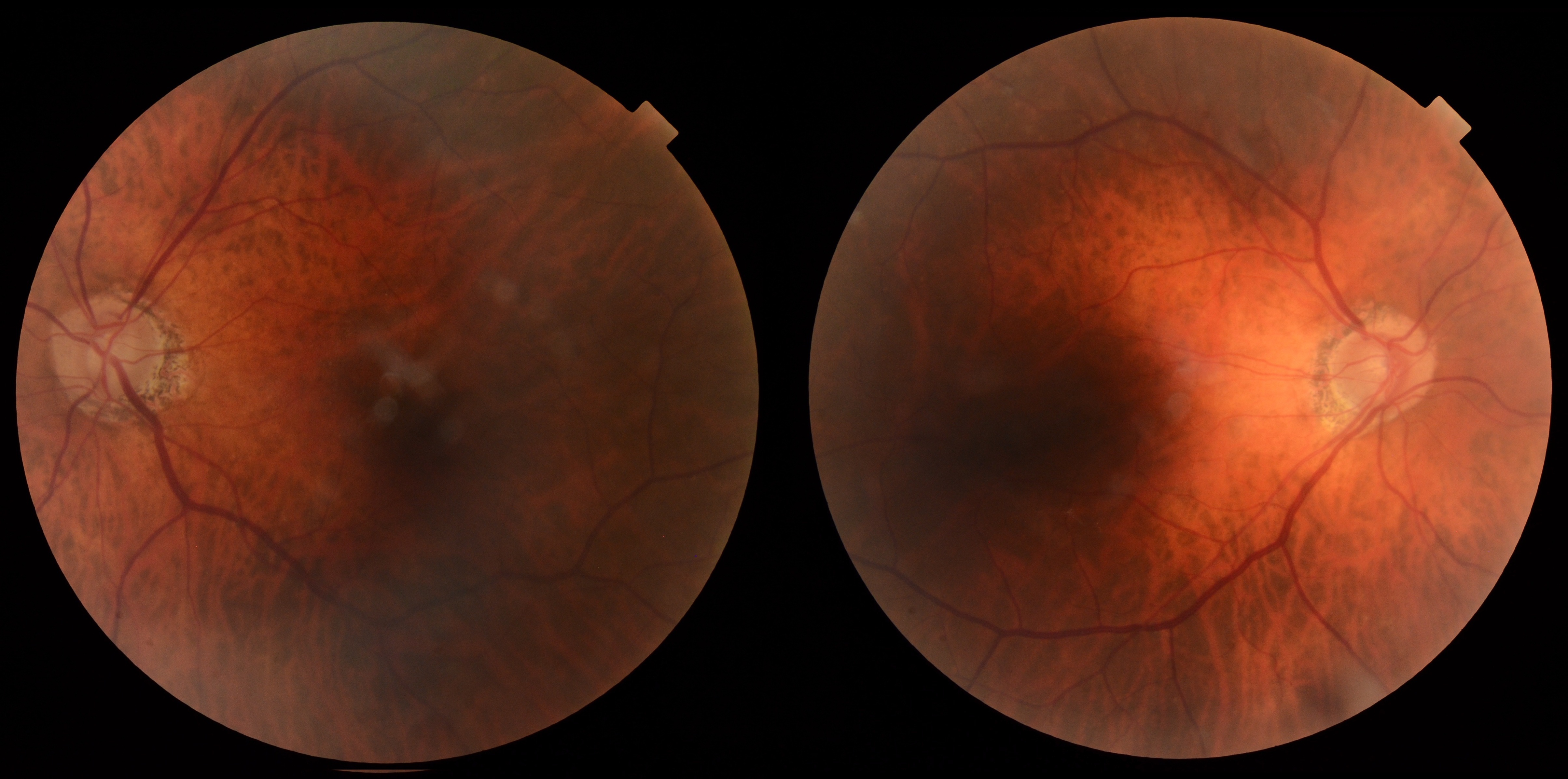
Dr. Arjmand illustrated the limitations of disease-specific AI through a case involving a patient whose AI screening result indicated no referable diabetic retinopathy.
Despite the negative AI result, the clinical examination revealed suspicious lesions that required further investigation.
The case reinforced an important principle: AI screening tools are designed to answer specific questions. They are not necessarily designed to identify every ocular abnormality.
This distinction is particularly important when evaluating patients with other retinal or ocular conditions that fall outside the algorithm's intended use.
5. Tele-Ophthalmology Can Help When the Referral Decision Is Uncertain
The discussion also explored the role of tele-ophthalmology and collaborative care.
Dr. Arjmand described Care1 as a pathway for cases where an optometrist is uncertain whether a patient requires referral, particularly when there is subtle interval change or an imaging finding that is difficult to interpret.
Examples include:
A patient with drusen showing a subtle change
An OCT finding that is difficult to classify
An imaging finding that may be benign but warrants another clinical opinion
Cases where the clinician is unsure whether a patient needs to see a retina specialist
The goal is not to replace appropriate urgent referrals. Emergencies and clearly sight-threatening findings still require timely in-person care.
Instead, collaborative tele-ophthalmology can help address the uncertainty that often sits between routine monitoring and referral.
6. Clinical Context Should Guide Referral Decisions
A recurring theme throughout the discussion was the importance of matching the imaging finding to the patient's clinical presentation.
For example, Dr. Arjmand discussed the difference between a patient with pathological myopia and atrophy who may require monitoring or low-vision support, versus a highly myopic patient with a myopic choroidal neovascular membrane and hemorrhage who requires prompt retinal assessment.
Similarly, small macular holes, vitreomacular traction, and other structural findings can require different levels of urgency depending on visual acuity, symptoms, and progression.
The appearance on the scan is only one part of the clinical picture.
The Role of Imaging in Collaborative Care
Dr. Arjmand emphasized that when submitting a case for collaborative review, the clinical question is just as important as the imaging itself.
A useful submission should include:
The primary clinical concern
Pertinent patient information
Visual acuity
The specific clinical question
Relevant prior imaging for comparison
Appropriate raw imaging
Annotations where an artifact or particular finding is in question
This helps the reviewing ophthalmologist understand not only what is visible on the scan, but also what the optometrist is trying to determine.
Practice at the highest level of medicine
✔ Specialist consults within 1-2 weeks ✔ AI-powered clinical support ✔ Greater confidence in complex decision making
OCT findings should always be interpreted in the context of the patient's clinical presentation
Not every cystic space represents edema, and not every abnormal-looking structure requires treatment
Several benign or non-urgent retinal findings can mimic more concerning pathology
AI can support screening and monitoring, but current systems remain disease-specific
A negative or ungradable AI result does not necessarily mean that an eye is normal
Clinicians should form their own impression before reviewing AI-generated interpretations
Patient history, medications, symptoms, visual acuity, and progression remain essential to clinical decision-making
Tele-ophthalmology can be valuable when the need for referral is uncertain
Clear clinical questions and relevant imaging strengthen collaborative case review
Notable Insights
“Form your own impression first.”
“Not every cyst on OCT is edema and not every edema leaks.”
“Just because something is approved doesn't mean that that approved technology is necessarily going to work in your clinic.”
The future of retinal care will continue to bring together advanced imaging, artificial intelligence, and specialist collaboration. But as Dr. Arjmand’s discussion demonstrated, these tools are most valuable when they strengthen clinical judgment rather than replace it.
Collaborative Care Keep patients connected to their optometrist while bringing ophthalmology expertise into the decision-making process.