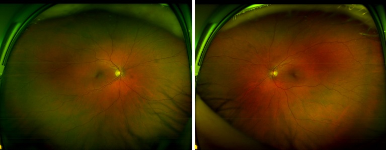
77-year-old patient with preserved 20/20 vision and persistent macular changes on OCT. What findings support continued observation?
Case Study: Macular Changes With 20/20 Vision
An optometrist uploaded this case to Care1.
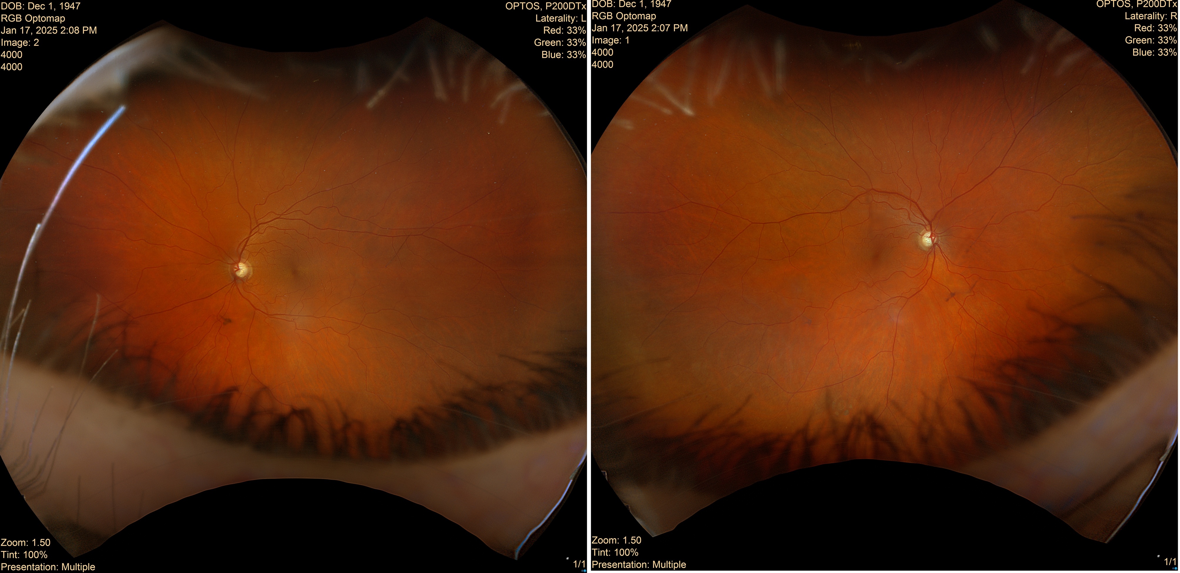
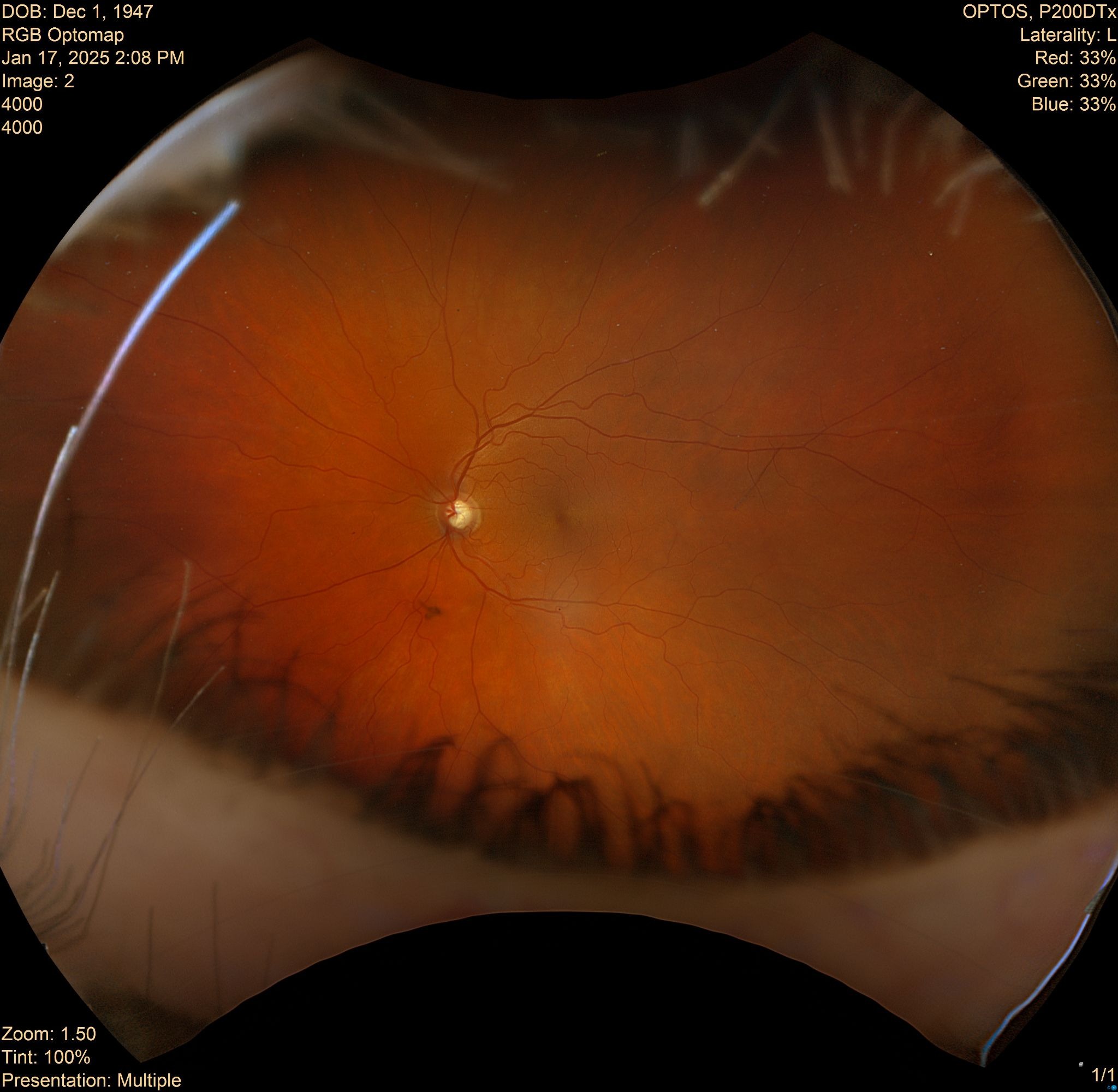
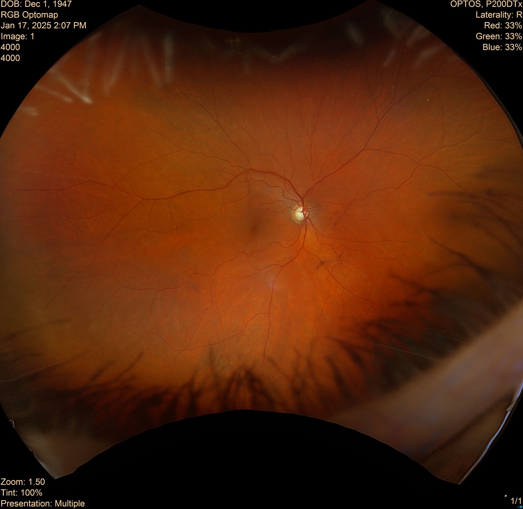
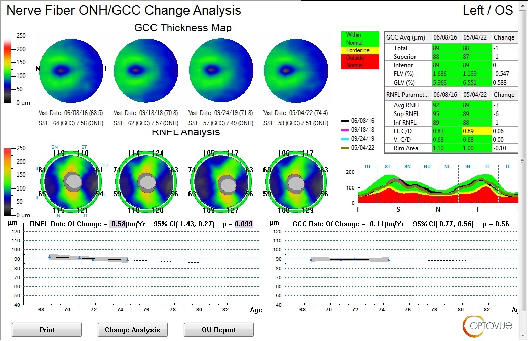
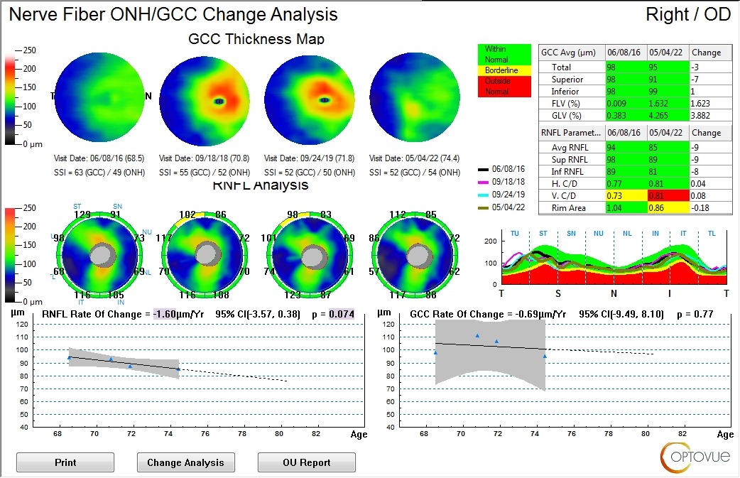
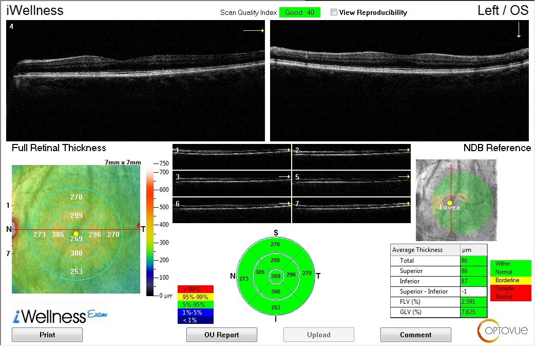
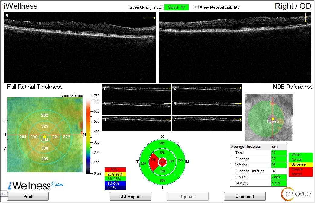
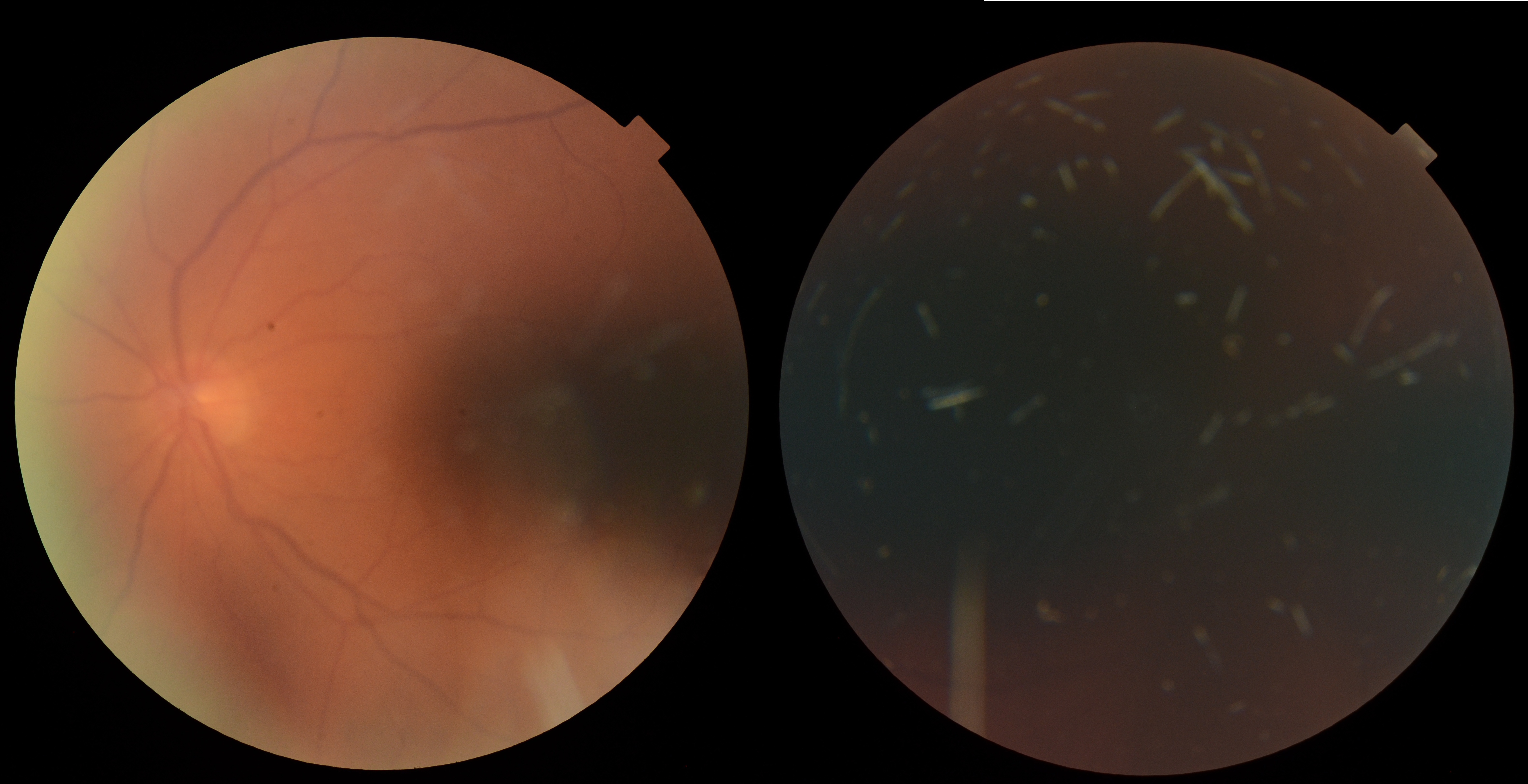
A 77-year-old patient has a history of right macular changes that have been monitored over several years. Best-corrected visual acuity is 20/20 in both eyes, with IOPs of 21 mmHg OD and 17 mmHg OS. Anterior segment examination shows early cataracts. Posterior segment imaging shows right macular structural changes associated with an epiretinal membrane, with recent OCT findings improved compared with prior imaging.
Given the preserved vision and longitudinal OCT findings, what factors should guide continued observation versus further retinal intervention?
A retina specialist provided a virtual consult within 1-2 weeks through Care1. Scroll below to see their diagnosis.
Care1 Ophthalmologist's Teleconsult
The patient has a longstanding right macular abnormality associated with an epiretinal membrane. Visual acuity remains excellent, and prior assessments support observation rather than surgical intervention.
The most recent review notes that the macular appearance is improved compared with previous imaging. Annual monitoring is recommended, with attention to any changes in visual function or macular structure.
Care1 AI's Clinical Insight
Optical coherence tomography (OCT) provides detailed cross-sectional imaging of the macula, allowing clinicians to assess foveal contour, retinal layer integrity, and vitreoretinal interface abnormalities. Careful interpretation of OCT morphology helps distinguish partial-thickness retinal defects from full-thickness defects and other conditions that can produce a similar macular appearance.
Practice at the highest level of medicine
✔ Specialist consults within 1-2 weeks ✔ AI-powered clinical support ✔ Greater confidence in complex decision making
A small OCT-based study identified four different traction-related pathways through which lamellar macular holes may develop. The study also found that visual acuity could remain relatively stable during formation and follow-up, highlighting why structural imaging and visual function should be assessed together.
Reference: Lee CY, Hsia Y, Yang CM. Formation and evolution of idiopathic lamellar macular hole: a pilot study. BMC Ophthalmol. 2022;22(1):432. doi:10.1186/s12886-022-02669-4.
Clinical Pearls
Compare serial OCT scans to identify structural changes.