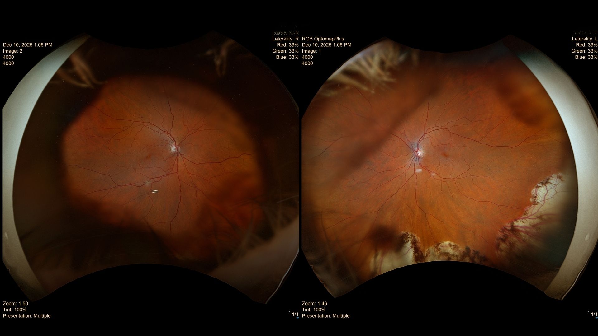
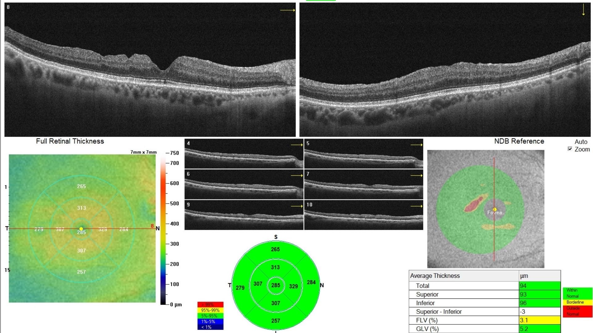
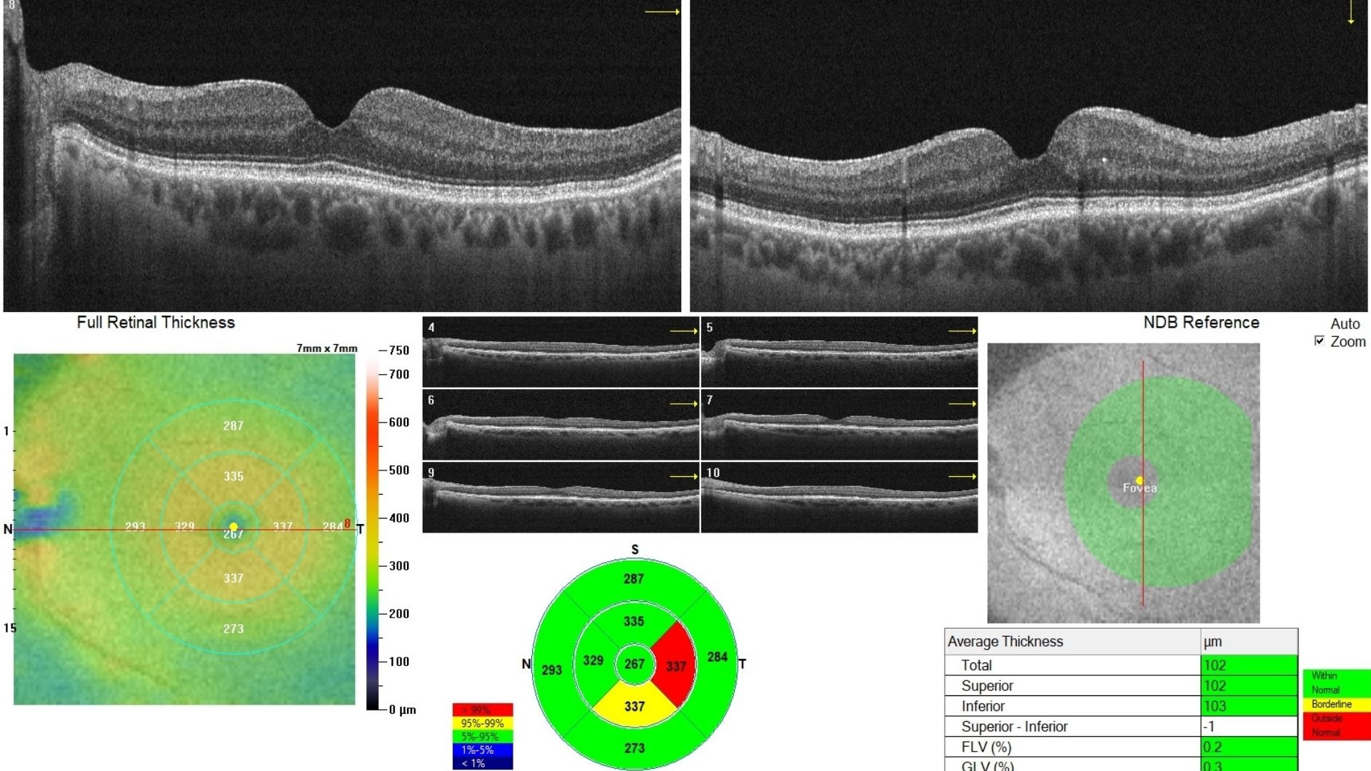
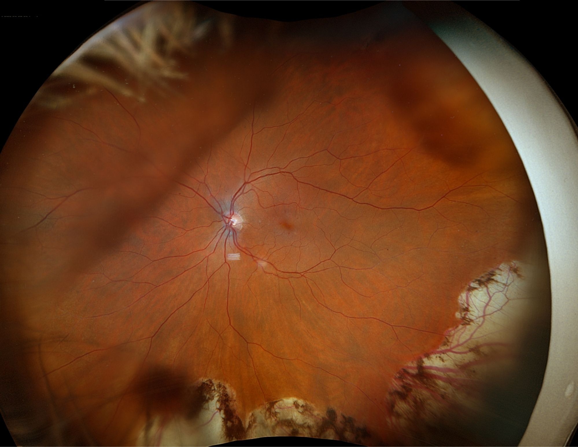
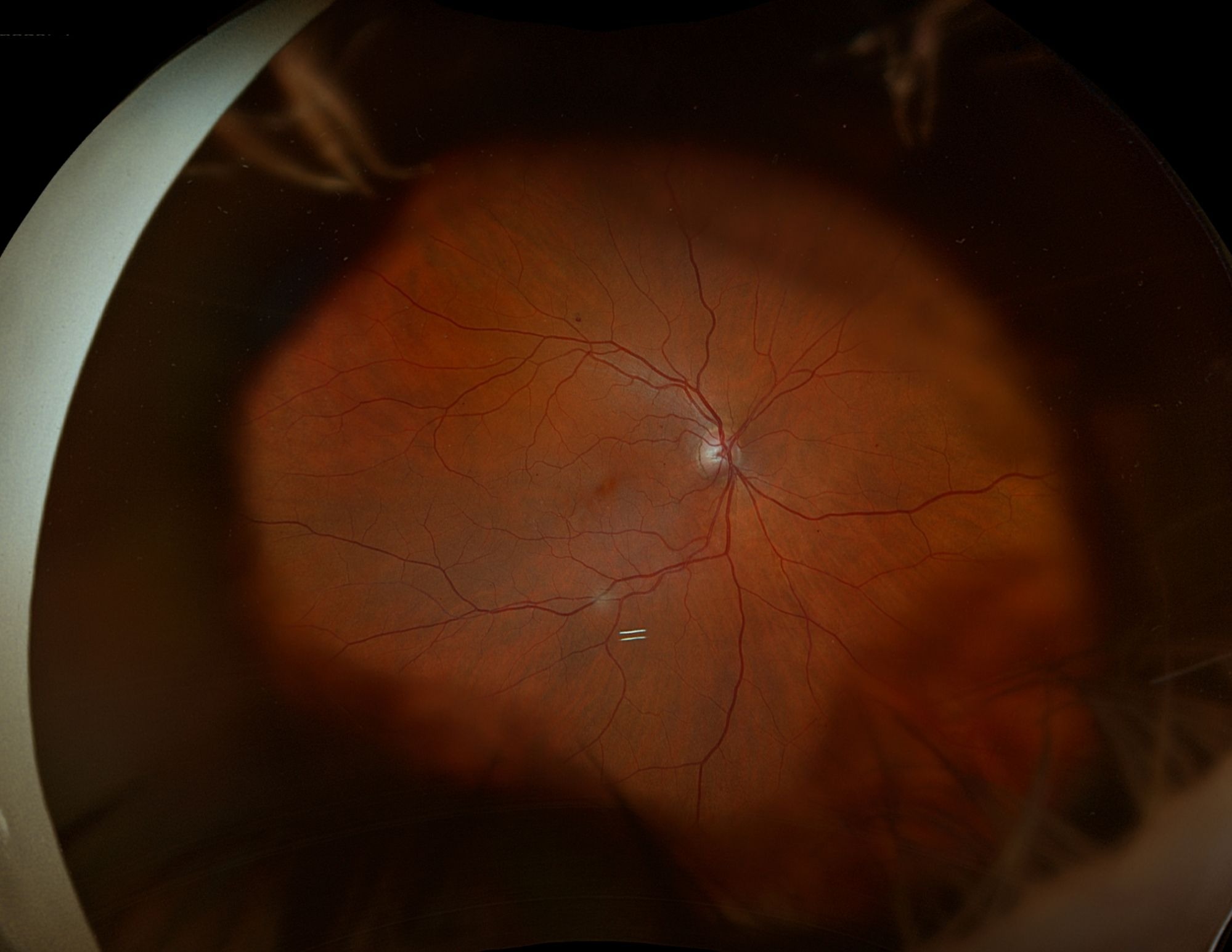
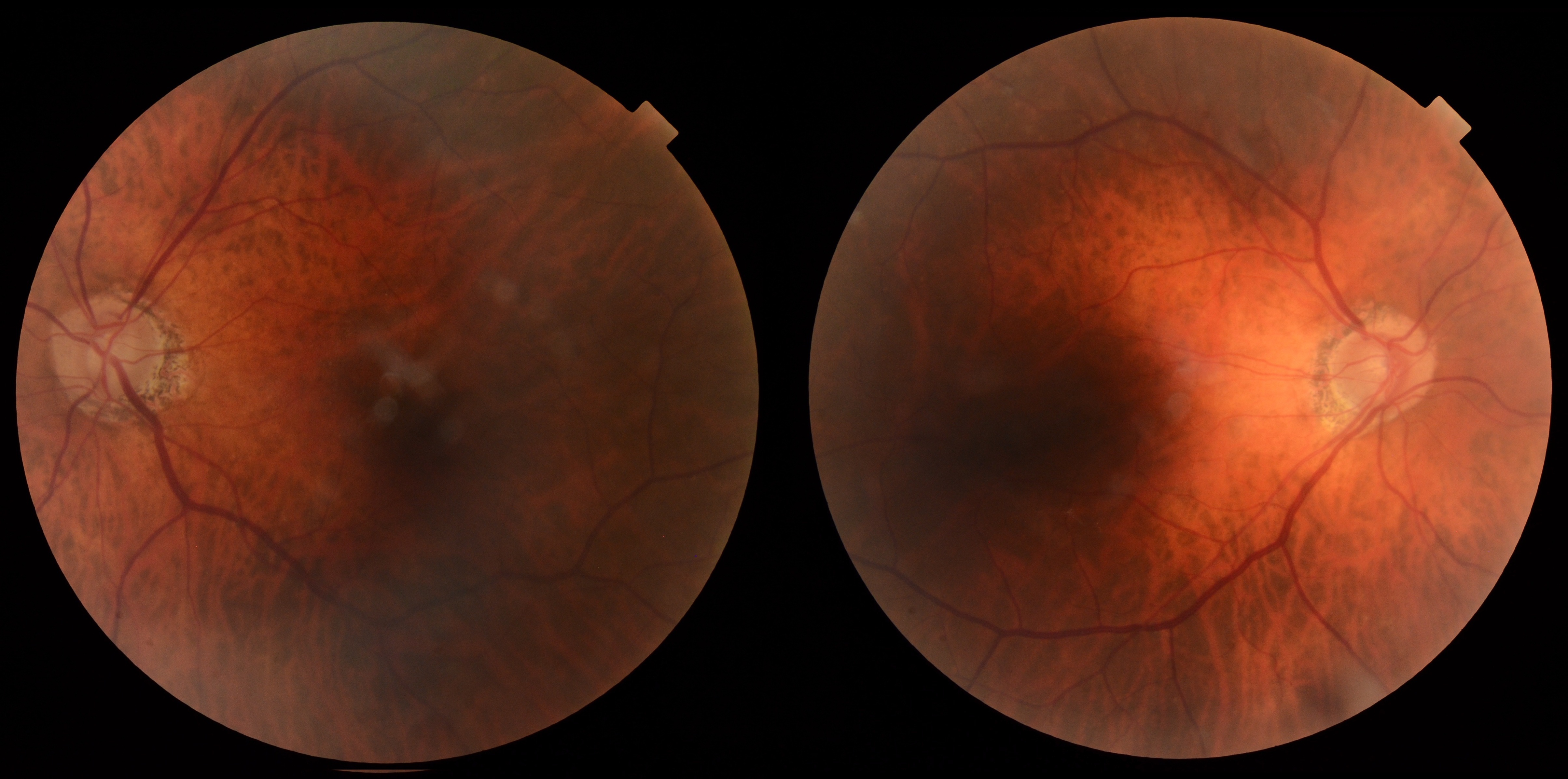
75-year-old woman with stable vision, diabetes, and OCT macular findings after previous retinal surgery requiring ongoing retinal assessment.
Case Study: Blurred Vision After Retinal Surgery
An optometrist uploaded this case to Care1.
An optometrist referred a 75-year-old woman with diabetes and a history of retinal surgery for ongoing retinal monitoring. Best corrected visual acuity was 20/30 OU and IOP measured 20 mmHg OD and 17 mmHg OS. The anterior segment showed pseudophakia OU with blepharitis, meibomian gland dysfunction, and superficial punctate keratitis. Posterior examination demonstrated mild diabetic retinopathy, stable peripheral retinal changes, and persistent macular OCT abnormalities following previous surgery.
What ongoing retinal follow-up or additional management would you recommend for these findings?
A retina specialist provided a virtual consult within 1-2 weeks through Care1. Scroll below to see their diagnosis.
Care1 Ophthalmologist's Teleconsult
The retinal findings remain stable, with no evidence of vision-threatening diabetic macular edema or high-risk diabetic retinopathy. Continued optimization of systemic diabetic control and routine retinal surveillance are appropriate. Previous retinal surgery appears stable on imaging, and no additional intervention is indicated based on the current examination.
Care1 AI's Clinical Insight
Vitreomacular interface disorders occur when traction develops between the vitreous and the macula, potentially altering retinal architecture and visual function. Optical coherence tomography plays a central role in identifying these changes, monitoring progression, and guiding referral decisions through detailed visualization of the macula.
Unlock your full revenue potential
✔ Specialist consults within 1-2 weeks ✔ AI-powered clinical support ✔ Greater confidence in complex decision making
Eyes with persistent vitreomacular traction can occasionally undergo spontaneous release without surgery, although reported rates vary considerably depending on OCT characteristics and symptom severity.
Reference: John V, Flynn HW Jr, Smiddy WE, et al. Clinical course of vitreomacular traction managed initially by observation. Ophthalmology. 2014;121(3):717-720. doi:10.1016/j.ophtha.2013.09.038
Clinical Pearls
Monitor OCT for vitreomacular interface changes.
Stable vision still warrants retinal surveillance.