Advances in diagnostic imaging continue to reshape how eye care professionals evaluate disease and communicate findings with patients. Care1’s latest update introduces a powerful new way to assess the optic nerve—by transforming standard fundus photos into a 3D view using AI.
From 2D Images to 3D Clinical Insight
Care1’s new AI-powered Stereoviewer uses AI to automatically detect the optic nerve head from uploaded fundus images. There are no manual steps, annotations, or additional workflows required. Simply upload your images, and Care1 handles the rest.
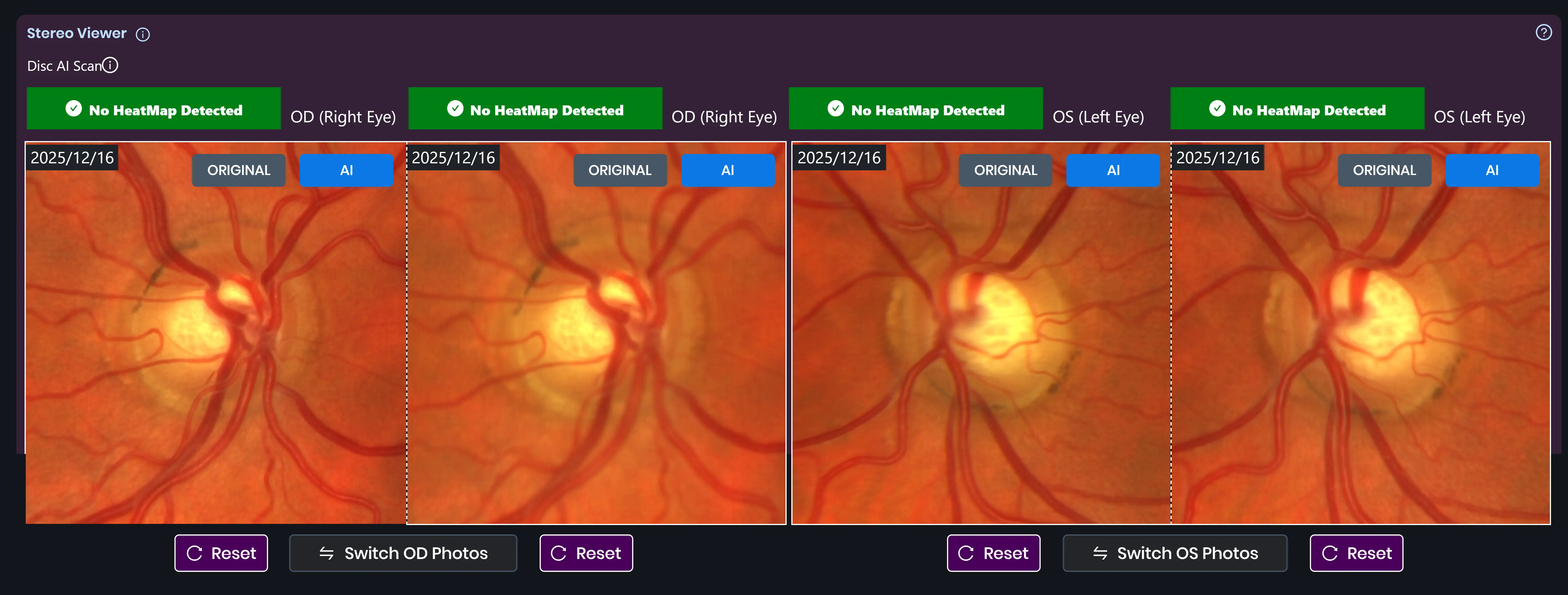
This automated optic nerve detection enables a three-dimensional visualization of the optic disc, offering a clearer and more intuitive way to assess structural changes that may be difficult to appreciate in traditional 2D images alone.
Why 3D Matters in Optic Nerve Evaluation
The optic nerve plays a critical role in the early detection and monitoring of glaucoma. Subtle changes in cupping or structure can be challenging to identify, particularly when comparing images across multiple visits.
With the Care1 Stereoviewer, clinicians can:
- Examine optic nerve cupping in a true 3D perspective
- Identify early glaucomatous changes with greater confidence
- Compare structure more effectively over time
- Use interactive visuals to improve patient understanding and engagement
By adding depth and context to fundus images, the Stereoviewer supports more informed clinical decision-making and clearer communication.