.avif)

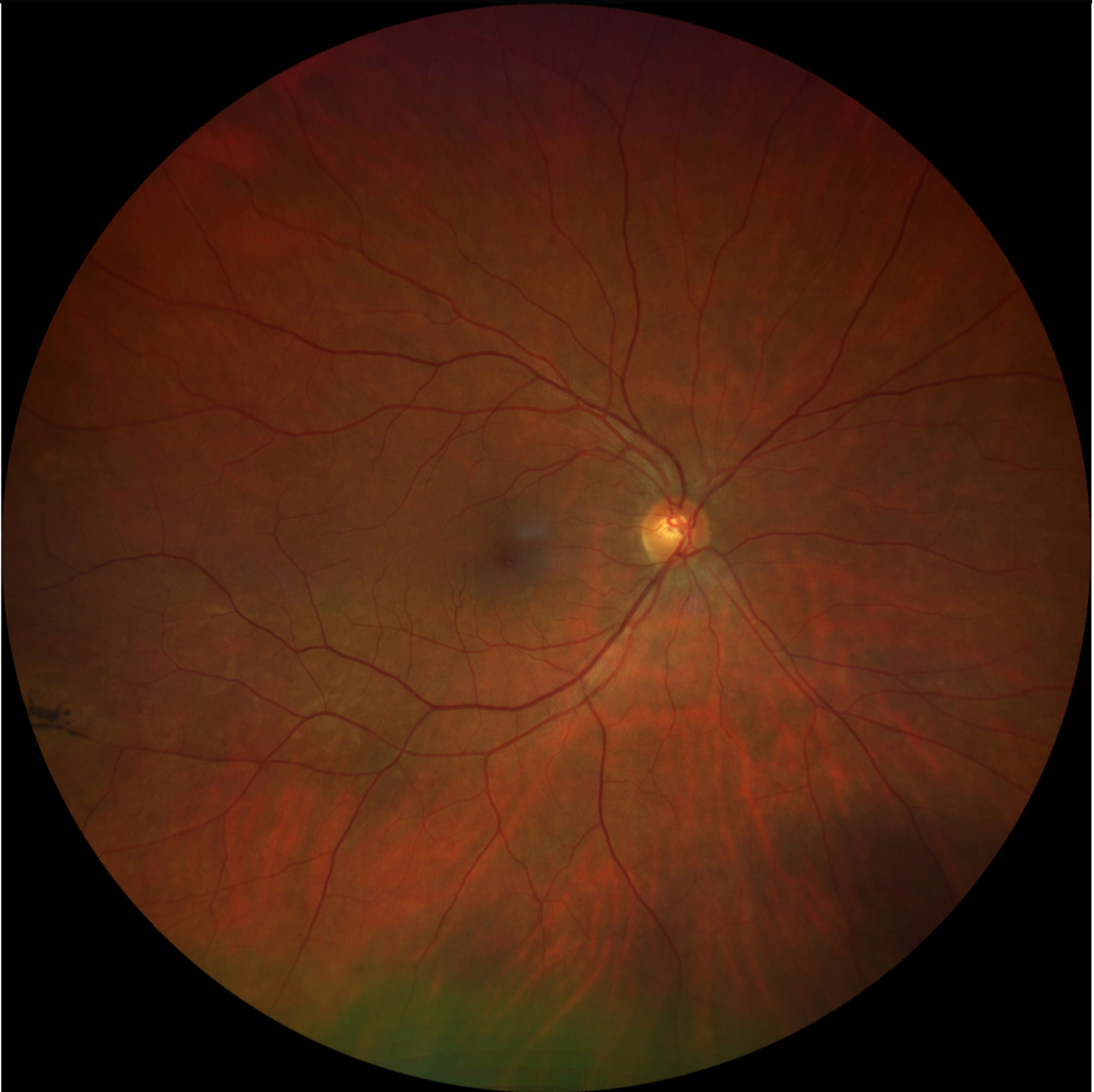
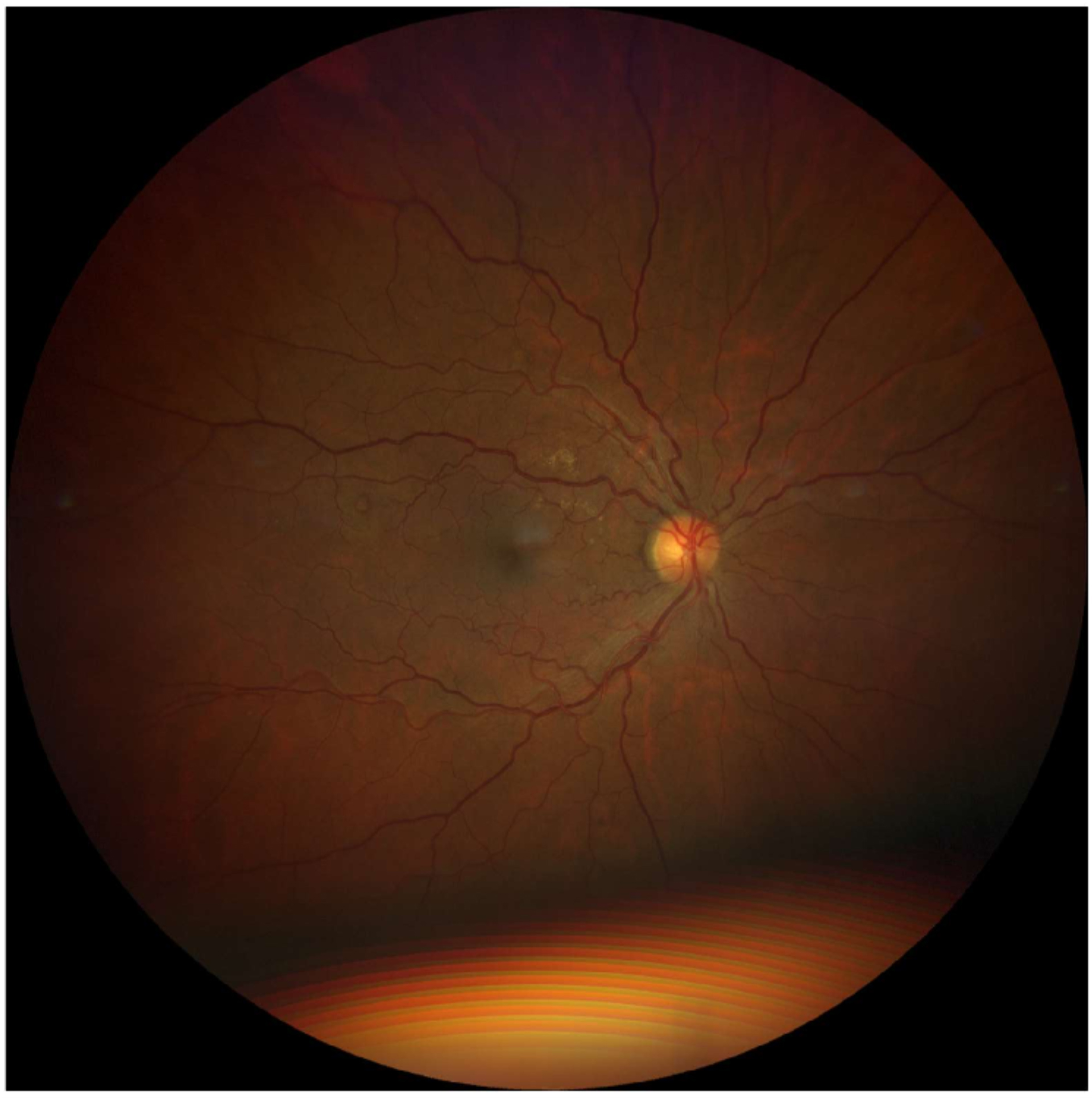
An adult patient presented for a routine eye exam with no visual complaints and no previous records available. Anisocoria was noted in the left eye (OS) greater than the right (OD), with no relative afferent pupillary defect (RAPD). A suspicious area of retinal atrophy or scarring was noted superior to the optic nerve head (ONH) in the left eye. Macular epiretinal membrane (ERM) was present in both eyes. No OCT imaging was performed at the time of the exam.
Note: A blurry area was observed over the superotemporal central retinal artery/vein, attributed to a linear floater artifact.
A retina specialist provided a virtual consult within 1-2 weeks through Care1. Scroll below to see their diagnosis.
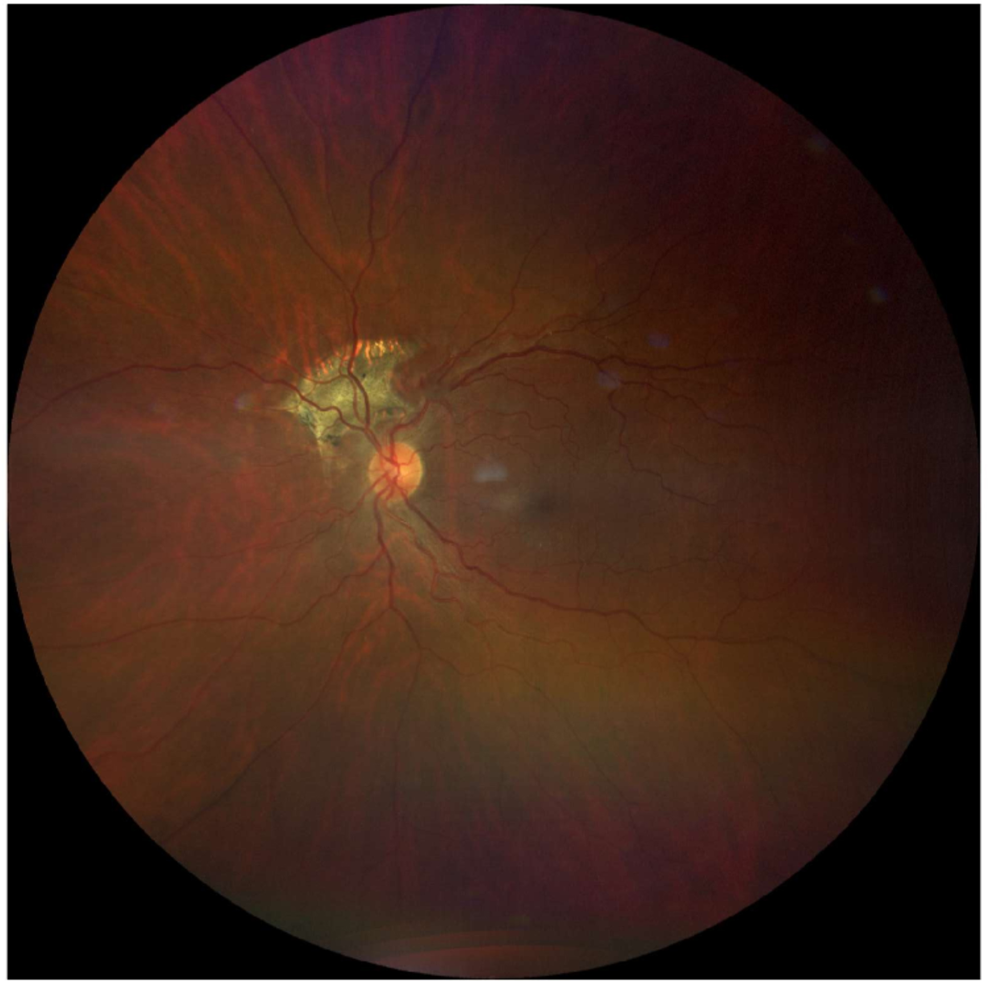
This case represents old peripapillary choroidal neovascularization (CNV) in the left eye. Most CNV cases resolve on their own without treatment, often leaving behind a disciform scar. In this instance, the scar appears longstanding and inactive, with no signs of fluid or hemorrhage. There are currently no concerns. The patient is advised to monitor vision changes using an Amsler grid. Continued observation and follow-up with the optometrist are recommended. Care1 OMDs will remain available for ongoing management through remote file and diagnostic reviews.
Peripapillary Choroidal Neovascularization (PP-CNV) is defined as choroidal neovascularization occurring within one disc diameter of the optic nerve head. It can be idiopathic or secondary to conditions such as age-related macular degeneration, angioid streaks, high myopia, or inflammatory chorioretinal diseases. The neovascular membrane originates from the choroid and may extend into the subretinal space, potentially causing fluid or hemorrhage. PP-CNV lesions are typically less aggressive than subfoveal CNV.
✔ Deliver the highest standards of care
✔ Increase patient satisfaction and retain patients
✔ Stimulate revenue
In a study of myopic CNV, most eyes experienced severe vision loss (worse than 20/200) within 10 years, primarily due to progressive chorioretinal atrophy around previous CNV sites.
Ikuno Y, Tano Y. Retinal and choroidal biometry in highly myopic eyes with spectral-domain optical coherence tomography. Ophthalmology. 2009;116(12):2315–2321. doi:10.1016/j.ophtha.2009.05.035